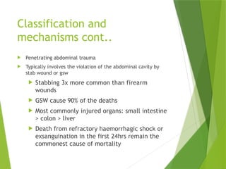
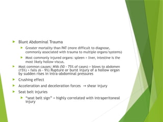
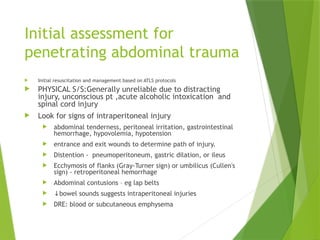
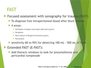
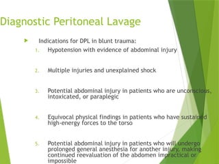
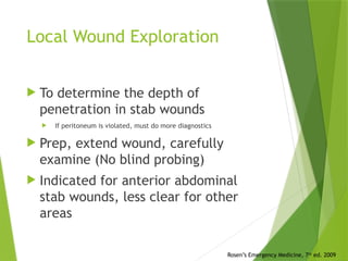
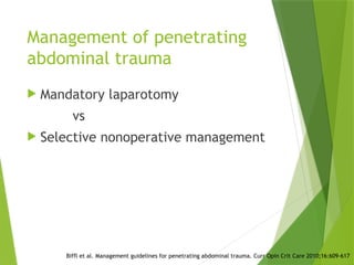
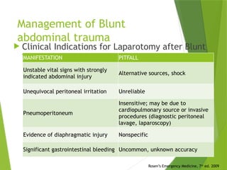
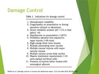
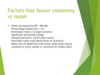
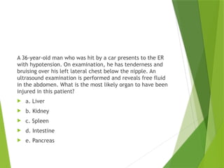
Management of abdominal
trauma
Classification
1. Penetrating abd trauma
2. Blunt abd trauma
Classification and
mechanisms cont..
 Penetrating abdominal trauma
 Typically involves the violation of the abdominal cavity by
stab wound or gsw
 Stabbing 3x more common than firearm
wounds
 GSW cause 90% of the deaths
 Most commonly injured organs: small intestine
> colon > liver
 Death from refractory haemorrhagic shock or
exsanguination in the first 24hrs remain the
commonest cause of mortality
 Blunt Abdominal Trauma
 Greater mortality than PAT (more difficult to diagnose,
commonly associated with trauma to multiple organs/systems)
 Most commonly injured organs: spleen > liver, intestine is the
most likely hollow viscus.
 Most common causes: MVA (50 - 75% of cases) > blows to abdomen
(15%) > falls (6 - 9%) Rupture or burst injury of a hollow organ
by sudden rises in intra-abdominal pressures
 Crushing effect
 Acceleration and deceleration forces shear injury
→
 Seat belt injuries
 “seat belt sign” = highly correlated with intraperitoneal
injury
Pathophysiology of injury
Penetrating Abdominal Trauma
 Stab Wounds
 Knives, screw drivers, pens,
coat hangers, broken bottles
 Liver, small bowel, spleen
 Gunshot wounds
 small bowel, colon and liver
 Often multiple organ
injuries, bowel perforations
 Other low velocity missiles-
arrows,spears
Initial assessment for
penetrating abdominal trauma
 Initial resuscitation and management based on ATLS protocols
 PHYSICAL S/S:Generally unreliable due to distracting
injury, unconscious pt ,acute alcoholic intoxication and
spinal cord injury
 Look for signs of intraperitoneal injury
 abdominal tenderness, peritoneal irritation, gastrointestinal
hemorrhage, hypovolemia, hypotension
 entrance and exit wounds to determine path of injury.
 Distention - pneumoperitoneum, gastric dilation, or ileus
 Ecchymosis of flanks (Gray-Turner sign) or umbilicus (Cullen's
sign) - retroperitoneal hemorrhage
 Abdominal contusions – eg lap belts
 ↓bowel sounds suggests intraperitoneal injuries
 DRE: blood or subcutaneous emphysema
Diagnostic studies
 Lab tests: not very helpful
 Do FHG, U/E/Cs, LFTs, lipase, tox screen
 Hct-serial to monitor ongoing haemorrhage,
interpret with hypotension and ongoing iv fluids in
mind
 Wbc-non specific-may be due to perforated viscus
or acute stress response
 Elevated lipase-suspect pancreatic injury,
collaborate with CT
 ABGs - Lactate and base deficit – degree of
haemorrhagic shock
Rosen’s Emergency Medicine, 7th
ed. 2009
Imaging
 Plain films:
 fractures – nearby visceral damage
 free intraperitoneal air
 Foreign bodies and missiles
Rosen’s Emergency Medicine, 7th
ed. 2009
Imaging
 CT
 Accurate for solid visceral lesions and intraperitoneal
hemorrhage
 guide nonoperative management of solid organ damage
 IV not oral contrast
 Disadvantages : insensitive for injury of the pancreas,
diaphragm, small bowel, and mesentery
Rosen’s Emergency Medicine, 7th
ed. 2009
Imaging
 Angiography
 To embolize bleeding vessels or solid
visceral hemorrhage from blunt trauma
in an unstable pt
 Rarely for diagnosing intraperitoneal
and retroperitoneal hemorrhage after
penetrating abdominal trauma
Rosen’s Emergency Medicine, 7th
ed. 2009
FAST
 Focused assessment with sonography for trauma (FAST)
 To diagnose free intraperitoneal blood after blunt trauma
 4 areas:
 Perihepatic & hepato-renal space (Morrison’s pouch)
 Perisplenic
 Pelvis (Pouch of Douglas/rectovesical pouch)
 Pericardium
 sensitivity 60 to 95% for detecting 100 mL - 500 mL of fluid
 Extended FAST (E-FAST):
 Add thoracic windows to look for pneumothorax and
pericardial tamponade
Trauma.org
Rosen’s Emergency Medicine, 7th
ed. 2009
Diagnostic Peritoneal Lavage
 Largely replaced by FAST and CT
 In blunt trauma, used to triage pt who is
HD unstable and has multiple injuries
with an equivocal FAST examination
 In stab wounds, for immediate dx of
hemoperitoneum, determination of
intraperitoneal organ injury, and
detection of isolated diaphragm injury
 In GSW, not used much
Rosen’s Emergency Medicine, 7th
ed. 2009
Diagnostic Peritoneal Lavage
 Indications for DPL in blunt trauma:
1. Hypotension with evidence of abdominal injury
2. Multiple injuries and unexplained shock
3. Potential abdominal injury in patients who are unconscious,
intoxicated, or paraplegic
4. Equivocal physical findings in patients who have sustained
high-energy forces to the torso
5. Potential abdominal injury in patients who will undergo
prolonged general anesthesia for another injury, making
continued reevaluation of the abdomen impractical or
impossible
Contraindications of DPL
 Absolute :
 Peritonitis
 Injured diaphragm
 Extraluminal air by x-ray
 Significant intraabdominal injury by CT scan
 Intraperitoneal perforation of the bladder by cystography
 Relative :
 Previous abdominal operations (because of adhesions)
 Morbid obesity
 Gravid Uterus
 Advanced cirrhosis (because of portal hypertension and the risk of
bleeding)
 Preexisting coagulopathy
Evaluation of DPL
 Fluid is sent for: cell count, amylase, alk phos, presence of bile
Index Positive value
Aspirate Blood >10 mL
Fluid Enteric content
Lavage RBC > 100,000/mL
WBC > 500/mL
Amylase >175 U/dL
Alk Phos > 3 IU
Bile Confirmed
Negative RBC < 50,000/mL
WBC < 100/mL
Amylase < 75 U/dL
Diagnostic Peritoneal Lavage
RBC Count Incidence of visceral damage
>100,000 95%
20,000-100,000 15-25% Warrant further investigation
<20,000 < 5%
 Complications of DPL: Perforation of small bowel,
mesentery, bladder and retroperitoneal vascular
structures.
 Limitation: offers no information about status of
retroperitoneal organs nor allow determination of
which organ has been injured.
Local Wound Exploration
 To determine the depth of
penetration in stab wounds
 If peritoneum is violated, must do more diagnostics
 Prep, extend wound, carefully
examine (No blind probing)
 Indicated for anterior abdominal
stab wounds, less clear for other
areas
Rosen’s Emergency Medicine, 7th
ed. 2009
Laparoscopy
 Most useful to eval penetrating wounds to
thoracoabdominal region in stable pt
 esp for diaphragm injury: Sens 87.5%, specificity 100%
 Can repair organs via the laparoscope
 diaphragm, solid viscera, stomach, small bowel.
 Disadvantages:
 poor sensitivity for hollow visceral injury,
retroperitoneum
 Complications from trocar misplacement.
 If diaphragm injury, PTX during insufflation
Rosen’s Emergency Medicine, 7th
ed. 2009
Management
 General trauma principles:
 airway management, 2 large bore IVs, cover
penetrating wounds and eviscerations with sterile
dressings
 Prophylactic antibiotics: decrease risk of intra-
abdominal sepsis due to intestinal perf/spillage
 (eg zosyn (piperacillin / tazobactam) 3.375 g IV)
 In general, leave foreign bodies in and remove
in the OR
Rosen’s Emergency Medicine, 7th
ed. 2009
Management of penetrating
abdominal trauma
 Mandatory laparotomy
vs
 Selective nonoperative management
Biffl et al. Management guidelines for penetrating abdominal trauma. Curr Opin Crit Care 2010;16:609-617
Management of Blunt
abdominal trauma
 Clinical Indications for Laparotomy after Blunt
Trauma
MANIFESTATION PITFALL
Unstable vital signs with strongly
indicated abdominal injury
Alternative sources, shock
Unequivocal peritoneal irritation Unreliable
Pneumoperitoneum
Insensitive; may be due to
cardiopulmonary source or invasive
procedures (diagnostic peritoneal
lavage, laparoscopy)
Evidence of diaphragmatic injury Nonspecific
Significant gastrointestinal bleeding Uncommon, unknown accuracy
Rosen’s Emergency Medicine, 7th
ed. 2009
Damage Control
 Patients with major exsanguinating
injuries may not survive complex
procedures
 Control hemorrhage and
contamination with abbreviated
laparotomy followed by
resuscitation prior to definitive
repair
Waibel et al. Damage control in trauma and abdominal sepsis. Crit Care Med 2010 38:S421-430
Damage Control
 0. initial resuscitation
 1. Control of hemorrhage and contamination
 Control injured vasculature, bleeding solid organs
 Abdominal packing
 2. back to the ICU for resuscitation
 Correction of hypothermia, acidosis,
coagulopathy
 3. Definitive repair of injuries
 4. Definitive closure of the abdomen
Waibel et al. Damage control in trauma and abdominal sepsis. Crit Care Med 2010 38:S421-430
Damage Control
Waibel et al. Damage control in trauma and abdominal sepsis. Crit Care Med 2010 38:S421-430
Damage Control
Resuscitation in the ICU
 IVF (crystalloid, not colloid)
 Transfusion
 ?1:1:1 PRBC/plt/FFP
 Recombinant activated factor VII
 Increased thromboembolic complications
 Rewarming if hypothermic
 Correction of metabolic abnormalities-electrolytes and
acidosis
 Low tidal volume ventilation recommended (4-6 ml/kg)
Waibel et al. Damage control in trauma and abdominal sepsis. Crit Care Med 2010 38:S421-430
Damage Control
Open abdominal wounds and definitive closure
 40-70% can’t have primary closure after definitive
repair.
 Temporary closure methods
Waibel et al. Damage control in trauma and abdominal sepsis. Crit Care Med 2010 38:S421-430
Conclusions
 Watch out for implements and missiles violating the
abdomen
 Laparotomy is mandatory if shock, evisceration, or
peritonitis
 Diagnostic studies used to determine need for laparotomy in
PAT and BAT
 FAST is noninvasive, quick and accurate way to evaluate for
intraperitoneal blood
 Damage Control is a principle of staged operative
management with control and resuscitation prior to
definitive repair
 Abdominal compartment syndrome is a common problem in
abdominal trauma
Factors that favour colostomy
vs repair
 Shock (preoperative BP < 80/60)
Hemorrhage (blood loss > 1L)
Multiorgan injury (>2 organ systems)
Significant peritoneal soilage
Delayed operation (>8 hrs post injury)
Nonviable colon (wall destruction or ischemia)
Major loss of abdominal wall (close range blast injury)
Location of injury (distal vs. proximal to middle colic)
A 36-year-old man who was hit by a car presents to the ER
with hypotension. On examination, he has tenderness and
bruising over his left lateral chest below the nipple. An
ultrasound examination is performed and reveals free fluid
in the abdomen. What is the most likely organ to have been
injured in this patient?
 a. Liver
 b. Kidney
 c. Spleen
 d. Intestine
 e. Pancreas
A 22-year-old woman who is 4 months pregnant presents
after a motor vehicle collision complaining of abdominal
pain and right leg pain. She has an obvious deformity of her
right femur. She is hemodynamically stable. Which of the
following is the best next step in her management?
 a. Observation with serial abdominal exams
 b. Diagnostic peritoneal lavage
 c. Plain film of the abdomen with a lead apron as a shield
 d. Focused assessment with sonography for trauma (FAST)
examination of the abdomen
 e. MRI of the abdomen
The patient shown in this chest x-ray film and contrast study was
hospitalized after a car collision 2 days ago in which he suffered
blunt trauma to the abdomen. He sustained several left rib
fractures, but was hemodynamically stable. Which of the following
is the appropriate next step in the patient’s management?
 a. Observation and serial abdominal exams
 b. Immediate left posterolateral thoracotomy and repair of the
injury
 c. Immediate exploratory laparotomy and repair of the injury
 d. Delayed left posterolateral thoracotomy and repair of the
injury
 e. Delayed exploratory laparotomy and repair of the injury
A 29-year-old woman was hit by a car while crossing the
street. She is hemodynamically unstable with a heart rate of
124 beats per minute and a systolic blood pressure of 82/45
mm Hg. The ultrasound machine is broken, and therefore a
diagnostic peritoneal lavage (DPL) is performed. Which of the
following findings on DPL is an indication for exploratory
laparotomy in this patient?
 a. Aspiration of 5 cc of gross blood initially
 b. Greater than 50,000/μL red blood cells (RBCs)
 c. Greater than 100,000/μL RBCs
 d. Greater than 100/μL white blood cells (WBCs)
 e. Greater than 250/μL WBCs

3.abdominal trauma (1).pptx notes

  • 1.
  • 2.
    Classification 1. Penetrating abdtrauma 2. Blunt abd trauma
  • 3.
    Classification and mechanisms cont.. Penetrating abdominal trauma  Typically involves the violation of the abdominal cavity by stab wound or gsw  Stabbing 3x more common than firearm wounds  GSW cause 90% of the deaths  Most commonly injured organs: small intestine > colon > liver  Death from refractory haemorrhagic shock or exsanguination in the first 24hrs remain the commonest cause of mortality
  • 4.
     Blunt AbdominalTrauma  Greater mortality than PAT (more difficult to diagnose, commonly associated with trauma to multiple organs/systems)  Most commonly injured organs: spleen > liver, intestine is the most likely hollow viscus.  Most common causes: MVA (50 - 75% of cases) > blows to abdomen (15%) > falls (6 - 9%) Rupture or burst injury of a hollow organ by sudden rises in intra-abdominal pressures  Crushing effect  Acceleration and deceleration forces shear injury →  Seat belt injuries  “seat belt sign” = highly correlated with intraperitoneal injury
  • 5.
    Pathophysiology of injury PenetratingAbdominal Trauma  Stab Wounds  Knives, screw drivers, pens, coat hangers, broken bottles  Liver, small bowel, spleen  Gunshot wounds  small bowel, colon and liver  Often multiple organ injuries, bowel perforations  Other low velocity missiles- arrows,spears
  • 6.
    Initial assessment for penetratingabdominal trauma  Initial resuscitation and management based on ATLS protocols  PHYSICAL S/S:Generally unreliable due to distracting injury, unconscious pt ,acute alcoholic intoxication and spinal cord injury  Look for signs of intraperitoneal injury  abdominal tenderness, peritoneal irritation, gastrointestinal hemorrhage, hypovolemia, hypotension  entrance and exit wounds to determine path of injury.  Distention - pneumoperitoneum, gastric dilation, or ileus  Ecchymosis of flanks (Gray-Turner sign) or umbilicus (Cullen's sign) - retroperitoneal hemorrhage  Abdominal contusions – eg lap belts  ↓bowel sounds suggests intraperitoneal injuries  DRE: blood or subcutaneous emphysema
  • 7.
    Diagnostic studies  Labtests: not very helpful  Do FHG, U/E/Cs, LFTs, lipase, tox screen  Hct-serial to monitor ongoing haemorrhage, interpret with hypotension and ongoing iv fluids in mind  Wbc-non specific-may be due to perforated viscus or acute stress response  Elevated lipase-suspect pancreatic injury, collaborate with CT  ABGs - Lactate and base deficit – degree of haemorrhagic shock Rosen’s Emergency Medicine, 7th ed. 2009
  • 8.
    Imaging  Plain films: fractures – nearby visceral damage  free intraperitoneal air  Foreign bodies and missiles Rosen’s Emergency Medicine, 7th ed. 2009
  • 9.
    Imaging  CT  Accuratefor solid visceral lesions and intraperitoneal hemorrhage  guide nonoperative management of solid organ damage  IV not oral contrast  Disadvantages : insensitive for injury of the pancreas, diaphragm, small bowel, and mesentery Rosen’s Emergency Medicine, 7th ed. 2009
  • 10.
    Imaging  Angiography  Toembolize bleeding vessels or solid visceral hemorrhage from blunt trauma in an unstable pt  Rarely for diagnosing intraperitoneal and retroperitoneal hemorrhage after penetrating abdominal trauma Rosen’s Emergency Medicine, 7th ed. 2009
  • 11.
    FAST  Focused assessmentwith sonography for trauma (FAST)  To diagnose free intraperitoneal blood after blunt trauma  4 areas:  Perihepatic & hepato-renal space (Morrison’s pouch)  Perisplenic  Pelvis (Pouch of Douglas/rectovesical pouch)  Pericardium  sensitivity 60 to 95% for detecting 100 mL - 500 mL of fluid  Extended FAST (E-FAST):  Add thoracic windows to look for pneumothorax and pericardial tamponade Trauma.org Rosen’s Emergency Medicine, 7th ed. 2009
  • 12.
    Diagnostic Peritoneal Lavage Largely replaced by FAST and CT  In blunt trauma, used to triage pt who is HD unstable and has multiple injuries with an equivocal FAST examination  In stab wounds, for immediate dx of hemoperitoneum, determination of intraperitoneal organ injury, and detection of isolated diaphragm injury  In GSW, not used much Rosen’s Emergency Medicine, 7th ed. 2009
  • 13.
    Diagnostic Peritoneal Lavage Indications for DPL in blunt trauma: 1. Hypotension with evidence of abdominal injury 2. Multiple injuries and unexplained shock 3. Potential abdominal injury in patients who are unconscious, intoxicated, or paraplegic 4. Equivocal physical findings in patients who have sustained high-energy forces to the torso 5. Potential abdominal injury in patients who will undergo prolonged general anesthesia for another injury, making continued reevaluation of the abdomen impractical or impossible
  • 14.
    Contraindications of DPL Absolute :  Peritonitis  Injured diaphragm  Extraluminal air by x-ray  Significant intraabdominal injury by CT scan  Intraperitoneal perforation of the bladder by cystography  Relative :  Previous abdominal operations (because of adhesions)  Morbid obesity  Gravid Uterus  Advanced cirrhosis (because of portal hypertension and the risk of bleeding)  Preexisting coagulopathy
  • 15.
    Evaluation of DPL Fluid is sent for: cell count, amylase, alk phos, presence of bile Index Positive value Aspirate Blood >10 mL Fluid Enteric content Lavage RBC > 100,000/mL WBC > 500/mL Amylase >175 U/dL Alk Phos > 3 IU Bile Confirmed Negative RBC < 50,000/mL WBC < 100/mL Amylase < 75 U/dL
  • 16.
    Diagnostic Peritoneal Lavage RBCCount Incidence of visceral damage >100,000 95% 20,000-100,000 15-25% Warrant further investigation <20,000 < 5%  Complications of DPL: Perforation of small bowel, mesentery, bladder and retroperitoneal vascular structures.  Limitation: offers no information about status of retroperitoneal organs nor allow determination of which organ has been injured.
  • 17.
    Local Wound Exploration To determine the depth of penetration in stab wounds  If peritoneum is violated, must do more diagnostics  Prep, extend wound, carefully examine (No blind probing)  Indicated for anterior abdominal stab wounds, less clear for other areas Rosen’s Emergency Medicine, 7th ed. 2009
  • 18.
    Laparoscopy  Most usefulto eval penetrating wounds to thoracoabdominal region in stable pt  esp for diaphragm injury: Sens 87.5%, specificity 100%  Can repair organs via the laparoscope  diaphragm, solid viscera, stomach, small bowel.  Disadvantages:  poor sensitivity for hollow visceral injury, retroperitoneum  Complications from trocar misplacement.  If diaphragm injury, PTX during insufflation Rosen’s Emergency Medicine, 7th ed. 2009
  • 19.
    Management  General traumaprinciples:  airway management, 2 large bore IVs, cover penetrating wounds and eviscerations with sterile dressings  Prophylactic antibiotics: decrease risk of intra- abdominal sepsis due to intestinal perf/spillage  (eg zosyn (piperacillin / tazobactam) 3.375 g IV)  In general, leave foreign bodies in and remove in the OR Rosen’s Emergency Medicine, 7th ed. 2009
  • 20.
    Management of penetrating abdominaltrauma  Mandatory laparotomy vs  Selective nonoperative management Biffl et al. Management guidelines for penetrating abdominal trauma. Curr Opin Crit Care 2010;16:609-617
  • 21.
    Management of Blunt abdominaltrauma  Clinical Indications for Laparotomy after Blunt Trauma MANIFESTATION PITFALL Unstable vital signs with strongly indicated abdominal injury Alternative sources, shock Unequivocal peritoneal irritation Unreliable Pneumoperitoneum Insensitive; may be due to cardiopulmonary source or invasive procedures (diagnostic peritoneal lavage, laparoscopy) Evidence of diaphragmatic injury Nonspecific Significant gastrointestinal bleeding Uncommon, unknown accuracy Rosen’s Emergency Medicine, 7th ed. 2009
  • 22.
    Damage Control  Patientswith major exsanguinating injuries may not survive complex procedures  Control hemorrhage and contamination with abbreviated laparotomy followed by resuscitation prior to definitive repair Waibel et al. Damage control in trauma and abdominal sepsis. Crit Care Med 2010 38:S421-430
  • 23.
    Damage Control  0.initial resuscitation  1. Control of hemorrhage and contamination  Control injured vasculature, bleeding solid organs  Abdominal packing  2. back to the ICU for resuscitation  Correction of hypothermia, acidosis, coagulopathy  3. Definitive repair of injuries  4. Definitive closure of the abdomen Waibel et al. Damage control in trauma and abdominal sepsis. Crit Care Med 2010 38:S421-430
  • 24.
    Damage Control Waibel etal. Damage control in trauma and abdominal sepsis. Crit Care Med 2010 38:S421-430
  • 25.
    Damage Control Resuscitation inthe ICU  IVF (crystalloid, not colloid)  Transfusion  ?1:1:1 PRBC/plt/FFP  Recombinant activated factor VII  Increased thromboembolic complications  Rewarming if hypothermic  Correction of metabolic abnormalities-electrolytes and acidosis  Low tidal volume ventilation recommended (4-6 ml/kg) Waibel et al. Damage control in trauma and abdominal sepsis. Crit Care Med 2010 38:S421-430
  • 26.
    Damage Control Open abdominalwounds and definitive closure  40-70% can’t have primary closure after definitive repair.  Temporary closure methods Waibel et al. Damage control in trauma and abdominal sepsis. Crit Care Med 2010 38:S421-430
  • 27.
    Conclusions  Watch outfor implements and missiles violating the abdomen  Laparotomy is mandatory if shock, evisceration, or peritonitis  Diagnostic studies used to determine need for laparotomy in PAT and BAT  FAST is noninvasive, quick and accurate way to evaluate for intraperitoneal blood  Damage Control is a principle of staged operative management with control and resuscitation prior to definitive repair  Abdominal compartment syndrome is a common problem in abdominal trauma
  • 28.
    Factors that favourcolostomy vs repair  Shock (preoperative BP < 80/60) Hemorrhage (blood loss > 1L) Multiorgan injury (>2 organ systems) Significant peritoneal soilage Delayed operation (>8 hrs post injury) Nonviable colon (wall destruction or ischemia) Major loss of abdominal wall (close range blast injury) Location of injury (distal vs. proximal to middle colic)
  • 29.
    A 36-year-old manwho was hit by a car presents to the ER with hypotension. On examination, he has tenderness and bruising over his left lateral chest below the nipple. An ultrasound examination is performed and reveals free fluid in the abdomen. What is the most likely organ to have been injured in this patient?  a. Liver  b. Kidney  c. Spleen  d. Intestine  e. Pancreas
  • 30.
    A 22-year-old womanwho is 4 months pregnant presents after a motor vehicle collision complaining of abdominal pain and right leg pain. She has an obvious deformity of her right femur. She is hemodynamically stable. Which of the following is the best next step in her management?  a. Observation with serial abdominal exams  b. Diagnostic peritoneal lavage  c. Plain film of the abdomen with a lead apron as a shield  d. Focused assessment with sonography for trauma (FAST) examination of the abdomen  e. MRI of the abdomen
  • 31.
    The patient shownin this chest x-ray film and contrast study was hospitalized after a car collision 2 days ago in which he suffered blunt trauma to the abdomen. He sustained several left rib fractures, but was hemodynamically stable. Which of the following is the appropriate next step in the patient’s management?  a. Observation and serial abdominal exams  b. Immediate left posterolateral thoracotomy and repair of the injury  c. Immediate exploratory laparotomy and repair of the injury  d. Delayed left posterolateral thoracotomy and repair of the injury  e. Delayed exploratory laparotomy and repair of the injury
  • 34.
    A 29-year-old womanwas hit by a car while crossing the street. She is hemodynamically unstable with a heart rate of 124 beats per minute and a systolic blood pressure of 82/45 mm Hg. The ultrasound machine is broken, and therefore a diagnostic peritoneal lavage (DPL) is performed. Which of the following findings on DPL is an indication for exploratory laparotomy in this patient?  a. Aspiration of 5 cc of gross blood initially  b. Greater than 50,000/μL red blood cells (RBCs)  c. Greater than 100,000/μL RBCs  d. Greater than 100/μL white blood cells (WBCs)  e. Greater than 250/μL WBCs

Editor's Notes

  • #5 Wounds from stabbing implements occur nearly three times more often than wounds from firearms, but the latter have a significantly greater associated mortality rate
  • #8 films in which the patient is in a lateral decubitus position, air is located in the superior flank and outlines the lateral liver edge Demonstration of free intraperitoneal air on left lateral decubitus film. This is the preferred decubitus position because it avoids confusion with the gastric bubble and splenic flexure Erect film demonstrates the soap bubble appearance of retroperitoneal air outlining the right kidney. Duodenal perforation is the responsible pathologic condition
  • #9 Grade 4 splenic laceration Grade 3 right renal laceration (encircled).  CT is particularly helpful in guiding nonoperative management of solid organ damage.[44-46] This includes as-needed follow-up studies of convalescing patients with these injuries. It has also proven effective when incorporated in delayed fashion for patients with decreasing hematocrit, increasing base deficit, or subtle examination changes. By minimizing the incidence of nontherapeutic laparotomies for self-limited injury to the liver or spleen, trauma centers are using CT with intravenous (IV) contrast only, as it has been shown that little additional information is provided by the addition of oral contrast, which delays scanning and may pose an aspiration risk for the patient.[48,49]
  • #10 Angioembolization of splenic laceration. Note coil in the splenic artery (white arrow) and blush representing active hemorrhage stemming from two branches
  • #11 Dependent portions of the intraperiton when time is precious in the critical patient, the FAST can provide rapid answers to the key question in the decision matrix, which is whether hemoperitoneum is present. Unlike DPL, the FAST can evaluate intrathoracic structures, is noninvasive, and can be performed serially and by multiple technicians. Unlike CT, it is not a potential radiation hazard and does not require administration of contrast agentseal cavity where blood is likely to accumulate
  • #12 GSW Because of the more serious nature and greater likelihood of an injury with abdominal gunshot wounds
  • #17 (many do not reach the peritoneum) If LWE indicates that the peritoneum is violated, further diagnostics are indicated. When the stab wound is documented to be superficial to the abdominal cavity, the patient can be safely discharged home after appropriate wound care.[85] Other areas: like back, flank, chest
  • #19 This is considered safest in the event that the implement is intravascular or in a highly vascularized organ.The accuracy of physical examination is limited in cases of blunt and penetrating trauma. It is rendered less reliable by distracting injury, altered sensorium (e.g., head trauma, alcohol or drug intoxication, mental retardation), and spinal cord injury. intestinal perf/spillage can occur afger blunt or PAT Cover anaerobes
  • #22 For more extensive abdominal trauma, a central concept is that of damage control
  • #24 When would you use the damage control strategy? Essentially if the pt is really sick
  • #25 These are all big topics, about general ICU management but management in the ICU involves: The best transfusion protocol is debated.. 11.2010 NEJM eval of off-label, prospective clinical trials -> increased arterial thromboembolic complications with rfvii Low tidal volume ventilation- extrapolation from ards studies Critical care med 2004 retrospective cohort study- found association between the initial tidal volume and the development of acute lung injury suggests that ventilator-associated lung injury may be an important cause of this syndrome
  • #26 The best transfusion protocol is debated.. Low tidal volume ventilation