INTRODUCTION
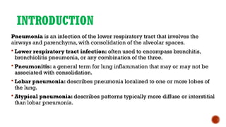
Pneumonia is aninfection of the lower respiratory tract that involves the
airways and parenchyma, with consolidation of the alveolar spaces.
Lower respiratory tract infection: often used to encompass bronchitis,
bronchiolitis pneumonia, or any combination of the three.
Pneumonitis: a general term for lung inflammation that may or may not be
associated with consolidation.
Lobar pneumonia: describes pneumonia localized to one or more lobes of
the lung.
Atypical pneumonia: describes patterns typically more diffuse or interstitial
than lobar pneumonia.
4.
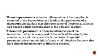
Bronchopneumonia refers toinflammation of the lung that is
centered in the bronchioles and leads to the production of a
mucopurulent exudate that obstructs some of these small airways
and causes patchy consolidation of the adjacent lobules.
Interstitial pneumonitis refers to inflammation of the
interstitium, which is composed of the walls of the alveoli, the
alveolar sacs and ducts, and the bronchioles. Interstitial
pneumonitis is characteristic of acute viral infections but may also
be a chronic inflammatory or fibrosing process.
5.
ETIOLOGY
Defects inhost defenses increase the risk of pneumonia.
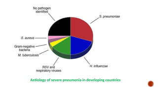
Infectious agents that commonly cause community-acquired pneumonia vary by age.
Streptococcus pneumoniae is the most common bacterial cause of pneumonia (particularly
lobar pneumonia) and occurs in children of any age outside the neonatal period.
Other common causes include
Respiratory Syncytial Virus (RSV) in infants
Parainfluenza Viruses
Influenza Viruses
Human Metapneumovirus
Adenoviruses in children younger than 5 years old
Mycoplasma pneumoniae in children older than age 5 years.
»»» M.pneumoniae and Chlamydophila pneumoniae are principal causes of Atypical
Pneumonia.
6.
Chlamydia trachomatis andless commonly Mycoplasma hominis,Ureaplasma urealyticum, and
cytomegalovirus (CMV) cause a similar respiratory syndrome in infants 2 weeks to 3 months of
age, with subacute onset of an afebrile pneumonia; cough and hyperinflation are the
predominant signs.
These infections are difficult to diagnose and distinguish from each other. In adults these
organisms are carried primarily as part of the genital mucosal flora.Women who harbor these
agents may transmit them perinatally to newborns.
Additional agents occasionallycause pneumonia.
Severe acute respiratory syndrome (SARS) is due to SARS-associated coronavirus (SARS-
CoV).
Avian influenza (bird flu) is a highly contagious viral disease of poultry and other birds
caused by influenza A (H5N1). There were outbreaks among humans in Southeast Asia in 1997
and 2003-2004, with high mortality rates.
A novel influenza A (H1N1) of swine origin began circulating in 2009.
Other etiological agents to consider, based on specific exposure history, include
Staphylococcus aureus and Streptococcus pyogenes (especially after influenza infection),
Mycobacterium tuberculosis,Francisella tularensis,Brucella spp.,Coxiella burnetii,
Chlamydophila psittaci,Legionella pneumophila, hantavirus, Histoplasma capsulatum,
Coccidioides immitis,Blastomyces dermatitidis, and oral flora or gram-negative bacilli (after
aspiration).
9.
Causes of pneumoniain immunocompromised persons include:
• Gram-negative enteric bacteria
• Mycobacteria (M. avium complex)
• Fungi (aspergillosis)
• Viruses (CMV)
• Pneumocystis jirovecii (formerly carinii)
Pneumonia in patients with cystic fibrosis is usually caused by Staph
aureus in infancy and Pseudomonas aeruginosa or Burkholderia
cepacia in older patients.
EPIDEMIOLOGY
Immunizations have markedlyreduced the incidence of pneumonia caused by
pertussis, diphtheria, measles, Haemophilus influenzae type b, and S.
pneumoniae.
Where used, Bacilli Calmette-Guérin (BCG) immunization for tuberculosis has
also had some impact.
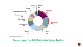
Pneumonia is the single largest contributor of childhood mortality worldwide,
killing an estimated 1 million children under 5 years of age annually.
Risk factors for lower respiratory tract infections include gastroesophageal
reflux, neurological impairment (aspiration), immunocompromised states,
anatomical abnormalities of the respiratory tract, residence in residential care
facilities.
Age isa determinant in the clinical manifestations of pneumonia.
Neonates may have fever or hypoxia only, with subtle or absent physical
examination findings.
With a young infant, apnea may be the first sign of pneumonia.
Fever, chills, tachypnea, cough, malaise, pleuritic chest pain, retractions, and
apprehension—because of difficulty breathing or shortness of breath—are common
in older infants and children.
Physical examination findings cannot reliably distinguish viral and bacterial
pneumonias, but complete physical examination may help identify other foci of
disease or associated findings to suggest an etiology.
14.
Viral pneumoniasare generally associated more often with cough, wheezing,
or stridor; fever is less prominent than with bacterial pneumonia.
»»»Mucosal congestion and upper airway inflammation suggest a viral infection.
Bacterial pneumonias are typically associated with higher fever, chills, cough,
dyspnea, and auscultatory findings of lung consolidation.
Atypical pneumonia in young infants is characterized by tachypnea, cough,
and crackles on auscultation.
Concomitant conjunctivitis may be present in infants with chlamydial
pneumonia.
Other signs of respiratory distress include nasal flaring, intercostal and
subcostal retractions, and grunting.
15.
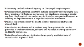
Asymmetry orshallow breathing may be due to splinting from pain.
Hyperexpansion, common in asthma but also frequently accompanying viral
lower respiratory infections, may cause a low diaphragm seen on a chest x-
ray. Poor diaphragmatic excursion may indicate hyperexpanded lungs or an
inability for expansion due to a large consolidation or effusion.
Dullness to percussion may be due to lobar or segmental infiltrates or
pleural fluid.
Auscultation may be normal in early or very focal pneumonia, but the
presence of localized crackles, rhonchi, and wheezes may help one detect
and locate pneumonia.
Distant breath sounds may indicate a large, poorly ventilated area of
consolidation or pleural fluid.
16.
LABORATORY AND IMAGINGSTUDIES
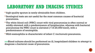
high-quality sputum is rarely obtainable from children.
Serological tests are not useful for the most common causes of bacterial
pneumonia.
The white blood cell (WBC) count with viral pneumonias is often normal or
mildly elevated, with a predominance of lymphocytes, whereas with bacterial
pneumonias the WBC count is elevated (>15-20,000/mm3), with a
predominance of neutrophils.
Mild eosinophilia is characteristic of infant C. trachomatis pneumonia.
»»»Blood cultures should be performed on ill, hospitalized children to attempt to
diagnose a bacterial cause of pneumonia.
17.
Viral respiratory pathogenscan be diagnosed using
polymerase chain reaction (PCR) or rapid viral antigen
detection, but neither can rule out concomitant bacterial
pneumonia.
18.
The need toestablish an etiological diagnosis of pneumonia is
greater in immunocompromised patients, patients with
recurrent pneumonia, or those with pneumonia unresponsive to
empirical therapy.
For these patients, bronchoscopy with bronchoalveolar lavage
and brush mucosal biopsy, needle aspiration of the lung, and
open lung biopsy are methods of obtaining material for
microbiologic diagnosis.
19.
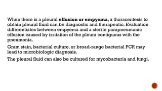
When there isa pleural effusion or empyema, a thoracentesis to
obtain pleural fluid can be diagnostic and therapeutic. Evaluation
differentiates between empyema and a sterile parapneumonic
effusion caused by irritation of the pleura contiguous with the
pneumonia.
Gram stain, bacterial culture, or broad-range bacterial PCR may
lead to microbiologic diagnosis.
The pleural fluid can also be cultured for mycobacteria and fungi.
20.
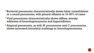
Bacterial pneumonia characteristicallyshows lobar consolidation
or a round pneumonia, with pleural effusion in 10-30% of cases
Viral pneumonia characteristically shows diffuse, streaky
infiltrates of bronchopneumonia and hyperinflation.
Atypical pneumonia, as with M. pneumoniae and C.pneumoniae,
shows increased interstitial markings or bronchopneumonia.
21.
Acute lobar pneumoniaof the right lower lobe in a 14-year-old boy with
fever and cough.
22.
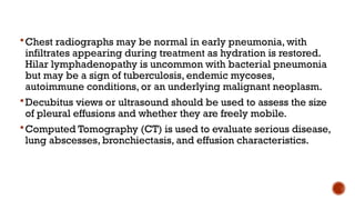
Chest radiographs maybe normal in early pneumonia, with
infiltrates appearing during treatment as hydration is restored.
Hilar lymphadenopathy is uncommon with bacterial pneumonia
but may be a sign of tuberculosis, endemic mycoses,
autoimmune conditions, or an underlying malignant neoplasm.
Decubitus views or ultrasound should be used to assess the size
of pleural effusions and whether they are freely mobile.
Computed Tomography (CT) is used to evaluate serious disease,
lung abscesses, bronchiectasis, and effusion characteristics.
23.
DIFFERENTIAL DIAGNOSIS
Pneumonia mustbe differentiated from
other acute pulmonary diseases, including allergic pneumonitis asthma, and cystic
fibrosis
cardiac diseases, such as pulmonary edema caused by heart failure
autoimmune diseases, such as certain vasculitides and systemic lupus
erythematosus.
Radiographically, pneumonia must be differentiated from lung trauma and
contusion, hemorrhage, foreign body aspiration, and sympathetic effusion due to
subdiaphragmatic inflammation.
24.
TREATMENT
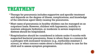
Therapy forpneumonia includes supportive and specific treatment
and depends on the degree of illness, complications, and knowledge
of the infectious agent likely causing the pneumonia.
Most cases of pneumonia in healthy children can be managed on an
outpatient basis. However, children with hypoxemia, inability to
maintain adequate hydration, or moderate to severe respiratory
distress should be hospitalized.
Hospitalization should be considered in infants under 6 months with
suspected bacterial pneumonia, those in whom there is a concern for
a pathogen with increased virulence (e.g., methicillin-resistant S.
aureus), or when concern exists about a family’s ability to care for the
child and to assess symptom progression.
25.
Because viruses causemany community-acquired pneumonias in young
children, not all children require empiric antibiotic treatment for pneumonia.
Exceptional situations include:
Lack of response to empiric therapy
Unusually severe presentations
Nosocomial pneumonia
Immunocompromised children susceptible to infections with opportunistic
pathogens.
Presumed pneumococcal pneumonia can be treated with high-dose
ampicillin therapy even with high-level penicillin resistance. Ceftriaxone
and/or vancomycin can be used if the isolate shows high-level resistance
and the patient is severely ill.
PREVENTION
Annual influenzavaccine is recommended for all children over 6 months of age.
Reducing the duration of mechanical ventilation and administering antibiotics
judiciously reduces the incidence of ventilator-associated pneumonias.
The head of the bed should be raised to 30-45 degrees for intubated patients to
minimize risk of aspiration.
All suctioning equipment and saline should be sterile.
Handwashing before and after every patient contact.
Use of gloves for invasive procedures are important measures to prevent nosocomial
transmission of infections.
Hospital staff with respiratory illnesses or who are carriers of certain organisms,
such as methicillin-resistant S. aureus, should comply with infection control policies
to prevent transfer of organisms to patients.
Treating sources of aerosols, such as air coolers, can prevent L.pneumonia.
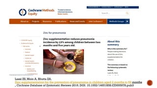
Lassi ZS, MoinA, Bhutta ZA.
Zinc supplementation for the prevention of pneumonia in children aged 2 months to 59 months
. Cochrane Database of Systematic Reviews 2016. DOI: 10.1002/14651858.CD005978.pub3
39.
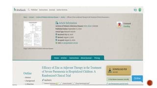
Objectives:
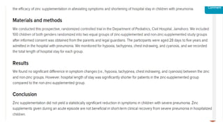
This studyevaluated the effect of zinc supplementation as adjuvant treatment on children with severe
pneumonia admitted to 17th Shahrivar Training Hospital in Rasht, Iran.
Methods:
In this double-blind placebo-controlled clinical trial, 120 children aged two to 60 months hospitalized for
pneumonia were randomly divided into 2 groups of size 60 each.The first group received zinc sulfate (20 mg
daily for children twelve months of age or older and 10 mg daily for children younger than 12 months), and the
second group received a placebo for seven days. All patients received standard antibiotic treatment for
pneumonia.The children were daily evaluated, and recovery time for fever and tachypnea (as primary
outcomes) and duration of hospitalization and mortality rate (as secondary outcomes) were compared
between the two groups.
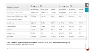
Results:
The mean age of patients was 14.1 months ± 13.9.The youngest and oldest patients were aged 2 and 60
months, respectively. No significant difference in age and sex distribution was found between the two groups.
The zinc receiving group experienced a considerably shorter time of fever (2.1 days vs. 2.84 days, P < 0.05)
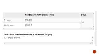
and tachypnea (1.75 days versus 2.1 days, P = 0.011).There was no significant difference in the duration of
admission between the two groups (P = 0.728), and no cases of death occurred in either group.
Conclusions:
This study showed that adjuvant treatment with zinc in children aged 2 to 60 months with severe pneumonia
accelerates recovery from pneumonia. Further studies are needed to investigate the effects of administering
zinc as adjunctive therapy for pneumonia in other age groups.
45.
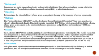
Background
Pneumonia isa major cause of morbidity and mortality of children. Zinc is known to play a central role in the
immune system.The deficiency of zinc increased susceptibility to infectious diseases.
Objective
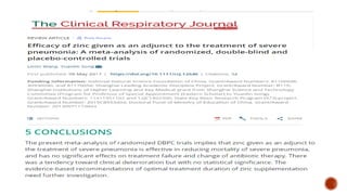
To investigate the clinical efficacy of zinc given as an adjunct therapy to the treatment of severe pneumonia.
Methods
The PubMed, Embase, MEDLINETM
and the Cochrane Central Register of Controlled Trials were searched to
identify all randomized, double-blind and placebo-controlled (DBPC) trials which evaluated the clinical efficacy
of zinc given as an adjunct in the treatment of severe pneumonia and published between January 1966 and
October 2015.
Results
Six randomized DBPC trials including 2216 patients with severe pneumonia were eligible.The results suggested
that zinc given as an adjunct therapy to the treatment of severe pneumonia had no significant improvement of
treatment failure (RR = 0.97, P = .71) and change of antibiotic therapy (RR = 1.09, P = .52).We also found a
favorable trend for clinical deterioration of severe pneumonia but with no statistical significance (RR = 0.88, P
= .55). Zinc produced a significant reduction in mortality caused by severe pneumonia (RR = 0.43, P = .01).
Conclusions
Zinc given as an adjunct to the treatment of severe pneumonia is effective in reducing the mortality of severe
pneumonia, and has no significant effects on treatment failure and change of antibiotic therapy.
46.
The World HealthOrganization (WHO) and the United Nations
Children’s Fund (UNICEF) recommend that the children living in
developing countries should take zinc supplement for 10 to 12
days as follows: 10 mg daily for infants younger than 6 months and
20 mg daily for infants older than 6 months.The purpose of this
treatment is to reduce the severity of acute diarrhea episodes and
hasten recovery from severe pneumonia in developing countries
47.
INPUT
1. Zinc shouldbe used as adjuvant therapy for hospitalized
pneumonia patients.
2. Multi-center studies with larger sample sizes would be useful to
confirm the results of the current study and applying them to other
age groups.
3. More studies should be performed to evaluate the effect of zinc
supplementation in the treatment of pneumonia in special patient
groups such as patients with malnutrition, failure to thrive, or
immune deficiency.
48.
REFERENCES
Nelson Essentialsof Paediatrics 2019
WHO pocket book for hospital care for children
Respiratory disease in children by Prof Addo Yobo
https://www.ncbi.nlm.nih.gov/pmc/articles/PMC3464689/#:~:text=A%20recent%
20clinical%20trial%20conducted,children%20with%20pneumonia%20%5B3%5D
.
https://www.ncbi.nlm.nih.gov/pmc/articles/PMC5638472/
https://methods.cochrane.org/equity/zinc-pneumonia
https://www.hindawi.com/journals/scientifica/2014/694193/
https://www.cureus.com/articles/18620-therapeutic-role-of-zinc-supplementation-
in-children-hospitalized-with-pneumonia
https://brieflands.com/articles/apid-105318.html
#5 Lower airways and secretions are sterile as a result of a multifactorial system. Airway contaminants are caught in the mucus secreted by the goblet cells. Cilia on epithelial surfaces, composing the ciliary elevator system, beat synchronously to move particles upward toward central airways and into the throat, where they are swallowed or expectorated. Polymorphonuclear neutrophils from the blood and tissue macrophages ingest and kill microorganisms. IgA secreted into the upper
airway fluid protects against invasive infections and facilitates viral neutralization.
#12 Diagnosis not always easy: from mild febrile illness to peripheral circulatory collapse with respiratory failure.
Referred pain to abdomen => usually Rt or Lt lower lobe involvement
neck stiffness => Rt upper lobe pneumonia
Coughing + poor peripheral perfusion, rapid pulse, High resp. rate, cardiomegaly, Hepato-(spleno)megaly is suggestive of Heart failure
#17 M. pneumoniae can be confirmed by Mycoplasma PCR. CMV pneumonitis can be diagnosed with PCR from bronchoalveolar lavage fluid. The diagnosis of M. tuberculosis is established by the tuberculin skin test, serum interferon-gamma release assay
#21 A) Posteroanterior and (B) lateral chest radiographs demonstrate right-lower-lobe airspace
consolidation, which obliterates the silhouette of the right heart border
#24 Management:
•Oxygen
•Antibiotics: choice depends on age
•Physiotherapy
•Although most pneumonias (40% or more) are viral in origin it is very difficult to exclude bacterial aetiology. Therefore, all given antibiotics.
#26 Antibiotics options in Management:
1st year:
•Streptococcal, Staphylococcal, E. coli ….chloramphenicol/ benzyl pen/ fluclox/(Cefuroxime) + Gent (as first line)
•Chlamydia trachealis ---Erythromycin
•PS: Chlamydia pneumonia ---dramatic X-ray changes in infants who are not that ill.
Beyond 1st year:
•Pneumococcus (over 90%) Amoxicillin /(Amoxiclav/Cefuroxime/? Co-trimoxazole for PCP)
•Remember Chlamydia, TB
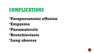
#28 Bacterial pneumonias frequently cause inflammatory fluid to collect in the adjacent pleural space, causing a parapneumonic effusion or, if grossly purulent, an empyema.
Air dissection within lung tissue results in a pneumatocele.
Scarring of the airways and lung tissue may leave dilated bronchi, resulting in bronchiectasis and increased risk for recurrent infection
Pneumonia that causes necrosis of lung tissue may evolve into a lung abscess.
Lung abscess is an uncommon problem in children and is usually caused by aspiration, infection behind an obstructed bronchus, or certain virulent organisms.
Anaerobic bacteria usually predominate, along with various streptococci, Escherichia coli, Klebsiella pneumoniae, P. aeruginosa, and S. aureus.
TREATMENT: Lung abscesses usually respond to appropriate antimicrobial therapy with clindamycin, penicillin G, or ampicillin-sulbactam.
#29 In a few children, symptoms may last longer than 1 month or may be recurrent. In such cases, the possibility of underlying disease must be investigated further