
Recommended
More Related Content
What's hot
What's hot (20)
Similar to Pediatric tuberculosis dr. anu
Similar to Pediatric tuberculosis dr. anu (20)
Recently uploaded
Recently uploaded (20)
Pediatric tuberculosis dr. anu
- 2. INTRODUCTION Causative agent- Mycobacterium tuberculosis Less common- Mycobacterium bovis, M. Avium intra cellulare Acid fast bacilli, non motile Obligate aerobe Discovered by Dr. Robert Koch, 1882 White plague of 18 th century
- 3. Mode of infection- droplet spread, air borne infection Primary site of infection- lung May remain dormant- latentTB infection (LTBI) May get activated – disease Affects almost all organs of the body
- 4. HOST FACTORS Age: no exemption for any age. Infants >> more vulnerable Sex: adolescent girls; around puberty Malnutrition: undernourished >> more susceptible; depressed immunological reaction. SAM child who doesn’t respond to nutritional therapy should be evaluated for TB
- 5. Immunodeficiency: both primary and secondary- HIV, DM, immuno suppressives Inter-current infections: post viral Incubation period: 4 to 8 weeks
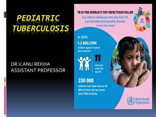
- 6. How prevalent is TB in children? In India, 3.42 lakh children getTB every year Male= female 31% global burden 6 % of cases reported to NTEP PulmonaryTB most common Extra pulmonary is common compared to adult
- 7. PATHOGENESIS
- 11. EXTRA PULMONARY TB Lymph nodeTB Pleural effusion Meningitis DisseminatedTB Renal Bone GenitourinaryTB
- 12. CLINICAL FEATURES Primary infection: Asymptomatic Mild fever, anorexia, weight loss, decreased activity Cough – inconsistent Diagnosed incidentally LatentTB(LTBI)- no symptoms/disease ;but shows only hypersensitivity toTB;
- 13. Progressive primary complex: High grade fever, cough- consolidation Endo-bronchial: troublesome cough, dyspnea, wheezing Miliary: Hematogenous spread ; infants Innumerable small foci Acute onset, high grade fever
- 14. Pleural effusion: Rupture of sub-pleural focus Occurs due to the hypersensitivity to tubercular proteins > 5years of age Insidious onset fever, cough , dyspnea, pleuritic chest pain (pleural rub) initially
- 15. Lymphadenitis: Extension of the primary lesions of the lung mainly cervical nodes; painless, matted Advances to caseating necrosis (cold abscess), rupture, draining sinus
- 16. CNS tuberculosis: Rich focus in the brain- lympho hematogenous spread Infants and children <4 years- vulnerable Infants- rapid progression- hydrocephalus, raised ICT, seizures Older child- sub acute course Headache, irritability, drowsiness, vomiting, focal signs
- 17. AbdominalTB: Hematogenous spread Non specific abdominal pain Doughy abdomen- omental involvement Rolled up omentum , mesenteric adenitis, thickened ileum- irregular nodular mass Ascites Hepato splenomegaly
- 18. DIAGNOSIS Clinical signs and symptoms CXR Tuberculin skin testing History of contact Microbiological confirmation- gold standard Contact : Any child who lives in a household with an adult taking ATT or has taken in the past 2 years.
- 19. COLLECTION OF SPECIMEN Gastric aspirate: In children who cant expectorate Early morning- after 4 to 6 hours fasting Pooled sample of swallowed sputum Induced sputum: Salbutamol nebulisation – hypertonic saline nebulisation- nasopharyngeal aspirate with suction Expectorated sputum
- 20. Bronchio-alveolar lavage fluid FNAC of lymph node Biopsy of the node CSF Pleural fluid Ascitic fluid
- 21. MICROBIOLOGICAL DIAGNOSIS Ziehl –Neelson technique Auramine Rhodamine staining Cartridge based Nucleic acid amplification technique (CB-NAAT)- 2 hours; identifies rifampicin (rif) sensitivity Tru- NAT- 45 minutes
- 22. Culture: Lowenstein Jensen medium- conventional- 6weeks Mycobacterial growth indicator tube(MGIT)- liquid culture- 2 weeks Line probe assay (LPA)- for detecting the drug sensitivity Interferon gamma release assay(IGRA)- alternative to Mantoux CopenhagenTB (CTb ) test- yet to come
- 23. MANTOUX TEST Demonstrate delayed type hypersensitivity 2TU of tuberculin PPDRT23 0r 5TU of PPD-S 0.1 ml of PPD ; intradermal; volar aspect of forearm Read after 48-72 hours Induration (not erythema) to be measured > 10 mm & >5 – in immuno compromised significant False positive- Atypical mycobacteria infection False negative- immuno compromised
- 24. RADIOLOGICAL DIAGNOSIS CHEST X RAY: Highly suggestive: Hilar or para tracheal lymph adenopathy Miliary Cavitary Non specific shadows Broncho pneumonia, consolidation, collapse, emphysematous change ,pleural effusion etc
- 25. DIAGNOSTIC ALGORITHM PRESUMPTIVETB DIAGNOSIS Persistent fever >2wk, without a known cause and/or Unremitting cough for >2wk and/or Weight loss of 5%; or, no weight gain in past 3 mo despite adequate nutrition; or, failure of nutritional rehabilitation in babies with severe acute malnutrition With or without contact with patient with pulmonaryTB in past 2 years
- 27. DIAGNOSTIC ALGORITHM FOR DRTB
- 29. NATIONAL TB ELIMINATION 2025
- 30. TREATMENT
- 31. Only 1 category (4HRZE+ 2HRE); No class 2 Total duration- 6 months; daily Neurological, bone, joint- continuation phase extended up to 10 months Fixed dose combinations(FDC) Second line drugs: Quinolones, Injectable aminoglycosides Newer- Bedaquiline; Delamanid
- 32. WEIGHT BANDS
- 33. MONITORING OF THERAPY Every 2-4 weeks initially; then every 4 -8 weeks Clinical resolution: maximum of 6 to 8 weeks Check for compliance Look for anthropometry, side effects of drugs If no response- think of DRTB Radiological- after 2weeks or based on clinical response; clearance occurs late Microbiological- not routinely
- 34. Notifiable disease Registered through NIKSHAY
- 35. NEONATES OF MOTHER WITH TB CongenitalTB Hematogenous spread through umblical vessels – primary foci- liver Ingestion of infected amniotic fluid- lung Post natal transmission possible Rule out active infection in neonate
- 36. Mother can breast feed (if she had completed 1 month of intensive phase) Cough etiquette to be followed BCG can be given immediately Isoniazid prophylaxis ( INH 10 mg/kg/day along with pyridoxine ) for 6 months Closely watch the baby for the development of disease
- 37. ISONIAZID PREVENTIVE THERAPY Children < 5 years with Contact with adult Children 5 to 15 years who have tested for LTBI Children with HIV Children on immunodeficiency or on immunosuppressive drugs INH should be given 10 mg/kg/day for 6 months along with pyridoxine after ruling out active disease
- 38. BACILLUS CALMITTE GUERIN (BCG) VACCINE 0.1 ml ; intra dermal; left arm; at the insertion of deltoid Live attenuated vaccine Given at birth; Can be given up to 1 year Prevents serious extra pulmonary form ofTB Not for primary complex Efficacy- 20-80% Complication : BCG abscess, BCG adenitis,
- 40. THANK YOU