young call girls in Rajiv Chowk🔝 9953056974 🔝 Delhi escort Service
Paper id 212014137
1. E-ISSN: 2321–9637
Volume 2, Issue 1, January 2014
International Journal of Research in Advent Technology
Available Online at: http://www.ijrat.org
413
FETAL ECG ANALYSIS BY REDUCTION OF
MUTUAL INFORMATION
Mayurkumar Nanda ,
Electronics And Telecommunication Department,
D.J.Sanghvi College Of Engineering,Mumbai
nanda.mayurkumar2@gmail.com
ABSTARCT:
Fetal electrocardiogram (FECG) signal contains potentially precise information that could assist
clinicians in making more appropriate and timely decisions during labor. The ultimate reason for the
interest in FECG signal analysis is in clinical diagnosis and biomedical applications. The extraction and
detection of the FECG signal from composite abdominal signals with powerful and advance
methodologies are becoming very important requirements in fetal monitoring. The signal is a mixture of
the fetal ECG, the maternal ECG and noise. The key idea is to project the signal into higher dimensions,
and then use an assumption of statistical independence between the components to separate them from
the mixtures using Independent Component Analysis
Keywords: Independent Component Analysisv; Kullback-Leiber Divergence; .Fetal ecg
1. THE MAIN TEXT
Heart defects are among the most common birth defects and the leading cause of birth defect-related deaths .
Every year about one out of 125 babies are born with some form of congenital heart defects . The defect may be
so slight that the baby appears healthy for many years after birth, or so severe that its life is in immediate
danger. Congenital heart defects originate in early stages of pregnancy when the heart is forming and they can
affect any of the parts or functions of the heart. Cardiac anomalies may occur due to a genetic syndrome,
inherited disorder, or environmental factors such as infections or drug misuse . However, except for during
labor, fetal electrocardiography has not proved an effective tool for imaging specific structural defects. Rather,
fetal electrocardiography has been concerned to more global issues such as general ischemia due to specific fetal
positioning that chokes the umbilical cord . The reason for this limitation is that the noninvasive fetal
electrocardiogram (ECG) is contaminated by fetal brain activity, myographic (muscle) signals (from both the
mother and fetus), movement artifacts and multiple layers of different dielectric biological media through which
the electrical signals must pass. When continuous electronic fetal heart rate monitoring was introduced into
clinical practice in the 1970s, there was enormous optimism that the widespread use of this technology would
reduce the incidence of intra-partum fetal injury and death. Unfortunately, fetal heart rate monitoring has not
lived up to its initial promise. A meta-analysis of nine randomized, controlled trials comparing fetal monitoring
to intermittent auscultation of the fetal heart rate showed that current monitoring techniques increase the use of
cesarean, forceps, and vacuum delivery, but do not reduce perinatal morbidity or mortality . Since the advent of
fetal heart rate monitoring 40 years ago, there have been no clinically significant advances in intra-partum fetal
monitoring. Moreover, continuous fetal monitoring is utilized in over 85% of labor episodes in the United
States, and represents the standard of care . Fetal monitoring today is based entirely on the fetal heart rate and
does not incorporate characteristics of the fetal ECG (fECG) waveform characteristics that are the cornerstone
of cardiac evaluation of both children and adults.
2. E-ISSN: 2321–9637
Volume 2, Issue 1, January 2014
International Journal of Research in Advent Technology
Available Online at: http://www.ijrat.org
414
2. PROBLEM DEFINITION
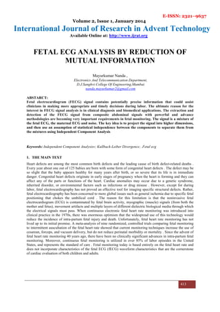
Fig.1 comparison of scalp ECG and Abdominal ECG (Adapted from 3)
Fig.1 illustrates an example of a short segment of fECG recorded both invasively (upper trace) through a fetal
scalp electrode, and non-invasively (lower two traces) through electrodes placed on the mother's abdomen. Four
fetal beats (labeled a, b, c and d) are circled on all three traces. Note the abdominal traces contain much smaller
fetal beats embedded in significant broadband noise, and much larger amplitude artifacts (transient oscillations)
which are due to the mother's heart .Note also that the artifacts manifest both in between and on top of fetal
heart beats, with a similar morphology to the fetal heart beats.
Fig.2 The amplitude and frequency range of different bio-signals, some of which interfere with fetal cardiac signals (Adapted from 3)
In Fig. 2 the amplitude and frequency range of fECG have been compared with other biosignals and artifacts.
Accordingly, the fECG is much weaker than the other biosignals. Moreover, from the signal processing
3. E-ISSN: 2321–9637
Volume 2, Issue 1, January 2014
International Journal of Research in Advent Technology
Available Online at: http://www.ijrat.org
415
perspective, there is no specific domain (time, space, frequency, or feature) in which the fECG can be totally
separated from the interfering signals.
Fig 3 A general representation of the signal, artifacts and noise present in the ECG in the frequency domain (Adapted from 3)
Fig. 3 illustrates the fact that the main part of the fetal heart beat on the electrocardiogram (the QRS complex)
lies in the same frequency domain as the adult QRS complex, as well as broadband muscle noise. Despite of the
richness of the literature, there are still several key areas that require further study in the field of fetal
electrocardiography, particularly in the domain of multichannel noninvasive maternal abdominal measurements.
3 . Methodology
In this paper noble technique of Independent component analysis is presented to separate fetal ecg analysis. ICA
problem was raised as a cocktail party problem, which demands separating different speaker’s voice from each
other and background music. In this study, three-channel signals are picked up using three different leads. Each
measured signal comprises multiple signal components from different sources. This mixing procedure is
depicted in Fig. 4 In this model, the sources s1(t) and s2(t) represent signals generated by maternal heart and
foetal heart respectively. s3(t) represents random noise. These signals are transmitted to the maternal body
surface through the body issues with unknown parameters aij, (i, j=1, 2, 3). The mixed signals are picked up as
x1(t), x2(t) and x3(t) via two abdominal leads and one chest lead respectively using cutaneous electrodes on the
maternal body surface. Analytical equations for the ICA model can be expressed in a matrix form as
x(t) = As(t ) (1)
where s(t)=
x(t)=
4. E-ISSN: 2321–9637
Volume 2, Issue 1, January 2014
International Journal of Research in Advent Technology
Available Online at: http://www.ijrat.org
416
A=
A is called a “mixing matrix”. Superscript T denotes vector or matrix transposition. The goal of ICA is to find a
“separating matrix” W, which is as close to A-1 as possible, based upon a proper statistical criteria, in order to
optimally recover the original source signals as
y(t ) =Wx(t ) =WAs(t) = s(t )
Fig4 simplified mixing of fetus ECG,Maternal ECG and Noise (Adapted from 5)
In a simplified ICA model, we suppose that matrix A is a time invariant (constant) matrix and the propagation
delays can be ignored. Signals s1, s2 and s3 are statistically independent means that their joint probability
density function is factorable. The mutual information I( ) is defined as the Kullback-Leibler divergence
between the joint density of all signals and the product of their marginal densities as
(2)
It is clear that I( ) will be equal to zero when the recovered signals y1, y2 and y3 are mutual independent. For
an invertible non-linear transformation
Y = g(v) = g(Wx), (3)
(4)
Satisfying above conditions we can apply different algorithms of Independent Component Analysis and extract
fetal ecg. Therefore, using observed signals x and selecting a proper contrast function g, y can be optimally
separated by minimizing I(py) with respect to W. If the joint probability density function of and marginal
density functions are known, i.e. a prior knowledge on signal model, maximum likelihood estimation can be
applied. If it is not the case, truncating Edgeworth expansion can be used to approximate probability densities.
A fixed-point algorithm for updating W was developed by A.Hyvarinen Its iterative steps are recapitulated
below
1. Choose an arbitrary (random) matrix as an initial W.
5. E-ISSN: 2321–9637
Volume 2, Issue 1, January 2014
International Journal of Research in Advent Technology
Available Online at: http://www.ijrat.org
417
2. Let = E{xg(Wx)}−E{g’ (Wx)}W
3. = /
4. If not converged, repeat iteration from step 2.
where g(Wx) is selected as a hyperbolic tangent function, i.e. g(Wx)=tanh(Wx). The convergence means that the
old and new values of W point in the same direction, i.e. their dotproduct is near to one.
4. Simulation Results
Fig5 Abdomen signals
Fig6 Estimated Sources
Fig 5 shows the abdomen signals taken through electrodes from different position of mother abdomen .fig6
indicates separated independent components separated by fixed point ica algorithm. First estimated component
indicates fetal ecg. Fixed point algorithm used , uses mutual information as measure of independence rather than
non-gaussianty.
6. E-ISSN: 2321–9637
Volume 2, Issue 1, January 2014
International Journal of Research in Advent Technology
Available Online at: http://www.ijrat.org
418
5. CONCLUSION
Fixed point algorithm which uses mutual information as measure of independence is used and showed
satisfactory results in separating fetal ecg from maternal ecg and other artifacts as compared to other methods
using reduction of gaussianty.
References
1. Congenital Heart Defects in Children Fact Sheet. American Heart Association; 2012. [Online]. Available:
http://www.americanheart.org/children
2. Congenital Heart Defects. March of Dimes. 2005. [Online]. Available:
http://www.marchofdimes.com/professionals/14332_1212.asp
3. Reza Sameni1 and Gari D. Clifford, A review of fetal ecg signal processing; issues and promising directions, Open
Pacing Electrophysiol Ther J . 2010 January 1; 3: 4–20. doi:10.2174/1876536X01003010004
4. A. Cichocki, S, Amari, K, Siwek, T. Tanaka, Anh Huy Phan, R. Zdunek, ICALAB – MATLAB Toolbox Ver. 3 for
signal processing.
5. Aapo Hyv¨arinen, Juha Karhunen, and Erkki Oja, Independent Component Analysis, A Wiley-Interscience Publication,
Final version of 7 March 2001
6. Ganesh R. Naik, Independent Component Analysis For Audio And Biosignal Applications, InTech Janeza Trdine 9,
51000 Rijeka, Croatia
7. http://www.physionet.org/cgi-bin/atm/ATM?database=mitdb&tool=plot_waveforms