This document provides an overview of pediatrics as a medical specialty. It discusses pediatrics as the branch of medicine dealing with the care of children and adolescents. Key points include:
- Pediatrics covers children and adolescents under age 18 and aims to enable their healthy growth, development and achievement.
- India has a large child population but also high child mortality, with over 1.7 million under-5 deaths annually.
- Pediatrics encompasses care from premature neonates to adolescents and includes many specialized fields like neonatology and pediatric critical care.
- The historical development and current challenges of pediatrics in India are outlined.
![Introduction to Pediatrics -
Future of Child Health
The nation is addressing child health challenges with
greaterdynamism than ever before. Investments are being
madeforhealthprograms and healthsystem strengthening.
Conditional cash transfers and entitlements are enshrined
to stimulate demand for maternal, newborn and child
health care. ICDS is being strengthened, particularly in high
burden districts to addresschildhood undernutrition. The
country is surging ahead with stronger economy and
accelerated development. India ispoisedtoattainlow child
mortality rate, and improve remarkably the health and
nutrition status of her children in near future.
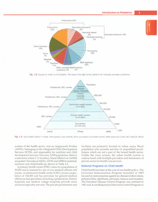
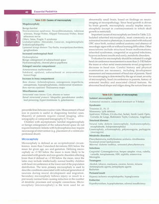
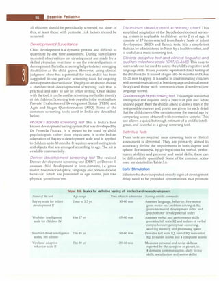
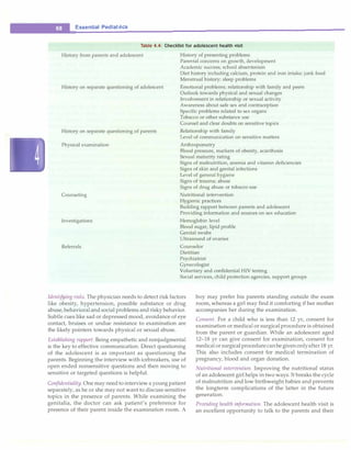
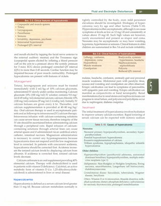
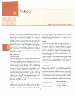
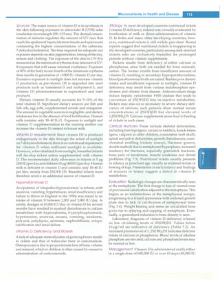
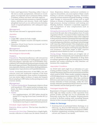
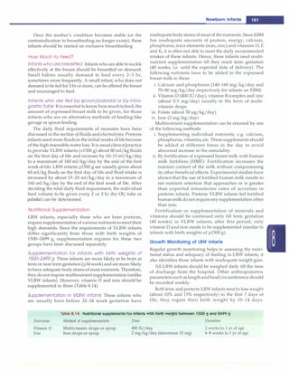
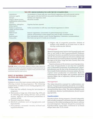
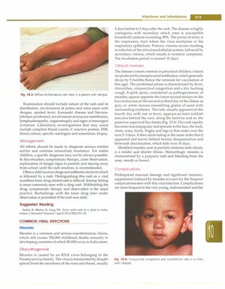
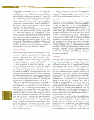
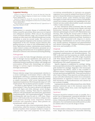
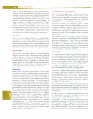
Table 1.4: Summary of maternal, newborn and child health services in the RMNCH+A Strategy under NRHM
Pregnancy, childbirth and Interventions
immediate newborn care
Skilled obstetric care and essential Package
newborn care including resuscitation Facility deliveries by skilled birth attendants
Neonatal resuscitation
Emergency obstetric and
newborn care (EmONC)
Postpartum care for mother and baby
Newborn and child care
Home-based newborn care
Facility-based newborn care
Essentialnewborn care (warmth, hygienic care, breastfeeding, extra care of small babies,
problem detection)
Linkages to facility-based newborn care for sick neonates
Program drivers: The schemes that drive uptake of the intervention packages
Janani Suraksha Yojana (]SY) that provides cash incentive to the woman (and to the
ASHA) for delivery in the facility
Janani Shishu Suraksha Karyakram (JSSK) that entitles the mother and less than one
month neonate to free delivery, medicines/blood, diet, pickup and drop in government
facilities
Navjat Shishu Suraksha Kan;akram that aims to train nurses and doctors in neonatal
resuscitation
Interventions
Home visits by ASHAs (six for facility born babies, on days 3, 7, 14, 21, 28 and 42; an
extra visit on day 1 for home births)
Interventions for infants
Examination; counsel for warmth; breastfeeding; hygiene; extra care of low birthweight
babies; detection of sickness, referral
Interventions for mother
Postpartum care and counseling for family planning
ASHA given cash incentive for home care, birthweight record, birth registration and
immunization (BCG, first dose OPV and DPT)
Special newborn care units (SNCU)
These specialized newborn units at district hospitals with specialized equipments
including radiant warmers. These units have a minimum of 12-16 beds with a staff of
3 physicians, 10 nurses and 4 support staff to provide round the clock services for new
born requiring special care, such as those with very low birthweight, neonatal sepsis/
pneumonia and common complications
Newborn stabilization units (NBSU)
These are step down units providing facilities for neonates from the periphery where
babies can be stabilized through effective care. These are set up in CHCs and provide
services, including resuscitation, provision of warmth, initiation of breastfeeding,
prevention of infection and cord care, supportive care: oxygen, IV fluids, provision for
monitoring of vital signs and referral
Newborn care comers (NBCC)
These are special corners within the labor room at all facilities (PHC, CHC, DH) where
deliveries occur. Services include resuscitation, provision of warmth, prevention of
infections and early initiation of breastfeeding
Program drivers: The schemes that drive uptake of the intervention packages
Janani Shishu Suraksha Kan;akram (JSSK) that entitles the mother and neonate to free
delivery, medicines/blood, diet, pickup and drop in government facilities
Contd...](https://image.slidesharecdn.com/paediatrics8theditiono-230807055545-451d483c/85/Paediatrics-8th-Edition-O-P-Ghai-pdf-10-320.jpg)
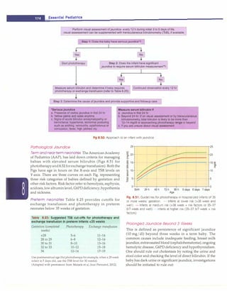
![acceleratedlymphoidgrowthis thefrequentfindingoflarge
tonsils and palpable lymph nodes in normal children
between 4 and 8 yr.
Growth of body fat and muscle mass. Body tissues can be
divided into fat and fat-free components. The lean body
mass includes muscle tissue, internal organs and skeleton
and contains only a small amount of fat. The growth in
leanbody massis primarilyduetoincrease in muscle mass.
Lean body mass correlates closely with stature. Taller
childrenhavegreaterleanbodymass thanshorter children
of the same age. After thepubertal growth spurt, boyshave
greater lean body mass compared to girls. Body fat is the
storehouse of energy. It is primarily deposited in the sub
cutaneous adipose tissue. Girls have more subcutaneous
adipose tissue than boys. Moreover, the sites and quantity
of adipose tissue differs in girls and boys. Girls tend to add
adiposetissue to breasts, buttocks, thighs and back of arms
during adolescence.
SOMATIC GROWTH
Skeletal Growth
Skeletal growth is a continuous process occurring during
the whole of childhood and adolescence. It is steady until
the pubertal growth spurt when it accelerates and
subsequently slows considerably. The skeleton is mature
oncetheepiphysis orgrowthplates at theend oflongbones
fuse to the shaft or diaphysis. This occurs by about 18 yr in
girls and 20-22 yr in boys.The degree of skeletal maturation
closely correlates with the degree of sexual maturation. A
child who has advanced sexual maturity will also have
earlier skeletal maturation.
Skeletalmaturationisassessedby notingthe appearance
and fusion of epiphysis at the ends of long bones. Apart
from this, bone mineraldensity can beascertainedby dual
energy X-ray absorptiometry [DXA]. This method allows
assessment of bonemineralcontentanddensityat different
ages.
Normal Growth and its Disorders
-
Bone Age Estimation
Assessment of bone agepostnatally is based on (i) number,
shape and size of epiphyseal centers and (ii) size, shape
and density of the ends of bones. Tanner and Whitehouse
described 8 to 9 stages of development of ossification
centers and gave them 'maturity scoring'.Fifty percent of
the score was given for carpal bones, 20% for radius, ulna
and 30% for phalanges. Twenty ossification centers are
generallyusedfor determining theboneage.Theseinclude:
(i) carpal bones, (ii) metacarpals, (iii) patella, (iv) distal and
proximal toes in both sexes; and (v) distal and middle
phalanges in boys and distal and proximal phalanges in
girls.Todeterminetheskeletalage ininfantsbetween3 and
9months, aradiographofshoulderismost helpful. Asingle
film of hands and wrists is adequate in children between
the ages of 1 and 13 yr. For children between 12 and 14 yr,
radiographs of elbow and hip give helpful clues.
Eruption of Teeth
Primary teeth. The teeth in the upper jaw erupt earlier than
those in the lower jaw, except for lower central incisors
and second molar (Table 2.2).
Permanent teeth. Theorder oferuptionis shown inTable 2.2.
The first molars are the first to erupt.
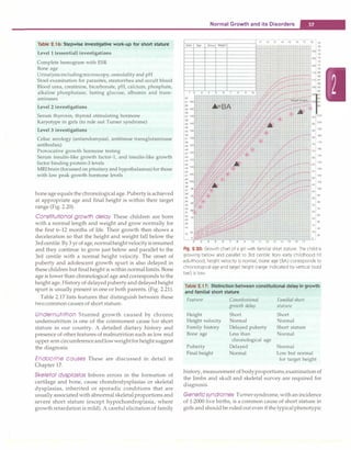
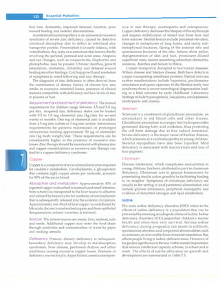
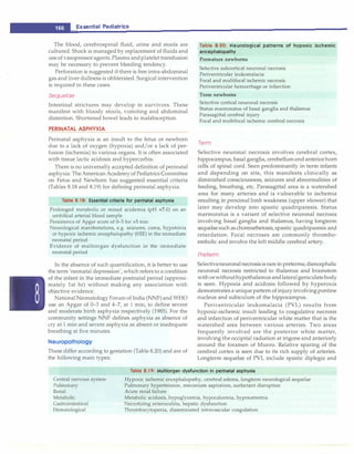
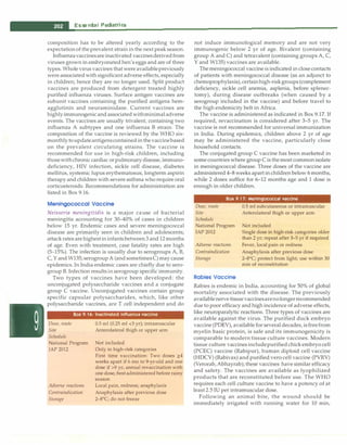
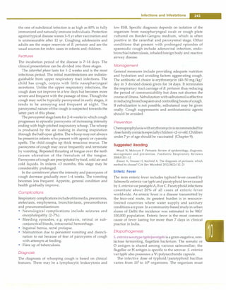
ASSESSMENT OF PHYSICAL GROWTH
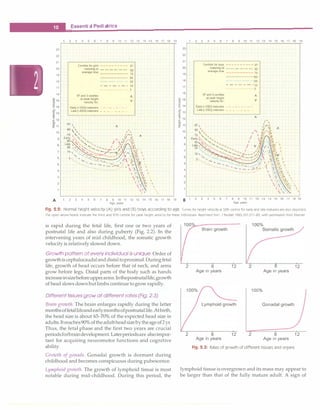
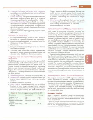
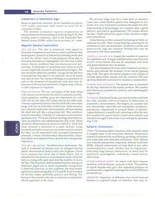
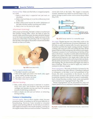
Weight. The weight of the child in the nude or minimal
light clothing is recordedaccuratelyon a lever or electronic
type of weighing scale (Fig. 2.4). Spring balances are less
accurate. The weighing scale should have a minimum unit
of 100 g. It is important that child beplacedinthe middle of
weighing pan. The weighing scale should be corrected for
any zero error before measurement. Serial measurement
should be done on the same weighing scale.
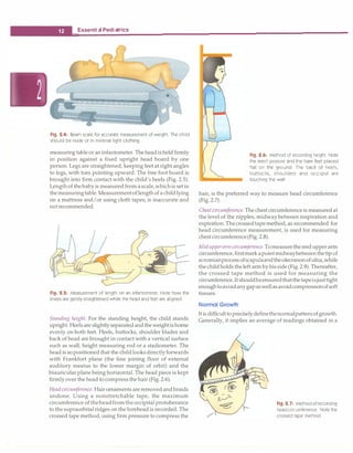
Length. Length is recorded for children under 2 yr of age.
Hairpins are removed and braids undone. Bulky diapers
should be removed. The child is placed supine on a rigid
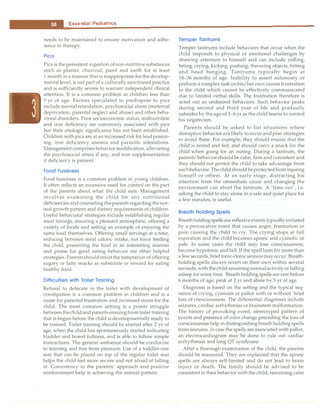
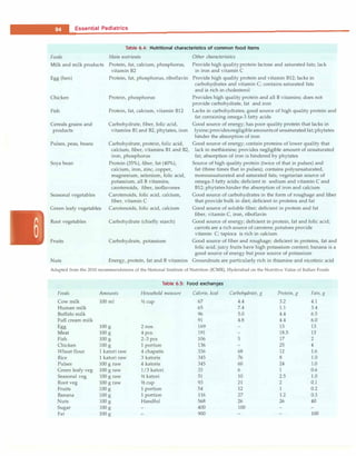
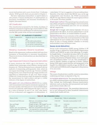
Table 2.2: Timing of dentition
Primary dentition Time of eruption, months Time of fall, years
Upper Lower Upper Lower
Central incisors 8-12 6-10 6-7 6-7
Lateral incisors 9-13 10-16 7-8 7-8
First molar 13-19 14-18 9-11 9-11
Canine 16-22 17-23 10-12 9-12
Second molar 25-33 23-31 10-12 10-12
Permanent teeth Time of eruption, years
Upper Lower Upper Lower
First molar 6-7 6-7 First premolar 10-11 10-12
Central incisors 7-8 6-7 Second premolar 10-12 10-12
Lateral incisors 8-9 7-8 Second molar 12-13 11-13
Canine 11-12 10-12 Third molar 17-21 17-21](https://image.slidesharecdn.com/paediatrics8theditiono-230807055545-451d483c/85/Paediatrics-8th-Edition-O-P-Ghai-pdf-16-320.jpg)
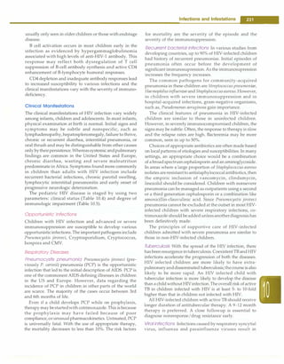
![__E
_
s
_
s
_
e
_
n
_
t
_
ia
_
l
_
P
_
e
_
d
_
i
_
a
_
tr
_
ic
_
s_________________________________
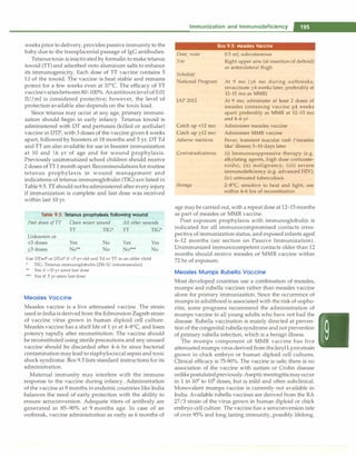
Table 5.5: Causes of hyponatremia
Hypovolemic hypona
tremia (sodium loss
in excess of free water)
Normovolemic hypo
natremia (conditions
that predispose to
SIADH)
Hypervolemic hypo
natremia (excess free
water retention)
Renal loss: Diuretic use,
osmotic diuresis, renal salt
wasting, adrenal insufficiency,
pseudohypoaldosteronism
Extra-renal loss: Diarrhea,
vomiting, drains, fistula
sweat (cystic fibrosis),
cerebral salt wasting
syndrome, third-spacing
(effusions, ascites)
Inflammatory central nervous
system disease (meningitis,
encephalitis), tumors
Pulmonary diseases (severe
asthma, pneumonia)
Drugs (cyclophosphamide,
vincristine)
Nausea, postoperative
Congestive heart failure,
cirrhosis, nephrotic syndrome,
acute or chronic renal failure
urine output and high urine sodium. The treatments are
different, as cerebral salt wasting requires replacement of
urinary salt-water losses while SIADHismanagedby fluid
restriction.
Hyperosmolality resulting from non-sodium molecules
(hyperglycemia, mannitol overdose) draws water from the
intracellular space to dilute the extracellular sodium
concentration. Factitious hyponatremia, reported when
hyperlipidemia (chylomicronemia) or hyperproteinemia
coexist, isuncommondue to use ofion-selectiveelectrodes.
Patients are generally symptomatic when serum sodium
concentrationfalls below 125 mEq/1 or the decline is acute
(<24 hr). Early features include headache, nausea,
vomiting, lethargy and confusion. Advanced mani
festations are seizures, coma, decorticate posturing, dilated
pupils, anisocoria, papilledema, cardiac arrhythmias,
myocardial ischemia and central diabetes insipidus.
Cerebral edema occurs at levels of 125 mEq/1 or less.
Hypo-osmolality causes influx of water into the
intracellular space, which results in cytotoxic cerebral
edema and increased intracranial pressure and can lead
to brain ischemia, herniation and death. The brain's
primary mechanism in adapting to hyponatremia is the
extrusion of intracellular electrolytes and organic
osmolytes. Some ofthese organic osmolytes are excitatory
amino acids, such as glutamate and aspartate that can
produce seizures in the absence of detectable cerebral
edema. Major risk factors for developing hyponatremic
encephalopathyareyoungage,hypoxemiaandneurological
disease. Children are at significantly higher risk than are
adults for developing hyponatremic encephalopathy due
to their relatively larger brain to intracranial volume ratio
compared with adults. Hyponatremia in association with
increased intravascular volume can result in pulmonary
edema, hypertension and heart failure. Asymptomatic
hyponatremia inpretermneonatesisassociatedwith poor
growth and development, sensorineural hearing loss and
a risk factor for mortality in neonates who suffered
perinatal birth asphyxia.
Treatment
The first step is to determine whether hyponatremia is
acute (<48 hr) or chronic (>48 hr), symptomatic or
asymptomatic and evaluate the volume status (hyper
volemic, euvolemic, hypovolemic) (Box 5.1). The under
lying cause should be corrected, where possible. Losses
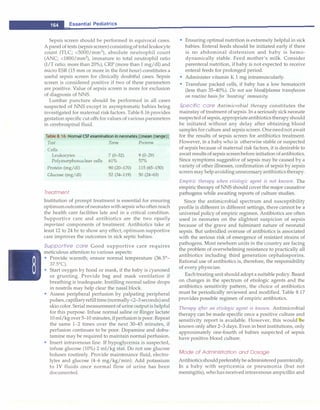
due to renal or adrenocortical disease are suggested by
urinary sodium concentration of more than 20 mEq/1 in
presence of clinically evident volume depletion (Fig. 5.5).
In case of hypovolemic hyponatremia the fluid and
sodium deficit is estimated and replaced over 24 to 48 hr.
The dose of sodium required is calculated using the
formula:
Sodium deficit (mEq) =
0.6 x body wt (kg) x [(desired Na+
) - (observed Na+
)] mEq/1
The optimal rate of correction is 0.6 to 1 mEq/1/hr until
the sodium concentration is 125 mEq/1 and then at a
Box 5.1: Treatment of hyponatremia
• Treat hypotensionfirst, regardless of serum sodium (normal
saline bolus, Ringer lactate, 5% albumin). WHO oral
rehydration solution is preferable for asymptomatic cases
with hypovolemia
• Correct deficit over 48 to 72 hr for chronic hyponatremia.
Rapid decline is associated with risk of central pontine
myelinosis. Recommended rate of increase is 0.5 mEq/
!/hr (8-10 mEq/1/day). Correction in the first 48 hr should
not exceed 15-20 mEq/1
• Acute and symptomatic hyponatremia may be corrected
rapidly. For symptomatic cases, immediate increase in
serum sodium level by 5-6 mEq/1 with hypertonic saline
(3% saline) is recommended. IV infusion at a dose of 3-5
ml/kg over 1-2 hr will raise serum sodium by 5-6 mEq/1.
Alternatively, 3% saline may be given as 1-3 boluses at 2
ml/kg/bolus over 10 min (maximum 100 ml/bolus)
• Stop further therapy with 3% saline when patient is either
symptom free and/or acute rise in sodium of 10 mEq/1 is
noted in first 5 hr.
• Increase of serum Na+
can be estimated using Adrogue
Madias formula (Box 5.2)
• Hypotonic infusates (0.45% dextrose normal saline) are used
as maintenancefluid. Normal saline should be avoided except
for correction of hypovolemia; can aggravate cerebral edema
in those with impaired free water clearance.
• Fluid restriction alone is needed for SIADH; sodium and
water restriction is required in hypervolemic hyponatremia.
Diuretics may be added in refractory cases.](https://image.slidesharecdn.com/paediatrics8theditiono-230807055545-451d483c/85/Paediatrics-8th-Edition-O-P-Ghai-pdf-60-320.jpg)
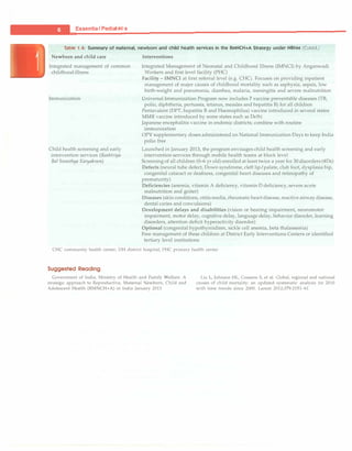
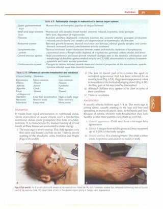
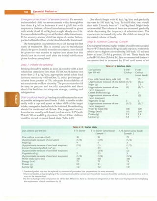
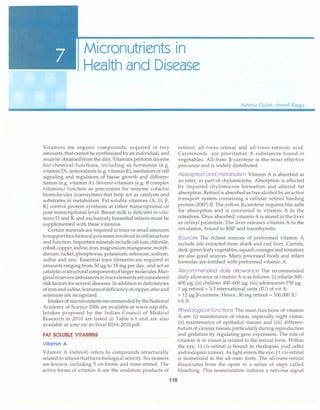
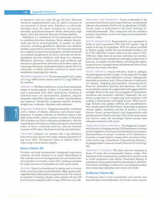
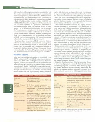
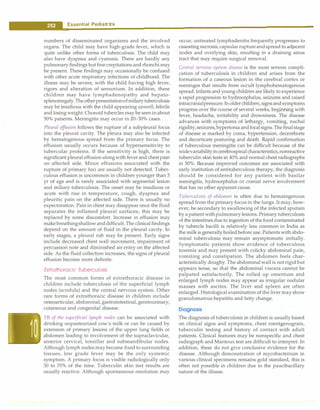
![Fluid and Electrolyte Disturbances
-
Assessment of volume status
=i
Hypovolemia
Total body water .J,
Total body sodium U
J -
Euvolemia
l
Total body water i
Total body sodium H
Hypervolemia
Total body water ii
Total body sodium i
• + • J
U [Na•] >20
r- !
U [Na] <20 U [Na•] >20 U [Na•] >20
.--t-
u [Na] <20
• • f • -.
Extrarenal SIADH Acute, chronic Nephrotic syndrome
Renal losses
Diuretics
Mineralocorticoid deficiency
Salt losing nephropathy
Cerebral salt wasting
losses
Vomiting
Diarrhea
Third spacing
Glucocorticoid deficiency
Hypothyroidism
renal failure Cirrhosis
Congestive heart failure
Stress
Drugs
Renal tubular acidosis
Fig. 5.5: Diagnostic approach to hyponatremia. U(Na•) urinary sodium, mEq/1; i increased; !decreased
slower rate. For symptomatic patients a combination of
intravenous infusion of hypertonic saline (3% sodium
chloride) and fluid restriction rapidly increase serum
sodium by 5-6 mEq/1 and ameliorates the symptoms.
Thereafter, if the symptoms remit, the remaining deficit
can be corrected slowly over the next 1 to 3 days. When
slow correction of hyponatremia in a volume-expanded
patient is indicated, water restriction alone, or if this is
unsuccessful, in combination with a loop-diuretic such as
furosemide is preferred. Serum sodium concentration
should be monitored every 2 to 4 hr and appropriate
adjustments made.
Aggressive therapy with hypertonic saline in patients
with chronic hyponatremia (where brain adaptation to
hypo-osmolality has set in by extrusion of intracellular
electrolytes and organic osmoles) can lead to osmotic
demyelination called central pontine myelinolysis. This
condition, which isoften irreversibleisreportedinpatients
with liver disease, severe malnutrition and hypoxia.
Patients generally become symptomatic 2 to 7 days
followingrapidcorrection (>25mEq/1inthefirst24-48 hr)
of chronic hyponatremia. Clinical features include
mutism, dysarthria, spastic quadriplegia, ataxia, pseudo
bulbar palsy, altered mental status, seizures and
hypotension.
Fluid restriction alone has no role in the management
of symptomatic hyponatremia. Normal saline is also
inappropriate for treating hyponatremic encephalopathy
due to non-hemodynamic states of vasopressin excess,
such as SIADH and postoperative hyponatremia, as it is
not sufficiently hypertonic toinduce reduction in cerebral
edema. In presence of elevated ADH levels there is
impaired ability to excrete free water with the urine
osmolality exceeding that of plasma. V2-receptor anta
gonists or vaptans that block the binding of ADH to its
V2 receptor, are yet not recommended for treatment of
hyponatremic encephalopathy. These agents may have
a role in treating euvolemic hyponatremia from SIADH
and hypervolemic hyponatremia in congestive heart
failure.
Hypernatremia
Hypernatremia is defined as increase in serum sodium
concentration to levels more than 150 mEq/1. It may be
accompanied by the presence of low, normal or high total
body sodium content. The major cause of hypernatremia
is loss of body water, inadequate intake of water, a lack of
antidiuretic hormone (ADH), or excessive intake of
sodium (e.g. solutions with high sodium such as sodium
bicarbonate) (Table 5.6).Diabetesinsipidusmayresultfrom
a deficiency of ADH or its end organ unresponsiveness.
In the presence of an intact thirst mechanism, a slight
increase in serum sodium concentration (3 to 4 mEq/1)
above normal elicits intense thirst. The lack of thirst in
the presence of hypernatremia in a mentally alert child
indicates adefectin eithertheosmoreceptors orthe cortical
thirst center. The most objective sign of hypernatremia is
lethargy or mentalstatuschanges,whichproceedsto coma
and convulsions. With acute and severe hypernatremia,
the osmotic shift of water from neurons leads to shrinkage
of the brain and tearing of the meningeal vessels and
intracranialhemorrhage;slowlydevelopinghypematremia
Table 5.6: Causes of hypernatremia
Net water loss
Pure water loss
Insensible losses
Diabetes insipidus
Inadequate breastfeeding
Hypotonicfluid loss
Renal: Loop, osmotic diuretics,postobstructive,polyuric phase
of acute tubular necrosis
Gastrointestinal: Vomiting, nasogastric drainage, diarrhea;
lactulose
Hypertonic sodium gain
Excess sodium intake
Sodium bicarbonate, saline infusion
Hypertonic feeds, boiled skimmed milk
Ingestion of sodium chloride
Hypertonic dialysis
Endocrine: Primary hyperaldosteronism, Cushing syndrome](https://image.slidesharecdn.com/paediatrics8theditiono-230807055545-451d483c/85/Paediatrics-8th-Edition-O-P-Ghai-pdf-61-320.jpg)
![__E_s_s_e_n_ti_a _i _P_e_d_ia_t_ri_cs--------------------------------
is generally well tolerated. The latter adaptation occurs
initially by movement of electrolytes into cells and later
by intracellular generation of organic osmolytes, which
counter plasma hyperosmolarity.
Treatment
Treatment involves restoring normal osmolality and
volume and removal of excess sodium through the
administration of diuretics and hypotonic crystalloid
solutions. The speed of correction depends on the rate of
development of hypernatrernia and associated symptoms
(Box 5.2). Because chronic hypernatrernia is well tolerated,
rapid correction offers no advantage and may be harmful
since it may result in brain edema. Usually a maximum of
10% of the serum sodium concentration or about 0.5 mEq/
1/hr should be the goal rate of correction. Seizures due to
hypernatremia are treated using 5-6 ml/kg infusion of
3% saline over 1-2 hr.
POTASSIUM
Physiology
------------------
Potassium being a predominantly intracellular cation, its
blood levels are unsatisfactory indicator of total body
stores. Normal serum concentration of potassium ranges
between 3.5 and 5 mEq/1. Common potassium-rich foods
include meats, beans, fruits and potatoes. Gastrointestinal
absorption is complete and potassium homeostasis is
Box 5.2: Treatment of hypernatrernia
• Treat hypotension first, regardless of serum sodium
(normal saline bolus, Ringer lactate, 5% albumin)
• Correct deficit over 48 to 72 hr. Rapid decline of chronic
(>48 hr) hypematremia is associated with risk of cerebral
edema. Recommended rate of drop in serum sodium is
0.5 mEq/1/hr (10-12 mEq/1/day)
• Oral solutions preferred to parenteral correction
• Generally hypotonic infusates are used (infusate sodium
of - 40 mEq/1, as N/4 or N/5 saline). Sodium free fluids
should be avoided (except in acute onset hypematremia,
e.g. sodium overload)
• Decline of serum Na+ can be estimated using Adrogue
Madias Formula:
D[Na
+
J= {[Na
+
Jinf
+ (K
+
Jinf- [Na
+L)
(TBW + 1)
Where �[Na+] expected change in serum sodium; [Na+]inf
sodium and [K+]infpotassium in 1 liter of the infusate, [Na+Js
serum sodium; TBW or total body water = 0.6 x body
weight
• Seizures due to hypematremia are treated using hypertonic
(3%) saline at 5-6 ml/kg infusion over 1-2 hr.
• Renal replacement therapy (peritoneal or hemodialysis,
hemofiltration) is indicated for significant hypematremia
(>180-200 mEq/1) with concurrent renal failure and/or
volume overload.
• Ensure correction of ongoing fluid losses; frequent bio
chemical and clinical reassessment is needed.
maintainedpredominantlythroughtheregulationof renal
excretion. The fractional excretion of potassium is about
10%,chieflyregulated byaldosteroneat thecollectingduct.
Renal adaptive mechanisms maintain potassium
homeostasis until theglomerularfiltration ratedropstoless
than 15-20 ml/min. Excretion is increased by aldosterone,
high sodium delivery to the collecting duct (e.g. diuretics),
urine flow (e.g. osmotic diuresis), blood potassium level,
glucocorticoids, ADH and delivery of negatively charged
ions to the collectingduct (e.g. bicarbonate). Inrenal failure,
the proportion of potassium excreted through the gut
increases, chiefly by the colon in exchange for luminal
sodium.
Aldosterone and insulin are two play important roles
inpotassiumhomeostasis. Insulin stimulatedbypotassium
ingestion increases uptake of potassium in muscle cells,
through increased activity of the sodium pump. High
potassium levels stimulate its renal secretion via aldos
terone-mediated enhancement of distal expression of
secretory potassium channels (ROMK). Insulin, beta
adrenergic stimuli and alkalosis enhance potassium entry
into cells. The reverse happens with glucagon, a-adrener
gic stimuli and acidosis.
Hypokalemia
Hypokalernia is defined as a serum potassium level below
3.5 mEq/1. The primary pathogenetic mechanisms result
ing in hypokalemia include increased losses, decreased
intake or transcellular shift (Table 5.7). Vomiting, a
Table 5.7: Causes of hypokalernia
Increased losses
Renal
Renal tubular acidosis (proximal or distal)
Drugs (loop and thiazide diuretics, amphotericin B,
aminoglycosides, corticosteroids)
Cystic fibrosis
Gitelman syndrome, Bartter syndrome, Liddle syndrome
Ureterosigmoidostomy
Mineralocorticoid excess (Cushing syndrome, hyperal
dosteronism, congenital adrenal hyperplasia (11 P-hydroxy
lase, 17 o:-hydroxylase deficiency)
High renin conditions (renin secreting tumors, renal artery
stenosis)
Extrarenal
Diarrhea, vomiting, nasogastric suction, sweating
Potassium binding resins (sodium polystyrene sulfonate)
Decreased intake or stores
Malnutrition, anorexia nervosa
Potassium-poor parenteral nutrition
Intracellular shift
Alkalosis, high insulin state, medications (P2-adrenergic
agonists, theophylline, barium, hydroxychloroquine),
refeeding syndrome, hypokalemic periodic paralysis,
malignant hyperthermia, thyrotoxic periodic paralysis](https://image.slidesharecdn.com/paediatrics8theditiono-230807055545-451d483c/85/Paediatrics-8th-Edition-O-P-Ghai-pdf-62-320.jpg)

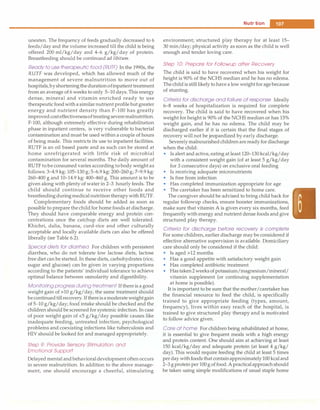
![__E_s_s_ e_n_t_ia_l_P_e _d_ia_t_ri_c_
s __________________________________
calcium absorption in the distal convoluted tubule. These
mechanisms help to maintain normal levels of serum
calcium.
Plasma calcium exists in 3 different forms: 50% as
biologically active ionized form, 45% bound to plasma
proteins (mainlyalbumin)and5%complexedtophosphate
and citrate. In the absence of alkalosis or acidosis, the
proportion of albumin-bound calcium remains relatively
constant. Metabolic acidosis leads to increased ionized
calcium from reduced protein binding and alkalosis has
the opposite effect. Plasma calcium is tightly regulated
despite its large movements across the gut, bone, kidney
and cells in the normal range of 9-11 mg/dl.
Because calcium bindsto albumin and onlythe unbound
(free or ionized) calcium is biologically active, the serum
level must be adjusted for abnormal albumin levels. For
every1 g/dldropin serumalbuminbelow4 g/dl,measured
serum calcium decreases by 0.8 mg/dl. Corrected calcium
can be calculated using the following formula:
Corrected Ca =
[4 - plasma albumin in g/dl] x 0.8 + measured serum calcium
Alternatively, serum free (ionized) calcium levels can
be directly measured, negating the need for correction
for albumin.
Hypocalcemia
Hypocalcemia is defined as serum calcium less than
8 mg/dl or ionized calcium below 4 mg/dl. The causes
and algorithm for investigating the etiology are shown in
Table 5.9 and Fig. 5.8. Hypocalcemia manifests as central
nervous system irritabilityandpoormuscular contractility.
Newborns present with nonspecific symptoms such as
lethargy, poor feeding, jitteriness, vomiting, abdominal
distension and seizures. Children may develop seizures,
twitching, cramps and rarely laryngospasm (Box 5.5).
Tetany and signs of nerve irritability may manifest as
musculartwitching, carpopedalspasmandstridor.Latent
tetany can be diagnosed clinically by clinical maneuvers
such as Chvostek sign (twitching of the orbicularis oculi
Table 5.9: Causes of hypocalcemia
Neonatal: Early (within 48-72 hr after birth) or late (3-7 days
after birth) neonatal hypocalcemia; prematurity; infant of
diabetic mother; neonates fed high phosphate milk
Parathyroid: Aplasia or hypoplasia of parathyroid glands,
DiGeorge syndrome, idiopathic; pseudohypoparathyroidism;
autoimmune parathyroiditis; activating mutations of calcium
sensing receptors
Vitamin D: Deficiency; resistance to vitamin D action; acquired
or inherited disorders of vitamin D metabolism
Others: Hypomagnesernia; hyperphosphatemia (excess intake,
renal failure); malabsorption syndromes; idiopathic
hypercalciuria; renal tubular acidosis; metabolic alkalosis;
hypoproteinemia; acute pancreatitis
Drugs: Prolonged therapy with frusernide, corticosteroid or
phenytoin
Low serum calcium
•
Correct for level of serum albumin;
measure ionized calcium
•
Normal
J_
Low
+
No action] Serum magnesium
+
High
•
Normal
Serum phosph�
=
LowJ
Correct hypomagnesemiaj
+
Low
.r
Parathormone l?5(0H)D3and 1,25(0H)2D3 levels
l
+L. --F •
+
Hi�
!
Renal failure
Pseudohypoparathyroidism
Tumor lysis
I
+
Low �OH)D3 low ll25(0H)D3 normal
I
1,25(0HhD3
r 1,25(0HhD3 low high
+ + +
Vitamin D
deficiency
VDDR type I
Renal failure
VDDR type II
Fig. 5.8: Algorithm for evaluation of hypocalcemia. VDDR vitamin D dependent rickets](https://image.slidesharecdn.com/paediatrics8theditiono-230807055545-451d483c/85/Paediatrics-8th-Edition-O-P-Ghai-pdf-66-320.jpg)
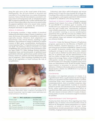
![for patients with severe hypermagnesemia and renal
impairment, or those with serious cardiovascular or
neuromuscular symptoms.
ACID-BASE DISORDERS
Regulation of Acid-Base Equilibrium
The body is sensitive to changes in blood pH level, as
disturbances in acid-base homeostasis can result in
denaturation of proteins and inactivation of enzymes that
may be potentially fatal. Strong mechanisms exist to
regulate acid-base balance and maintain arterial pH (7.35
to7.45), pC02 (35 to 45 mm Hg) and HC03-(20 to 28 mEq/1)
within a narrow range.
Acidemia is defined as H+
concentration exceeding
45 nmol/1 (pH <7.35); values below 35 nmol/1 (pH>7.45)
define alkalemia. The disorders that cause acidemia or
alkalemia are termed as acidosis and alkalosis respectively.
Metabolic activity results in production of two types of
acids, carbonic acid (a volatile acid, derived from carbon
dioxide) and nonvolatile acids (including sulfuric acid,
organic acids, uric acid and inorganic phosphates).
Accumulation of H+
ions of nonvolatile acidsdueto excess
production or inadequate buffering, failure to excrete H+
Fluid and Electrolyte Disturbances -
or loss of bicarbonate results in metabolic acidosis. If the
reverse occurs, it results in metabolic alkalosis. The
principle mechanism for carbon dioxide handling is by
the lungs. Hyperventilation results its CO2 washout and
drop in arterial pC02 (respiratory alkalosis), hypo
ventilation has the opposite effect (respiratory acidosis).
When only oneprimaryacid-baseabnormality occurs and
its compensatory mechanisms are activated, the disorder
is classified as simple acid-base disorder. A simple
algorithmfordefining simpleacid-base disordersis shown
in Fig. 5.9. When a combination of disturbances occurs,
the disorder is classified as a mixed acid-base disorder.
The latter aresuspectedwhenthecompensationin a given
patient differs from the predicted values in Table 5.11.
In order to maintain body homeostasis, changes in pH
are resisted by a complex system of intracellular and
extracellular buffers. The first line of defense, are the
chemical buffers. In metabolic disorders the extracellular
buffersrapidlytitrate theaddition of strong acids or bases.
Intracellular buffers chiefly accomplish the buffering of
respiratory disorders. Secondary respiratory compen
sations to metabolic acid-base disorders occur within
minutes and is completed by 12 to 24 hr. In contrast
secondary metabolic compensation of respiratory
Blood pH
Acidemia
(l pH)
--,
t
�
Lr�
IRespiratory acidosis Metabolic acido� Respiratory alkalosis Metaboli��
Respiratory acidosis
with compensatory
metabolic alkalosis
Metabolic acidosis
with compensatory
respiratory alkalosis
Metabolic alkalosis
with compensatory
respiratory acidosis
Fig. 5.9; Algorithm for simple acid-base disorders
Table 5.11: Compensation for primary acid-base disorders
Disorder Primary event Compensation Expected Compensation
Metabolic acidosis l [HC03
-J l pC02 pC02 l by 1-1.5 mm Hg for 1 mEq/l l[HC03]
Metabolic alkalosis t [HC03
-J t pC02 pC02 t by 0.5-1 mm Hg for 1 mEq/1 t [HC03]
Respiratory acidosis
Acute (<24 hr) tpC02 t[HC03-J [HC03] t by 1 mEq/1 for 10 mm Hg t pC02
Chronic (3-5 days) tpC02 ft [HC03-J [HC03
-J t by 4 mEq/1 for 10 mm Hg t pC02
Respiratory alkalosis
Acute (<24 hr) lpC02 l[HC03
-J [HC03
-J l by 1-3 mEq/1 for 10 mm Hg l pC02
Chronic (3-5 days) lpC02 U[HC03
-J [HC03
-J l by 2-5 mEq/1 for 10 mm Hg l pC02](https://image.slidesharecdn.com/paediatrics8theditiono-230807055545-451d483c/85/Paediatrics-8th-Edition-O-P-Ghai-pdf-69-320.jpg)
![include phosphates, sulfates and proteins (e.g. albumin).
Under typical conditions, unmeasured anions exceed
unmeasured cations; this is referred to as the anion gap
and can be represented by the following formula:
Anion gap = (Na+) - (Cl-+ HC03)
The anion gap is normally8 to 16 mEq/1. When a strong
acidisaddedtoorproducedinthebody, hydrogenionsare
neutralized by bicarbonate, resulting in a fall in bicarbo
nate. These acids include inorganic (e.g. phosphate or
sulfate), organic (e.g. ketoacids or lactate) or exogenous
(e.g. salicylate) acids incompletely neutralized by bicar
bonate. The accompanying unmeasured anion results in
increasedaniongapproportionaltothefallinbicarbonate.
Incontrast, when the bicarbonate is lost from the body, no
new anion is generated; therefore there is a reciprocal
increaseinchlorideions (proportionaltothefallinbicarbo
nate) resulting in normal anion gap. Hypoalbuminemia is
the most common cause of a low anion gap. Albumin
represents about half of the total unmeasured anion pool;
foreverydecreaseof 1 g/dlofplasmaalbumin, theplasma
anion gap decreases by 2.5 mEq/1.
Metabolic Acidosis
Metabolic acidosis is an acid-base disorder characterized
by a decrease in serum pH that results from either a loss
in plasma bicarbonate concentration or an increase in
hydrogen ion concentration (Table 5.12). Primary meta
bolic acidosis is characterized by an arterial pH of less
than 7.35 due to a decrease in plasma bicarbonate in the
absence of an elevated PaC02. If the measured PaC02 is
Table 5.12: Causes of metabolic acidosis
Normal anion gap (hyperchloremic acidosis)
Renal loss ofbicarbonate
Proximal (type 2) renal tubular acidosis, carbonic anhydrase
inhibitors (e.g. acetazolamide), tubular damage due to drugs
or toxins
Gastrointestinal bicarbonate loss
Diarrhea, ureteral sigmoidostomy, rectourethral fistula,
fistula or drainage of small bowel or pancreas
Decreased renal hydrogen ion excretion
Renal tubular acidosis type 1 and type 4 (aldosterone
deficiency)
Potassium sparing diuretics
Increased hydrogen chloride production
Parenteral alimentation, increased catabolisrn of lysine and
arginine
Ammonium chloride ingestion
Elevated anion gap
Increased acid production/accumulation: Sepsis, shock, poisonings
(ethanol, methanol, ethylene glycol); inborn errors of
metabolism
Ketoacidosis: Diabetic ketoacidosis, starvation
Exogenous acids: salicylates, iron, isoniazid, paraldehyde
Failure ofacid excretion: Acute or chronic renal failure
Fluid and Electrolyte Disturbances
-
higher thantheexpectedPaCO2, aconcomitant respiratory
acidosis is also present (caused by a depressed mental
state, airway obstruction or fatigue). Acutely, medullary
chemoreceptors compensate for metabolic acidosis
through increase in alveolar ventilation, which results in
tachypnea and hyperpnea that washes off CO2 and
corrects pH.
Calculation of plasma anion gap helps to classify
metabolic acidosis into thosewithelevated anion gap (i.e.
>12 mEq/1 as in increased acid production or decreased
losses) and those with normal anion gap (i.e. 8-12 mEq/1
as in gastrointestinal or renal loss of bicarbonate or when
hydrogen ions cannot be secreted because of renal failure)
(Table 5.12).
Another useful tool in the evaluation of metabolic
acidosis with normal anion gap is urinary anion gap.
Urinary anion gap = urinary [Na+]+ [K+] - [Cl-]
Urinary anion gap is negative in patients with diarrhea
regardless of urinarypH, andurinaryaniongapis positive
in renal tubular acidosis. An elevated osmolal gap (>20
mOsm/kg) with metabolic acidosis suggests the presence
of osmotically active agents such as methanol, ethylene
glycol or ethanol.
Clinical Features
Initially, patients with a metabolic acidosis develop a
compensatory tachypnea and hyperpnea, which may
progress if theacidemiaissevere, and the child can present
with significant work of breathing and distress (Kussmaul
breathing). An increase in H+ concentration results in
pulmonary vasoconstriction, which raises pulmonary
artery pressure and pulmonary vascular resistance.
Tachycardia is the most common cardiovascular effect
seen with mild metabolic acidosis. Cerebral vasodilation
occurs as a result ofmetabolicacidosis and maycontribute
to an increase in intracranial pressure. Acidosis shifts the
oxygen-hemoglobin dissociation curve to the right,
decreasing hemoglobin's affinity for oxygen. During
metabolic acidosis, excess hydrogen ionsmove toward the
intracellularcompartment andpotassium moves out of the
cell into the extracellular space. Untreated severe
metabolicacidosis maybeassociated with life-threatening
arrhythmias, myocardial depression, respiratory muscle
fatigue, seizures, shock and multiorgan failure.
Treatment
It is important to identify the cause of metabolic acidosis
as most cases resolve with correction of the underlying
disorder. The role of alkali therapy in acute metabolic
acidosis is limited. It is definitely indicated in some
situations, e.g. salicylate poisoning, inborn errors of
metabolism, or in those with pH below or equal to 7.0 or
[HC03-J less than 5 mEq/1, as severe acidosiscanproduce
myocardial dysfunction. The amount of bicarbonate
required is: Body weight (kg) x base deficit x 0.3.](https://image.slidesharecdn.com/paediatrics8theditiono-230807055545-451d483c/85/Paediatrics-8th-Edition-O-P-Ghai-pdf-71-320.jpg)
![__E_s_s_ e_n_t_ia_i_P_e _d_ia_t_ri_c_
s __________________________________
One ml of 7.5% sodium bicarbonate provides 0.9 mEq
bicarbonate. The recommendation is to replace only half
of the total bicarbonate deficit during the first few hours
of therapy. This amount is given as continuous infusion
over two hours. Rapid correction of acidosis with sodium
bicarbonate can lead to extracellular volume expansion,
exacerbating pulmonary edema in patients with cardiac
failure. In the latter, the rate of infusion should be slower
or sodium bicarbonate replaced by THAM [dose (ml) =
weight (kg) x base deficit] which is infused over 3-6 hr. If
hypernatremia is a concern, sodium bicarbonate may be
used as part of the maintenance intravenous solution.
During correction of acute metabolic acidosis, the effect
of sodium bicarbonate in lowering serum potassium and
ionized calcium concentrations must also be considered
and monitored. Since bicarbonate therapy generates large
amount of CO2, ventilation should increase proportion
ately otherwise this might worsen intracellular acidosis.
The inability to compensate may be especially important
in patients with diabetic ketoacidosis who are at risk for
cerebral edema. In diabetic ketoacidosis, insulin therapy
generally corrects the acidosis.
In newborns, frequent administration of hypertonic
solutionssuchassodiumbicarbonatehaveledtointracranial
hemorrhage resulting from hyperosmolality and resultant
fluid shifts from the intracellular space. Children with
inherited metabolicabnormalities,poisoning,orrenalfailure
may require hemodialysis.
Mild to moderate acidosis in renal failureor renal tubular
acidosis improves on oral alkali therapy, the dose being 0.5
to 2 mEq/kg/day of bicarbonate in 3-4 divided doses. In
cases of acidosisduetovolumedepletion,thevolumedeficit
should be corrected.
Metabolic Alkalosis
Metabolic alkalosis (pH >7.45) is an acid-base disturbance
caused by elevation in the plasma bicarbonate (HC03)
concentration in the extracellular fluid that results from a
net loss of acid, net gain of base or loss of fluid with more
chloride than bicarbonate. There are 2 types of metabolic
alkalosisclassified based on the amount of chloride in the
urine, i.e. chloride-responsive or chloride resistant
(Table 5.13). Chloride-responsive metabolic alkalosis
shows urine chloride levels of less than 10 mEq/1 and is
characterized by decreased ECF volume and low serum
chloride levels, such as occurs with vomiting or use of
diuretics.This type responds to administration of chloride
salt (usually as normal saline). Chloride resistant metabolic
alkalosis is characterized by urine chloride levels of more
than 20 mEq/l. Primary aldosteronism is an example of
chloride-resistant metabolic alkalosis and this type resists
administration of therapy with chloride.
The body compensates for metabolic alkalosis through
buffering of excessbicarbonateandhypoventilation. Intra
cellular buffering occurs through sodium-hydrogen and
potassium-hydrogen ion exchange, with eventual
Table 5.13: Causes of metabolic alkalosis
Chloride responsive
Gastric fluid loss (e.g. vomiting, nasogastric drainage)
Volume contraction (e.g. loop or thiazide diuretics, metolazone)
Congenital chloride diarrhea, villous adenoma
Cystic fibrosis
Post-hypercapnia syndrome (mechanically ventilated patients
with chronic lung disease)
Chloride resistant
Primary aldosteronism (adenoma, hyperplasia)
Renovascular hypertension, renin secreting tumor
Bartter and Gitelman syndromes
Apparent mineralocorticoid excess
Glucocorticoid remediable aldosteronism
Congenital adrenal hyperplasia (11�- and 17a-hydroxylase
deficiency)
Liddle syndrome
Excess bicarbonate ingestion
formation of CO2 and water from HC03.Within several
hours, elevated levels of HC03- and metabolic alkalosis
inhibit the respiratorycenter, resulting in hypoventilation
and increased pC02 levels. This mechanism produces
a rise in pC02 of as much as 0.7 to 1 mm Hg for each
1 mEq/1 increase in HC03.
Clinical Features
Signs and symptoms observed with metabolic alkalosis
usuallyrelatetothespecificdisease processthatcausedthe
acid-base disorder.Increased neuromuscular excitability
(e.g. from hypocalcemia), sometimes causes tetany or
seizures.Generalizedweaknessmaybenotedif thepatient
also has hypokalemia. Patients who develop metabolic
alkalosis from vomiting can have symptoms related to
severe volume contraction, with signs of dehydration.
Although diarrhea typically produces a hyperchloremic
metabolic acidosis, diarrheal stools may rarely contain
significant amountsofchloride,asinthecaseofcongenital
chloride diarrhea. Children with this condition present at
birthwithwatery diarrhea,metabolic alkalosis, andhypo
volemia. Weight gain and hypertension may accompany
metabolic alkalosis that results from a hyper
mineralocorticoid state.
Treatment
The overall prognosis in patients with metabolic alkalosis
depends on the underlying etiology. Prognosis is good
with prompt treatment and avoidance of hypoxemia. Mild
or moderate metabolic alkalosis or alkalemia rarely
requires correction.Forsevere metabolic alkalosis, therapy
should address the underlying disease state, in addition
to moderating the alkalemia. The initial target pH and
bicarbonate level in correcting severe alkalemia are
approximately 7.55 and 40 mEq/1, respectively.
Therapy with diuretics (e.g. furosemide, thiazides)
should be discontinued. Chloride-responsive metabolic
alkalosis responds to volume resuscitation and chloride](https://image.slidesharecdn.com/paediatrics8theditiono-230807055545-451d483c/85/Paediatrics-8th-Edition-O-P-Ghai-pdf-72-320.jpg)
![__E_s_s_e_n_ t_ia_l_P_e_d_ i_a _tr_ic_s_________________________________
with chronic hypervitaminosis may have dermatitis,
alopecia, hepatosplenomegalyand/or hyperostosis.When
taken by pregnant women in early gestation at daily levels
ofmorethan 7500 µg,fetal anomalies andpoor reproductive
outcomes are reported. The WHO recommends that
vitamin A intake during pregnancy should not exceed
3000 µg daily or 7500 µg every week.
Vitamin D
Vitamin D is the generic term for secosteroids,which have
an important role in maintaining calcium andphosphorus
homeostasis. Secosteroids have three intact rings and one
open ringwithconjugateddouble bonds. Various vitamin
D metabolites differ in the side chains attached to the
fourth ring. Vitamin D is a group of precursors of a
hormone, 1,25-dihydroxycholecalciferol, synthesized and
secreted by the kidneys under thecontrol of parathormone
and tissue phosphate levels (Fig. 7.2). Dietary vitamin D
is essential if the cutaneous synthesis of vitamin D3 is
insufficient. When deficient, disease manifestations
include rickets, with defective mineralization of growing
bone, and osteomalacia with impaired mineralization of
non-growing bones.
Absorption, metabolism and mechanism of action
Vitamin D refers to two prohormones, vitamin D2 (ergo
calciferol; derived from plants) and D3 (cholecalciferol,
available from animal sources) and their derivatives.
Vitamin D is absorbed in the duodenum by an active
transport system. In the enterocyte, vitamin D is incor
porated into chylomicrons and transported to the liver,
where its hydroxylation take place to form 25-hydroxy
vitaminD2 [250HD2]and25-hydroxyvitaminD3 [250HD3],
known as ercalcidiol and calcidiol respectively. This
hydroxylation is substrate dependent and without any
negative feedback control. Calcidiol is released into the
bloodstreamandhasa biological half-life ofapproximately
3 weeks. Subsequent hydroxylation by la-hydroxylase in
the proximal renal tubule leads to formation of 1,25-
dihydroxyvitamin D2 [l,25(0H)iD2] or ercalcitriol and
1,25-dihydroxyvitamin D3 [l,25(0H)iD3] or calcitriol(Fig.
7.2). While circulating l,25(0H)iD almost exclusively
results from renal production, extrarenal conversion also
takes place in the skin, colon, macrophages, vascular
smooth muscle cells, bone and parathyroid glands. Renal
conversion is regulated by parathormone, calcitonin,
calcium and phosphate; and inhibited by calcitriol and
the phosphaturic hormone, fibroblast growth factor 23
(FGF-23). A negative relationship exists between serum
250HD and parathormone levels (Fig. 7.2). Vitamin D
supplementation suppresses serum parathormone and
increases bone mineral density.
Active vitamin D affects calcium homeostasis through
its action on the intestine, kidney and bones. In the
intestine,thehormone induces calciumtransport proteins
and an intracellular calcium-binding protein (calbindin)
which aid in transport of calcium across the enterocyte.
In the kidney, the hormone enhances calcium resorption
in the tubule by a similar mechanism. It also inhibits the
activity of la-hydroxylase and stimulates renal 24-
hydroxylaseactivitythat inactivates both the substrate and
calcitrio1.
Dehydrocholesterol
�
Dietary (vitamin 02, 03)
�V�mioD/
l;�!:�roxylase
24,25-dihydroxyvitamin D .__
24_
·h
-
yd_rox_y1_as_e
_ 25-hydroxyvitamin D
(inactive)
Bone
Increased mineralization
-
I
1a·hydroxylase Parathormone
+l Kidney
1 + Low phosphorus
I •::.-------Fibroblast growth factor
1,25-dihydroxyvitamin D ----• Decrease parathormone
Kidney
Increased calcium and
phosphate reabsorption
Intestine
Increased calcium and
phosphate absorption
Fig. 7.2. Vitamin D metabolism. Serum levels of fibroblast growth factor (FGF) 23 are elevated in response to increased serum phosphate and
also inhibit the production of parathormone](https://image.slidesharecdn.com/paediatrics8theditiono-230807055545-451d483c/85/Paediatrics-8th-Edition-O-P-Ghai-pdf-98-320.jpg)
![__
e_s_s_e_n_ti_a
_1_P_e_d_1a_t_ri
_cs-------------------------------
Fig. 7.3: A 5-yr-
old child with rickets with wide wrists and bow legs
Fig. 7.4: Radiograph of wrist in 4-yr-old boy with rickets. Note
widening, cupping and fraying at the metaphyseal ends of forearm
bones
Table 7.2: Vitamin D levels in serum
Deficient
Insufficient
Optimal
High
Toxic
25-hydroxyvitamin D level (ng/ml)
Less than 10
10-20
20-60
60-90
Greater than90
daily for 10 days) followed by a maintenance dose of
400-800 IU/day and oral calcium supplements (30-
75mg/kg/day)for2 months. Followingadequatetherapy,
most patients with vitamin D deficiency rickets show
radiological evidence of healing (Fig. 7.5)within 4 weeks.
Reduction in blood levels of alkaline phosphatase and
resolution of clinical signs occur slowly. If radiologic
healing cannotbedemonstrated, despite 1 -
2large dosesof
vitamin D, patients should be evaluated for refractory
rickets (Fig. 7.6).
Familial Hypophosphatemic Rickets
This is the most commonly inherited form of refractory
rickets, being inheritedas X-linked dominantwithvariable
penetrance. Sporadic instances are frequent and an
autosomal recessive inheritance has also been reported.
Pathogenesis The gene for X-linked hypophosphatemic
rickets is termed the PHEX gene (phosphate regulating
gene with homology to endopeptidases on the X chromo
some). Theunderlying defect involves impairedproximal
tubular reabsorption of phosphate. Despite hypophos
phatemia the blood levels of 1,25(0H)z03 are low, which
Fig. 7,5: Healing of the growth plate after vitamin D therapy
Refractory rickets
Serum phosphate]
I
+
Low or Normal High
+
Blood pH]
I
Chronic kidney disease
Lowl Normal
...
Renal tubular acidosis Serum PTH, calcium
i i
High PTH, low or normal calcium] Normal PTH, normal calcium
+ +
Vitamin D dependent rickets Hypophosphatemic rickets
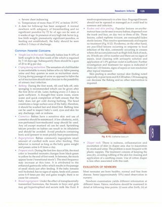
Fig. 7.6: Biochemical evaluation of a child with refractory rickets](https://image.slidesharecdn.com/paediatrics8theditiono-230807055545-451d483c/85/Paediatrics-8th-Edition-O-P-Ghai-pdf-100-320.jpg)


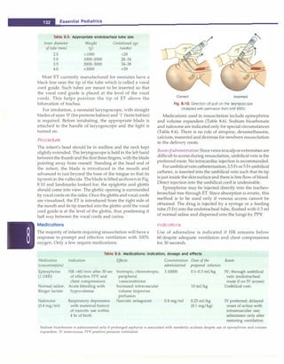
![some instances, the trachea in non-vigorous baby born
through meconium stained liqor). If necessary, insert an
endotracheal (ET) tube to ensure an open airway.
B-Breathing: Tactile stimulation to initiate respirations,
positive-pressure breaths using either bag and mask or
bag and ET tube when necessary.
C-Circulation: Stimulate and maintain the circulation of
blood with chest compressions and medications as
indicated.
Resuscitation Algorithm
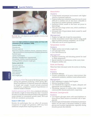
Figure 8.2 presents the algorithm of neonatal resuscitation.
At the time of birth, one should ask three questions about
the newborn:
i. Term gestation?
ii. Breathing or crying?
iii. Good muscle tone? (flexed posture and active
movement of baby denotes good tone)
Birth
I
I
I
I
I
I
I
I
I
I
I
30 sec
I
I
I
I
I
I
I
60 sec
Term gestation?
Breathing or crying?
Good tone?
No
Provide warmth, clear airway
if necessary, dry, stimulate
Heart rate below 100,
gasping or apnea?
Yes
1
[
Positive pressure ventilation
]
SpO, monitoring
!
1Heart rate below 100?1
Yes
[ Take ven� �ective step7")
_!
Heart rate below 60?
Yes!
___ -
r-- Consider intubation ::i
Chest compressions
Coordinate with positive pressure ventilation
!
L!:!.eart rate below 60?J
Yesu
IV epinephrine
Yes
No
No
Newborn Infants -
If answers to all the three questions are 'Yes', the baby
does not require any active resuscitation and "Routine
care" should be provided. Routine care consists of four
steps:
i. Warmth: Provided by putting the baby directly on the
mother's chest in skin-to-skin contact.
ii. Clearingofairway ifrequired: Done bywipingthe baby's
mouth and nose using a clean cloth. No need to suction
routinely.
iii. Dry the baby
iv. Ongoing evaluation for vital parameters. Helping
mother in breastfeeding will facilitate easy transition
to extrauterine environment.
If answer to any of the three questions is "No", the baby
requires resuscitation. After cutting the cord, the baby
should be subjected to a set of interventions known as
Initial steps.
Routine care
Provide warmth
Clear airway, if necessary Assessment
Dry
Keep with mother
Ongoing evaluation
rNo
,"Labored breathing Evaluation
�� pe2!.stent cyanosis?
Yes
Clear airway, SpO, monitoring
]
Co�erCPAP
Evaluation
care
Targeted SpO,after birth
1 minute: 60-65%
2 minutes: 65-70%
3 minutes: 70-75% Evaluation
4 minutes: 75-80%
5 minutes: 80-85%
10 minutes: 85-95%
Fig. 8.2: The algorithm of neonatal resuscitation. CPAP continuous positive airway pressure; PPV positive ventilation; Sp02 saturation of oxygen.
(Adapted with permission from American Academy of Pediatrics 2010)](https://image.slidesharecdn.com/paediatrics8theditiono-230807055545-451d483c/85/Paediatrics-8th-Edition-O-P-Ghai-pdf-113-320.jpg)
![- Essential Pediatrics
antibodies (IgM and IgG) develop at the time symptoms
occur, usually before the development of jaundice. IgM
anti-HEY titer declines rapidly during early convale
scence,whileIgG anti-HEYpersistsfor long duration and
provides protection against subsequent infections.
Treatment
As no specific therapy is capable of altering the course of
acute hepatitis E infection, prevention is the most effective
approach against the disease.
Prevention
Good personal hygiene, high quality standards for public
water supplies and proper disposal of wastehave resulted
in a low prevalence of HEV infections in developed
countries. At present, thereare no commercially available
vaccines for the prevention of hepatitis E.
Hepatitis due to other Viruses
Certain cases of post-transfusion (10%) and community
acquired hepatitis (20%) are of unknown origin. Three
viruses are potentially associated with liver disease but
no conclusive evidence exists to support them as a cause
for these cases. These viruses are HGV/GB virus C, TT
virus and SEN virus.
Suggested Reading
Aggarwal R, Jameel S. Hepatitis E. Hepatology 2011 Dec;54:2218-26
Dengue Infections
Dengue fever is an acute illness characterized by fever,
myalgia, arthralgia and rash. Severe dengue infection is
characterized by abnormalities in hemostasis and by
marked leakage of plasma from the capillaries; the latter
may lead to shock (dengue shock syndrome).
Epidemiology
The global prevalence of dengue has grown dramatically
in recent decades. The disease is now endemic in more
than 100 countries in Africa, the Americas, the Eastern
Mediterranean, South-East Asia and the Western Pacific.
WHO currently estimates there may be 50 million cases
of dengue infection worldwide every year. During
epidemics of dengue, attack rates among susceptible are
often40-50%,but mayreach80-90%. An estimated 500,000
cases of severe dengue infection require hospitalization
each year, of which very large proportions are children.
Without proper treatment in severe dengue infection
[earlier called dengue hemorrhagic fever (DHF) dengue
shock syndrome (DSS)] case fatality rates can exceed 20%.
The spread of dengue is attributed to expanding
geographic distribution of the four dengue viruses andof
their mosquito vectors, the most important of which is
the predominantly urban species Aedes aegypti. A rapid
rise in urbanpopulationsis bringing evergreaternumbers
of people into contact with this vector, especially in areas
that are favorable for mosquito breeding, e.g. where
household water storage is common and where solid
waste disposal services are inadequate.
Virus. Dengue fever is causedbyinfectiondue to any of the
four serotypes of dengue viruses. Dengue viruses are
arboviruses that belong to the family Flaviviridae. The
envelop protein bears epitopes that are unique to the
serotypes;theantibodiestotheseuniqueepitopesneutralize
by interfering with the entry of the virus into the cells.
Transmission. Dengue viruses are transmitted to humans
through the bites of infected female Aedes mosquitoes.
Mosquitoes generally acquire the virus while feeding on
the blood of an infected person. After incubation for 8-10
days, an infected mosquito is capable, during probing and
blood feeding, of transmitting the virus, to susceptible
individuals for the rest of its life. Infected female
mosquitoes may also transmit the virus to their offspring
by transovarial transmission, but the role of this in
sustaining transmission of virus to humans has not yet
been delineated.Humans are the main amplifying host of
the virus, although studies have shown that in some parts
of the world monkeys may become infected and perhaps
serve as a source of virus for mosquitoes. The virus
circulates inthebloodof infected humans fortwo to seven
days, at approximately the same time as they have fever;
Aedes mosquitoes may acquire the virus when they feed
on an individual during this period.
Pathophysiology
The major pathophysiologic changes that determine the
severity of disease in severe dengue infection and
differentiate it from dengue fever are plasma leakage and
abnormal hemostasisleading to rising hematocrit values,
moderate to marked thrombocytopenia and varying
degrees ofbleeding manifestations. Thecause of abnormal
leakage of plasma is not entirely understood. However,
rapid recovery without residual abnormality in vessels
suggests it to be the result of release and interaction of
biological mediators, which are capable of producing
severe illness with minimal structural injury.
It has been observed that sequential infection with any
two of the four serotypes of dengue virus results in severe
dengue infections in an endemic area. How a second
dengue infection causes severe disease andwhyonlysome
patients get severe disease remains unclear.It is suggested
that the residual antibodies produced during the first
infection areabletoneutralize a secondviralinfection with
the same serotype. However, when no neutralizing
antibodies are present (i.e. infection due to another
serotype of dengue virus), the second infection is under
the influence of enhancing antibodies and the resulting
infection and disease are severe. An alternative expla
nation is that certain strains (South-East Asian) of the
dengue virus may be inherently capable of supporting](https://image.slidesharecdn.com/paediatrics8theditiono-230807055545-451d483c/85/Paediatrics-8th-Edition-O-P-Ghai-pdf-210-320.jpg)

![__
E_s_s_e _n_t.ia•l•P•e•d-i
a_ t_r.ic_s_________________________________
Severe dengue fever
[A�ssme;;t° of shock
Hypotension
�+
loss 1111
..
[u'n'rec"ord� blood pressure j
+
DSSIV
Ringer lactate 20 ml/kg bolus; up to 3 boluses
Ringer lactate (RL) 10-20 ml/kg/hr]
,-- • Assessment1------------�
�Noimp�
Gradually decrease �ocrit increasedJ<14------'
jHe
matocrit !ecreasedj
Ringer lactate infusion with 1 -- T
monitoring as in Fig. 10.9 't 't
-r Colloids 10 ml/kg [Blood transfusio�
I •fAs-;;sment
1
4 I
I
.�---------'------..
Improved � improveme-;it]
Look for anemia, acidosis, myocardial dysfunction; treat accordingly
Fig. 10.10: Algorithm for management of severe dengue fever. DSS dengue shock syndrome
illness. Heart rate, respiratory rate, blood pressure and
pulse pressure should be monitored every 30 min till the
patient is stable, thereafter every 2-4 hr should be
continued as long as the child is in the hospital. In critically
ill children, central venous pressure and accurate urine
output with an indwelling urinary catheter should be
monitored. Absolute platelet counts should be checked
once a day till it shows a rising trend.
Prognosis
Dengue fever is a self-limited disease but the mortality in
severe dengue may be as high as 20-30% if left untreated.
Early recognition of illness, careful monitoring and
appropriate fluid therapy alone has resulted in consi
derable reduction ofcase fatality rate to less than 1 percent.
Early recognition of shock is of paramount importance as
the outcome of a patient with DSS depends on the duration
of shock. If shock is identified when pulse pressure starts
getting narrow and intravenous fluids are administered
at this stage, the outcome is excellent.
Prevention
Preventive measures are directed towards elimination of
adult mosquitoes and their larvae. During epidemics aerial
spraying or fogging with malathion is recommended for
control of adult mosquitoes. However, larval control
measures by source reduction anduse of larvicides are even
more crucial. Aedes aegypti mosquitoes breed in and around
human dwellings and flourish in fresh water. Special drives
should be launched during and soon after the rainy season
to interrupt breeding cycle of mosquitoes. There should be
no opportunity for stagnation of water in the bathroom,
kitchen, terrace,lawnandotheropenplaces. The stored water
should be kept covered. Cooperation from every house
owner and public establishment is crucial for the success of
control program. Strong motivationandcommitment on the
part of government and its employees are fundamental pre
requisites for the success of control measures.
Mesocyclops, the shellfish are credited to eat and
effectively eliminate larvae of Aedes aegypti. The strategy
has been used with success by Australian scientists
working in Vietnam by growing shellfish in ponds and
water traps. A live attenuated quadruple vaccine is
undergoing clinical trials but there are concerns whether
vaccine may predispose to development of severe
dengue.
Suggested Reading
Anonymous. Dengue Guidelines for Diagnosis, Treatment,
Prevention and Control. A joint publication of the World Health
Organization (WHO) and the Special Programme for Research and
Training in Tropical Diseases (TOR), 2009
Kabra SK, Jain Y, Pandey RM, et al. Dengue hemorrhagic fever in
children in the 1996 Delhi epidemic. Trans Royal Society Trop Med
Hygiene 1999;93:294-8](https://image.slidesharecdn.com/paediatrics8theditiono-230807055545-451d483c/85/Paediatrics-8th-Edition-O-P-Ghai-pdf-214-320.jpg)
![---E•s•s•e•n•t-ia•l•P-e.d.ia.t.ri•c•
s----------------------------------
p9, p6; these are derived from the precursor p55), the pol
region, which encodes the viral enzymes (reverse
transcriptase [p51], protease [plO], and integrase [p32]);
and the env region, which encodes the viral envelope
proteins (gp120 and gp41).
The major external viral protein of HIV-1 is a heavily
glycosylated gp120 protein which contains the binding
site for the CD4 molecule, the most common T lymphocyte
surface receptor for HIV. Most HIV strains have a specific
tropism for one of the chemokines: the fusion-inducing
molecule, CXCR-4, which has been shown to act as a co
receptor for HIV attachment to lymphocytes, and CCR-5,
a � chemokine receptor that facilitates HIV entry into
macrophages.
Following viral attachment, gp120 and the CD4
molecule undergo conformational changes, allowing gp41
to interact with the fusion receptor on the cell surface. Viral
fusion with the cell membrane allows entry of viral RNA
into the cell cytoplasm. Viral DNA copies are then trans
cribed from the virion RNA through viral reverse
transcriptase enzyme activityandduplicationof the DNA
copies produces double-stranded circular DNA. Because
the HIV-1 reverse transcriptase is error-prone, many
mutations arise, creating wide genetic variation in HIV-1
isolates even within an individual patient. The circular
DNA is transported into the cell nucleus where it is
integrated into chromosomal DNA; this is called as the
provirus. The provirus can remain dormant for extended
periods.
HIV-1transcriptionis followed bytranslation. A capsid
polyproteinis cleaved to produce, among other products,
the virus-specific protease (plO). This enzyme is critical
forHIV-1 assembly. TheRNAgenomeisthen incorporated
into the newly formed viral capsid. As the new virus is
formed, itbudsthrough thecellmembrane andisreleased.
HIV-2 is a rare cause of infection in children. It is most
prevalent in Western and Southern Africa. If HIV-2 is
suspected, a specific test that detects antibody to HIV-2
peptides should be used.
Transmission. Transmission of HIV-1 occurs via sexual
contact,parenteralexposuretoblood,orverticaltransmission
from mother to child. The primary route of infection in the
pediatric population is vertical transmission. Most large
studies in the United States and Europe have documented
mother-to-child transmission rates in untreated women
between 12 and 30%. In contrast, transmission rates in
Africa and Asia are higher, up to 50%.
Vertical transmission of HIV can occur during the
intrauterine or intrapartum periods, or through breast
feeding. Up to 30% of infected newborns are infected in
utero. The highest percentages of HIV-infected children
acquire the virus intrapartum. Breastfeeding is an
important route of transmission, especially in the
developing countries. The risk factors for vertical
transmission include preterm delivery (<34 week
gestation), a low maternal antenatal CD4 count, use of
illicit drugsduringpregnancy, >4 hr durationofruptured
membranes and birthweight <2500 g.
Transfusions of infected blood or blood products have
accounted for a variable proportion of all pediatric AIDS
cases. Heat treatment of factor VIII concentrate and HIV
antibody screening of donors hasvirtually eliminatedHIV
transmission to children with hemophilia. Blood donor
screening has dramatically reduced, but not eliminated,
the risk of transfusion-associated HIV infection. Sexual
contact is a major route of transmission in the adolescent
population.
Natural History
Before highly active antiretroviral therapy (HAART) was
available, three distinct patterns of disease were described
in children. Approximately 10-20% of HIV-infected
newborns in developed countries presented with a rapid
disease course, with onset of AIDS and symptoms during
the first few months of life and, if untreated, death from
AIDS-related complications by 4 yr of age. In resource
poor countries, >85% of the HIV-infected newborns may
have such a rapidly progressing disease.
It has been suggested that if intrauterine infection
coincides withthe period of rapid expansion of CD4 cells
in the fetus, it could effectively infect the majority of the
body's immunocompetent cells. Most children in this
group have a positive HIV-1 culture and/or detectable
virus in the plasma in the first 48 hr of life. This early
evidence of viral presence suggests that the newborn was
infected in utero. In contrast to the viralload in adults, the
viral load in infants stays high for at least the first 2 yr of
life.
The majority ofperinatallyinfectednewborns (60-80%)
present with a second pattern-that of a much slower
progression of disease with a median survivaltimeof 6 yr.
Many patients in this group have a negative viral culture
or PCR in the 1st week of life and are therefore considered
to be infected intrapartum. In a typical patient, the viral
load rapidly increases by 2-3 months of age (median
100,000copies/ml) andthenslowlydeclines over a period
of 24 months. This observation can be explained partially
by theimmaturity of theimmune system in newborns and
infants. The thirdpatternofdisease (i.e. longterm survivors)
occurs in a small percentage (<5%) of perinatally infected
children who have minimal or no progression of disease
with relatively normal CD4 counts and very low viral
loads for longer than 8 yr.
HIV-infected children have changes in the immune
system that are similar to those in HIV-infected adults.
CD4 cell depletion may be less dramatic because infants
normally have a relative lymphocytosis. Therefore, for
example, a value of 1,500 CD4 cells/mm3
in children <1
yr of age is indicative of severe CD4 depletion and is
comparableto<200CD4 cells/mm3
inadults. Lymphopenia
is relatively rare in perinatally infected children and is](https://image.slidesharecdn.com/paediatrics8theditiono-230807055545-451d483c/85/Paediatrics-8th-Edition-O-P-Ghai-pdf-216-320.jpg)
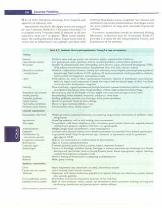
![Infections and Infestations -
HIV-infected
pregnant woman
Delivery HIV-exposed infant
(breastfed and nonbreastfed)
J�-::�·
Symptomatic HIV-exposed
child <18 months of age
(not previously diagnosed)
First HIV DNA PCR
ie
Repeat HIV DNA PCR]
to confirm _J
G 8
I
8 l
C --;;;;ative �
PCR tes�
Second PCR after 6-8 weeks I
of stopping breastfeeding or
I
earlier if symptomatic __J
[°s;cond PCR at 6 months
l
l to confirm status J
I
G
Repeat test and refer for
followup
8
Report
HIV negative
Repeat test
and refer for
followup
Fig. 10.11: Diagnosis of infection with HIV in children <18 months. If child is >18-month-old, adult testing strategies may be used
alternative regimen is a combination of stavudine,
lamivudine and nevirapine. The details of the anti
retroviral drugs are shown in Table 10.6. Pediatric fixed
dose combinations have been developed, and these are
administered using a weight-band based dosing system
(NACO guidelines).
Cotrimoxazole Prophylaxis
In resource-limited settings, cotrimoxazole prophylaxis is
recommended for all HIV exposed infants starting at
4-6 weeks of age and continued until HIV infection can be
excluded. Cotrimoxazole is also recommended for HIV
exposed breastfeeding children of any age and cotrimo
xazole prophylaxis should be continued until HIV infection
canbeexcluded byHIVantibodytesting (beyond 18months
of age) or virological testing (before 18 months of age) at
least six weeks after complete cessation of breastfeeding.
All children younger than one yr of age documented to
be living with HIV should receive cotrimoxazole pro
phylaxisregardlessofsymptomsor CD4percentage. After
one yr of age, initiation of cotrimoxazole prophylaxis is
recommended for symptomatic children (WHO clinical
stages 2, 3 or 4 for HIV disease) or children with CD4
<25%. All children who begin cotrimoxazole prophylaxis
(irrespective of whether cotrimoxazole was initiatedin the
first yr of life or after that) should continue until the age
of five yr, when they can be reassessed.
Nutrition
It is important to provide adequate nutrition to HIV
infected children. Many of these children have failure to
thrive. These children will need nutritional rehabilitation.
In addition, micronutrients like zinc may be useful.
Immunization
The vaccines that are recommended in the national
schedule can be administered to HIV infected children
except that symptomatic HIVinfectedchildrenshould not
be given the oral polio and BCG vaccines.
Prevention of Mother to Child Transmission (PMTCT)
The risk of MTCT can be reduced to under 2% by
interventions that include antiretroviral (ARV) prophylaxis
given to women during pregnancy and labor and to the
infant in the first weeks of life, obstetrical interventions
including elective cesarean delivery (prior to the onset of
labor and rupture of membranes)and complete avoidance
of breastfeeding.
Antiretroviral drug regimens for treating pregnant
women For HIV-infected pregnant women in need of
ART for their own health, ART should be administered
irrespective of gestational age and is continued through
out pregnancy, delivery and thereafter (recommendedfor
all HIV-infected pregnant women with CD4 cell count
<350 cells/mm3
, irrespective of WHO clinicalstaging;and
for WHO clinical stage 3 or 4, irrespective of CD4 cell
count).
Recommended regimen for pregnant women with
indication for ART is combination of zidovudine (AZT),
lamivudine (3TC) and nevirapine (NVP) or efavirenz
(EFV) during antepartum, intrapartum and postpartum](https://image.slidesharecdn.com/paediatrics8theditiono-230807055545-451d483c/85/Paediatrics-8th-Edition-O-P-Ghai-pdf-221-320.jpg)
![- Essential Pediatrics
Fig. 10.20: Miliary shadows with right paratracheal adenopathy
is most often made by combination of a positive tuberculin
skin test, chest radiograph, physical examination and
history of contact with adult patient with tuberculosis.
Newerdiagnostic methods such as PCR and serodiagnosis
have not given encouraging results. Newer staining and
culture methods have found their place in the manage
ment of tuberculosis. There is a need to develop better
techniques for diagnosis of tuberculosis in children. A
suggested algorithm for diagnosis of pulmonary tuber
culosis is given in Fig. 10.21.
Treatment
The principles of therapy in children with tuberculosis
are similar to that of adults. The drugs used for treatment
of tuberculosis in children are given in Table 10.9.
Drug Regimens
During the last few yr, changes have occurred in the
therapeutic approach to childhood tuberculosis as a result
of large number of treatment trials for children and
concern about the development of resistance to anti
tuberculosis drugs. Short-course chemotherapy, with the
Fever and/or cough for >2 weeks
+/- loss of weight or no weight gain
+/- history of contact with suspected or
confirmed case of active tuberculosis
Sputum examination
AFB present
Mantoux positive
l
and abnormal
chest X-ray
Treat for tuberculosis
ISputum negative
..
Oral antibiotics for 7-10 days
!Symptoms persist
Chest X-ray and Mantoux test
Mantoux negative or
Mantoux positive, chest X-ray normal]
•
Investigate further for
tuberculosis and other diagnosis
Fig. 10.21: Proposed diagnostic algorithm for pulmonary tuberculosis
treatment duration of 6 months, has become the standard
practice. Extrapulmonary TB (osteoarticular TB, neuro
logical TB) and disseminated TB are treated for longer
durations of 9-12 months or more.
The major problem in inclusion of children in directly
observed treatment short-course (DOTS) program has
been a difficulty in demonstration of AFB andclassification
of different clinical manifestations according to categories
described for adults. There have been efforts to develop
classification of different types of childhood tuberculosis
into 2 categories similar to those for adults. Recently a
consensus statement jointly prepared by Indian Academy
of Pediatrics and Revised National Tuberculosis Control
Program (RNTCP) has also proposed a classification of
different types of tuberculosis in children into two
categories (Table 10.10). In the RNTCP, thrice weekly
intermittent directly observed therapy is recommended.
Corticosteroids
Corticosteroids, in addition to antitubercular drugs, are
useful in treatment of patients with CNS tuberculosis and
occasionally pulmonary tuberculosis. These are useful in
Table 10.9: Doses and side effects of antitubercular drugs
Drug Dose (mg/kg/
day; frequency)
Isoniazid 10-15; q 24 hr
Rifampicin 10-20; q 24 hr
Streptomycin 20-25; q 24 hr
Ethambutol 15-25; q 24 hr
Pyrazinamide 25-35; q 24 hr
Ethionamide 15-20; q 12 hr
Cycloserine 15-20; q 12 hr
Side effects
Hepatotoxicity, hypersensitivity rash, fever, peripheral or optic neuritis, psychosis, seizures
Nausea, vomiting, hepatotoxicity, flu-like syndrome, blood dyscrasia, arthralgia, wheezing
Ototoxicity (vestibular or hearing loss), rash, fever, arthralgia, neuromuscular blockade,
peripheral neuritis, anaphylaxis
Hypersensitivity (rash, fever), joint pain, optic neuritis, GI upset, confusion, dizziness
GI upset, hepatotoxicity, hyperuricemia, photosensitivity, dysuria, arthralgia, fever,
thrombocytopenia
GI upset, hepatotoxicity, peripheral neuropathy, gynecomastia, rash, alopecia, headache,
diplopia, blurred vision, tremors, hypothyroidism
Seizures, psychosis, peripheral neuritis](https://image.slidesharecdn.com/paediatrics8theditiono-230807055545-451d483c/85/Paediatrics-8th-Edition-O-P-Ghai-pdf-242-320.jpg)
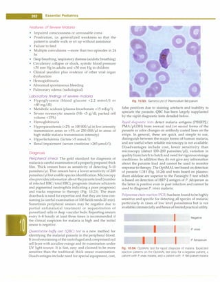
![Paediatrics (8th Edition) [O.P. Ghai] .pdf](https://image.slidesharecdn.com/paediatrics8theditiono-230807055545-451d483c/85/Paediatrics-8th-Edition-O-P-Ghai-pdf-249-320.jpg)
![Paediatrics (8th Edition) [O.P. Ghai] .pdf](https://image.slidesharecdn.com/paediatrics8theditiono-230807055545-451d483c/85/Paediatrics-8th-Edition-O-P-Ghai-pdf-250-320.jpg)
![Paediatrics (8th Edition) [O.P. Ghai] .pdf](https://image.slidesharecdn.com/paediatrics8theditiono-230807055545-451d483c/85/Paediatrics-8th-Edition-O-P-Ghai-pdf-251-320.jpg)
![Paediatrics (8th Edition) [O.P. Ghai] .pdf](https://image.slidesharecdn.com/paediatrics8theditiono-230807055545-451d483c/85/Paediatrics-8th-Edition-O-P-Ghai-pdf-252-320.jpg)
![Paediatrics (8th Edition) [O.P. Ghai] .pdf](https://image.slidesharecdn.com/paediatrics8theditiono-230807055545-451d483c/85/Paediatrics-8th-Edition-O-P-Ghai-pdf-253-320.jpg)
![Paediatrics (8th Edition) [O.P. Ghai] .pdf](https://image.slidesharecdn.com/paediatrics8theditiono-230807055545-451d483c/85/Paediatrics-8th-Edition-O-P-Ghai-pdf-254-320.jpg)
![Paediatrics (8th Edition) [O.P. Ghai] .pdf](https://image.slidesharecdn.com/paediatrics8theditiono-230807055545-451d483c/85/Paediatrics-8th-Edition-O-P-Ghai-pdf-255-320.jpg)
![Paediatrics (8th Edition) [O.P. Ghai] .pdf](https://image.slidesharecdn.com/paediatrics8theditiono-230807055545-451d483c/85/Paediatrics-8th-Edition-O-P-Ghai-pdf-256-320.jpg)
![Paediatrics (8th Edition) [O.P. Ghai] .pdf](https://image.slidesharecdn.com/paediatrics8theditiono-230807055545-451d483c/85/Paediatrics-8th-Edition-O-P-Ghai-pdf-257-320.jpg)
![Paediatrics (8th Edition) [O.P. Ghai] .pdf](https://image.slidesharecdn.com/paediatrics8theditiono-230807055545-451d483c/85/Paediatrics-8th-Edition-O-P-Ghai-pdf-258-320.jpg)
![Paediatrics (8th Edition) [O.P. Ghai] .pdf](https://image.slidesharecdn.com/paediatrics8theditiono-230807055545-451d483c/85/Paediatrics-8th-Edition-O-P-Ghai-pdf-259-320.jpg)
![Paediatrics (8th Edition) [O.P. Ghai] .pdf](https://image.slidesharecdn.com/paediatrics8theditiono-230807055545-451d483c/85/Paediatrics-8th-Edition-O-P-Ghai-pdf-260-320.jpg)
![Paediatrics (8th Edition) [O.P. Ghai] .pdf](https://image.slidesharecdn.com/paediatrics8theditiono-230807055545-451d483c/85/Paediatrics-8th-Edition-O-P-Ghai-pdf-261-320.jpg)
![Paediatrics (8th Edition) [O.P. Ghai] .pdf](https://image.slidesharecdn.com/paediatrics8theditiono-230807055545-451d483c/85/Paediatrics-8th-Edition-O-P-Ghai-pdf-262-320.jpg)
![Paediatrics (8th Edition) [O.P. Ghai] .pdf](https://image.slidesharecdn.com/paediatrics8theditiono-230807055545-451d483c/85/Paediatrics-8th-Edition-O-P-Ghai-pdf-263-320.jpg)
![Paediatrics (8th Edition) [O.P. Ghai] .pdf](https://image.slidesharecdn.com/paediatrics8theditiono-230807055545-451d483c/85/Paediatrics-8th-Edition-O-P-Ghai-pdf-264-320.jpg)
![Paediatrics (8th Edition) [O.P. Ghai] .pdf](https://image.slidesharecdn.com/paediatrics8theditiono-230807055545-451d483c/85/Paediatrics-8th-Edition-O-P-Ghai-pdf-265-320.jpg)
![Paediatrics (8th Edition) [O.P. Ghai] .pdf](https://image.slidesharecdn.com/paediatrics8theditiono-230807055545-451d483c/85/Paediatrics-8th-Edition-O-P-Ghai-pdf-266-320.jpg)
![Paediatrics (8th Edition) [O.P. Ghai] .pdf](https://image.slidesharecdn.com/paediatrics8theditiono-230807055545-451d483c/85/Paediatrics-8th-Edition-O-P-Ghai-pdf-267-320.jpg)
![Paediatrics (8th Edition) [O.P. Ghai] .pdf](https://image.slidesharecdn.com/paediatrics8theditiono-230807055545-451d483c/85/Paediatrics-8th-Edition-O-P-Ghai-pdf-268-320.jpg)
![Paediatrics (8th Edition) [O.P. Ghai] .pdf](https://image.slidesharecdn.com/paediatrics8theditiono-230807055545-451d483c/85/Paediatrics-8th-Edition-O-P-Ghai-pdf-269-320.jpg)
![Paediatrics (8th Edition) [O.P. Ghai] .pdf](https://image.slidesharecdn.com/paediatrics8theditiono-230807055545-451d483c/85/Paediatrics-8th-Edition-O-P-Ghai-pdf-270-320.jpg)
![Paediatrics (8th Edition) [O.P. Ghai] .pdf](https://image.slidesharecdn.com/paediatrics8theditiono-230807055545-451d483c/85/Paediatrics-8th-Edition-O-P-Ghai-pdf-271-320.jpg)
![Paediatrics (8th Edition) [O.P. Ghai] .pdf](https://image.slidesharecdn.com/paediatrics8theditiono-230807055545-451d483c/85/Paediatrics-8th-Edition-O-P-Ghai-pdf-272-320.jpg)
![Paediatrics (8th Edition) [O.P. Ghai] .pdf](https://image.slidesharecdn.com/paediatrics8theditiono-230807055545-451d483c/85/Paediatrics-8th-Edition-O-P-Ghai-pdf-273-320.jpg)
![Paediatrics (8th Edition) [O.P. Ghai] .pdf](https://image.slidesharecdn.com/paediatrics8theditiono-230807055545-451d483c/85/Paediatrics-8th-Edition-O-P-Ghai-pdf-274-320.jpg)
![Paediatrics (8th Edition) [O.P. Ghai] .pdf](https://image.slidesharecdn.com/paediatrics8theditiono-230807055545-451d483c/85/Paediatrics-8th-Edition-O-P-Ghai-pdf-275-320.jpg)
![Paediatrics (8th Edition) [O.P. Ghai] .pdf](https://image.slidesharecdn.com/paediatrics8theditiono-230807055545-451d483c/85/Paediatrics-8th-Edition-O-P-Ghai-pdf-276-320.jpg)
![Paediatrics (8th Edition) [O.P. Ghai] .pdf](https://image.slidesharecdn.com/paediatrics8theditiono-230807055545-451d483c/85/Paediatrics-8th-Edition-O-P-Ghai-pdf-277-320.jpg)
![Paediatrics (8th Edition) [O.P. Ghai] .pdf](https://image.slidesharecdn.com/paediatrics8theditiono-230807055545-451d483c/85/Paediatrics-8th-Edition-O-P-Ghai-pdf-278-320.jpg)
![Paediatrics (8th Edition) [O.P. Ghai] .pdf](https://image.slidesharecdn.com/paediatrics8theditiono-230807055545-451d483c/85/Paediatrics-8th-Edition-O-P-Ghai-pdf-279-320.jpg)
![Paediatrics (8th Edition) [O.P. Ghai] .pdf](https://image.slidesharecdn.com/paediatrics8theditiono-230807055545-451d483c/85/Paediatrics-8th-Edition-O-P-Ghai-pdf-280-320.jpg)
![Paediatrics (8th Edition) [O.P. Ghai] .pdf](https://image.slidesharecdn.com/paediatrics8theditiono-230807055545-451d483c/85/Paediatrics-8th-Edition-O-P-Ghai-pdf-281-320.jpg)
![Paediatrics (8th Edition) [O.P. Ghai] .pdf](https://image.slidesharecdn.com/paediatrics8theditiono-230807055545-451d483c/85/Paediatrics-8th-Edition-O-P-Ghai-pdf-282-320.jpg)
![Paediatrics (8th Edition) [O.P. Ghai] .pdf](https://image.slidesharecdn.com/paediatrics8theditiono-230807055545-451d483c/85/Paediatrics-8th-Edition-O-P-Ghai-pdf-283-320.jpg)
![Paediatrics (8th Edition) [O.P. Ghai] .pdf](https://image.slidesharecdn.com/paediatrics8theditiono-230807055545-451d483c/85/Paediatrics-8th-Edition-O-P-Ghai-pdf-284-320.jpg)
![Paediatrics (8th Edition) [O.P. Ghai] .pdf](https://image.slidesharecdn.com/paediatrics8theditiono-230807055545-451d483c/85/Paediatrics-8th-Edition-O-P-Ghai-pdf-285-320.jpg)
![Paediatrics (8th Edition) [O.P. Ghai] .pdf](https://image.slidesharecdn.com/paediatrics8theditiono-230807055545-451d483c/85/Paediatrics-8th-Edition-O-P-Ghai-pdf-286-320.jpg)
![Paediatrics (8th Edition) [O.P. Ghai] .pdf](https://image.slidesharecdn.com/paediatrics8theditiono-230807055545-451d483c/85/Paediatrics-8th-Edition-O-P-Ghai-pdf-287-320.jpg)
![Paediatrics (8th Edition) [O.P. Ghai] .pdf](https://image.slidesharecdn.com/paediatrics8theditiono-230807055545-451d483c/85/Paediatrics-8th-Edition-O-P-Ghai-pdf-288-320.jpg)
![Paediatrics (8th Edition) [O.P. Ghai] .pdf](https://image.slidesharecdn.com/paediatrics8theditiono-230807055545-451d483c/85/Paediatrics-8th-Edition-O-P-Ghai-pdf-289-320.jpg)
![Paediatrics (8th Edition) [O.P. Ghai] .pdf](https://image.slidesharecdn.com/paediatrics8theditiono-230807055545-451d483c/85/Paediatrics-8th-Edition-O-P-Ghai-pdf-290-320.jpg)
![Paediatrics (8th Edition) [O.P. Ghai] .pdf](https://image.slidesharecdn.com/paediatrics8theditiono-230807055545-451d483c/85/Paediatrics-8th-Edition-O-P-Ghai-pdf-291-320.jpg)
![Paediatrics (8th Edition) [O.P. Ghai] .pdf](https://image.slidesharecdn.com/paediatrics8theditiono-230807055545-451d483c/85/Paediatrics-8th-Edition-O-P-Ghai-pdf-292-320.jpg)
![Paediatrics (8th Edition) [O.P. Ghai] .pdf](https://image.slidesharecdn.com/paediatrics8theditiono-230807055545-451d483c/85/Paediatrics-8th-Edition-O-P-Ghai-pdf-293-320.jpg)
![Paediatrics (8th Edition) [O.P. Ghai] .pdf](https://image.slidesharecdn.com/paediatrics8theditiono-230807055545-451d483c/85/Paediatrics-8th-Edition-O-P-Ghai-pdf-294-320.jpg)
![Paediatrics (8th Edition) [O.P. Ghai] .pdf](https://image.slidesharecdn.com/paediatrics8theditiono-230807055545-451d483c/85/Paediatrics-8th-Edition-O-P-Ghai-pdf-295-320.jpg)
![Paediatrics (8th Edition) [O.P. Ghai] .pdf](https://image.slidesharecdn.com/paediatrics8theditiono-230807055545-451d483c/85/Paediatrics-8th-Edition-O-P-Ghai-pdf-296-320.jpg)
![Paediatrics (8th Edition) [O.P. Ghai] .pdf](https://image.slidesharecdn.com/paediatrics8theditiono-230807055545-451d483c/85/Paediatrics-8th-Edition-O-P-Ghai-pdf-297-320.jpg)
![Paediatrics (8th Edition) [O.P. Ghai] .pdf](https://image.slidesharecdn.com/paediatrics8theditiono-230807055545-451d483c/85/Paediatrics-8th-Edition-O-P-Ghai-pdf-298-320.jpg)
![Paediatrics (8th Edition) [O.P. Ghai] .pdf](https://image.slidesharecdn.com/paediatrics8theditiono-230807055545-451d483c/85/Paediatrics-8th-Edition-O-P-Ghai-pdf-299-320.jpg)
![Paediatrics (8th Edition) [O.P. Ghai] .pdf](https://image.slidesharecdn.com/paediatrics8theditiono-230807055545-451d483c/85/Paediatrics-8th-Edition-O-P-Ghai-pdf-300-320.jpg)
![Paediatrics (8th Edition) [O.P. Ghai] .pdf](https://image.slidesharecdn.com/paediatrics8theditiono-230807055545-451d483c/85/Paediatrics-8th-Edition-O-P-Ghai-pdf-301-320.jpg)
![Paediatrics (8th Edition) [O.P. Ghai] .pdf](https://image.slidesharecdn.com/paediatrics8theditiono-230807055545-451d483c/85/Paediatrics-8th-Edition-O-P-Ghai-pdf-302-320.jpg)
![Paediatrics (8th Edition) [O.P. Ghai] .pdf](https://image.slidesharecdn.com/paediatrics8theditiono-230807055545-451d483c/85/Paediatrics-8th-Edition-O-P-Ghai-pdf-303-320.jpg)
![Paediatrics (8th Edition) [O.P. Ghai] .pdf](https://image.slidesharecdn.com/paediatrics8theditiono-230807055545-451d483c/85/Paediatrics-8th-Edition-O-P-Ghai-pdf-304-320.jpg)
![Paediatrics (8th Edition) [O.P. Ghai] .pdf](https://image.slidesharecdn.com/paediatrics8theditiono-230807055545-451d483c/85/Paediatrics-8th-Edition-O-P-Ghai-pdf-305-320.jpg)
![Paediatrics (8th Edition) [O.P. Ghai] .pdf](https://image.slidesharecdn.com/paediatrics8theditiono-230807055545-451d483c/85/Paediatrics-8th-Edition-O-P-Ghai-pdf-306-320.jpg)
![Paediatrics (8th Edition) [O.P. Ghai] .pdf](https://image.slidesharecdn.com/paediatrics8theditiono-230807055545-451d483c/85/Paediatrics-8th-Edition-O-P-Ghai-pdf-307-320.jpg)
![Paediatrics (8th Edition) [O.P. Ghai] .pdf](https://image.slidesharecdn.com/paediatrics8theditiono-230807055545-451d483c/85/Paediatrics-8th-Edition-O-P-Ghai-pdf-308-320.jpg)
![Paediatrics (8th Edition) [O.P. Ghai] .pdf](https://image.slidesharecdn.com/paediatrics8theditiono-230807055545-451d483c/85/Paediatrics-8th-Edition-O-P-Ghai-pdf-309-320.jpg)
![Paediatrics (8th Edition) [O.P. Ghai] .pdf](https://image.slidesharecdn.com/paediatrics8theditiono-230807055545-451d483c/85/Paediatrics-8th-Edition-O-P-Ghai-pdf-310-320.jpg)
![Paediatrics (8th Edition) [O.P. Ghai] .pdf](https://image.slidesharecdn.com/paediatrics8theditiono-230807055545-451d483c/85/Paediatrics-8th-Edition-O-P-Ghai-pdf-311-320.jpg)
![Paediatrics (8th Edition) [O.P. Ghai] .pdf](https://image.slidesharecdn.com/paediatrics8theditiono-230807055545-451d483c/85/Paediatrics-8th-Edition-O-P-Ghai-pdf-312-320.jpg)
![Paediatrics (8th Edition) [O.P. Ghai] .pdf](https://image.slidesharecdn.com/paediatrics8theditiono-230807055545-451d483c/85/Paediatrics-8th-Edition-O-P-Ghai-pdf-313-320.jpg)
![Paediatrics (8th Edition) [O.P. Ghai] .pdf](https://image.slidesharecdn.com/paediatrics8theditiono-230807055545-451d483c/85/Paediatrics-8th-Edition-O-P-Ghai-pdf-314-320.jpg)
![Paediatrics (8th Edition) [O.P. Ghai] .pdf](https://image.slidesharecdn.com/paediatrics8theditiono-230807055545-451d483c/85/Paediatrics-8th-Edition-O-P-Ghai-pdf-315-320.jpg)
![Paediatrics (8th Edition) [O.P. Ghai] .pdf](https://image.slidesharecdn.com/paediatrics8theditiono-230807055545-451d483c/85/Paediatrics-8th-Edition-O-P-Ghai-pdf-316-320.jpg)
![Paediatrics (8th Edition) [O.P. Ghai] .pdf](https://image.slidesharecdn.com/paediatrics8theditiono-230807055545-451d483c/85/Paediatrics-8th-Edition-O-P-Ghai-pdf-317-320.jpg)
![Paediatrics (8th Edition) [O.P. Ghai] .pdf](https://image.slidesharecdn.com/paediatrics8theditiono-230807055545-451d483c/85/Paediatrics-8th-Edition-O-P-Ghai-pdf-318-320.jpg)
![Paediatrics (8th Edition) [O.P. Ghai] .pdf](https://image.slidesharecdn.com/paediatrics8theditiono-230807055545-451d483c/85/Paediatrics-8th-Edition-O-P-Ghai-pdf-319-320.jpg)
![Paediatrics (8th Edition) [O.P. Ghai] .pdf](https://image.slidesharecdn.com/paediatrics8theditiono-230807055545-451d483c/85/Paediatrics-8th-Edition-O-P-Ghai-pdf-320-320.jpg)
![Paediatrics (8th Edition) [O.P. Ghai] .pdf](https://image.slidesharecdn.com/paediatrics8theditiono-230807055545-451d483c/85/Paediatrics-8th-Edition-O-P-Ghai-pdf-321-320.jpg)
![Paediatrics (8th Edition) [O.P. Ghai] .pdf](https://image.slidesharecdn.com/paediatrics8theditiono-230807055545-451d483c/85/Paediatrics-8th-Edition-O-P-Ghai-pdf-322-320.jpg)
![Paediatrics (8th Edition) [O.P. Ghai] .pdf](https://image.slidesharecdn.com/paediatrics8theditiono-230807055545-451d483c/85/Paediatrics-8th-Edition-O-P-Ghai-pdf-323-320.jpg)
![Paediatrics (8th Edition) [O.P. Ghai] .pdf](https://image.slidesharecdn.com/paediatrics8theditiono-230807055545-451d483c/85/Paediatrics-8th-Edition-O-P-Ghai-pdf-324-320.jpg)
![Paediatrics (8th Edition) [O.P. Ghai] .pdf](https://image.slidesharecdn.com/paediatrics8theditiono-230807055545-451d483c/85/Paediatrics-8th-Edition-O-P-Ghai-pdf-325-320.jpg)
![Paediatrics (8th Edition) [O.P. Ghai] .pdf](https://image.slidesharecdn.com/paediatrics8theditiono-230807055545-451d483c/85/Paediatrics-8th-Edition-O-P-Ghai-pdf-326-320.jpg)
![Paediatrics (8th Edition) [O.P. Ghai] .pdf](https://image.slidesharecdn.com/paediatrics8theditiono-230807055545-451d483c/85/Paediatrics-8th-Edition-O-P-Ghai-pdf-327-320.jpg)
![Paediatrics (8th Edition) [O.P. Ghai] .pdf](https://image.slidesharecdn.com/paediatrics8theditiono-230807055545-451d483c/85/Paediatrics-8th-Edition-O-P-Ghai-pdf-328-320.jpg)
![Paediatrics (8th Edition) [O.P. Ghai] .pdf](https://image.slidesharecdn.com/paediatrics8theditiono-230807055545-451d483c/85/Paediatrics-8th-Edition-O-P-Ghai-pdf-329-320.jpg)
![Paediatrics (8th Edition) [O.P. Ghai] .pdf](https://image.slidesharecdn.com/paediatrics8theditiono-230807055545-451d483c/85/Paediatrics-8th-Edition-O-P-Ghai-pdf-330-320.jpg)
![Paediatrics (8th Edition) [O.P. Ghai] .pdf](https://image.slidesharecdn.com/paediatrics8theditiono-230807055545-451d483c/85/Paediatrics-8th-Edition-O-P-Ghai-pdf-331-320.jpg)
![Paediatrics (8th Edition) [O.P. Ghai] .pdf](https://image.slidesharecdn.com/paediatrics8theditiono-230807055545-451d483c/85/Paediatrics-8th-Edition-O-P-Ghai-pdf-332-320.jpg)
![Paediatrics (8th Edition) [O.P. Ghai] .pdf](https://image.slidesharecdn.com/paediatrics8theditiono-230807055545-451d483c/85/Paediatrics-8th-Edition-O-P-Ghai-pdf-333-320.jpg)
![Paediatrics (8th Edition) [O.P. Ghai] .pdf](https://image.slidesharecdn.com/paediatrics8theditiono-230807055545-451d483c/85/Paediatrics-8th-Edition-O-P-Ghai-pdf-334-320.jpg)
![Paediatrics (8th Edition) [O.P. Ghai] .pdf](https://image.slidesharecdn.com/paediatrics8theditiono-230807055545-451d483c/85/Paediatrics-8th-Edition-O-P-Ghai-pdf-335-320.jpg)
![Paediatrics (8th Edition) [O.P. Ghai] .pdf](https://image.slidesharecdn.com/paediatrics8theditiono-230807055545-451d483c/85/Paediatrics-8th-Edition-O-P-Ghai-pdf-336-320.jpg)
![Paediatrics (8th Edition) [O.P. Ghai] .pdf](https://image.slidesharecdn.com/paediatrics8theditiono-230807055545-451d483c/85/Paediatrics-8th-Edition-O-P-Ghai-pdf-337-320.jpg)
![Paediatrics (8th Edition) [O.P. Ghai] .pdf](https://image.slidesharecdn.com/paediatrics8theditiono-230807055545-451d483c/85/Paediatrics-8th-Edition-O-P-Ghai-pdf-338-320.jpg)
![Paediatrics (8th Edition) [O.P. Ghai] .pdf](https://image.slidesharecdn.com/paediatrics8theditiono-230807055545-451d483c/85/Paediatrics-8th-Edition-O-P-Ghai-pdf-339-320.jpg)
![Paediatrics (8th Edition) [O.P. Ghai] .pdf](https://image.slidesharecdn.com/paediatrics8theditiono-230807055545-451d483c/85/Paediatrics-8th-Edition-O-P-Ghai-pdf-340-320.jpg)
![Paediatrics (8th Edition) [O.P. Ghai] .pdf](https://image.slidesharecdn.com/paediatrics8theditiono-230807055545-451d483c/85/Paediatrics-8th-Edition-O-P-Ghai-pdf-341-320.jpg)
![Paediatrics (8th Edition) [O.P. Ghai] .pdf](https://image.slidesharecdn.com/paediatrics8theditiono-230807055545-451d483c/85/Paediatrics-8th-Edition-O-P-Ghai-pdf-342-320.jpg)
![Paediatrics (8th Edition) [O.P. Ghai] .pdf](https://image.slidesharecdn.com/paediatrics8theditiono-230807055545-451d483c/85/Paediatrics-8th-Edition-O-P-Ghai-pdf-343-320.jpg)
![Paediatrics (8th Edition) [O.P. Ghai] .pdf](https://image.slidesharecdn.com/paediatrics8theditiono-230807055545-451d483c/85/Paediatrics-8th-Edition-O-P-Ghai-pdf-344-320.jpg)
![Paediatrics (8th Edition) [O.P. Ghai] .pdf](https://image.slidesharecdn.com/paediatrics8theditiono-230807055545-451d483c/85/Paediatrics-8th-Edition-O-P-Ghai-pdf-345-320.jpg)
![Paediatrics (8th Edition) [O.P. Ghai] .pdf](https://image.slidesharecdn.com/paediatrics8theditiono-230807055545-451d483c/85/Paediatrics-8th-Edition-O-P-Ghai-pdf-346-320.jpg)
![Paediatrics (8th Edition) [O.P. Ghai] .pdf](https://image.slidesharecdn.com/paediatrics8theditiono-230807055545-451d483c/85/Paediatrics-8th-Edition-O-P-Ghai-pdf-347-320.jpg)
![Paediatrics (8th Edition) [O.P. Ghai] .pdf](https://image.slidesharecdn.com/paediatrics8theditiono-230807055545-451d483c/85/Paediatrics-8th-Edition-O-P-Ghai-pdf-348-320.jpg)
![Paediatrics (8th Edition) [O.P. Ghai] .pdf](https://image.slidesharecdn.com/paediatrics8theditiono-230807055545-451d483c/85/Paediatrics-8th-Edition-O-P-Ghai-pdf-349-320.jpg)
![Paediatrics (8th Edition) [O.P. Ghai] .pdf](https://image.slidesharecdn.com/paediatrics8theditiono-230807055545-451d483c/85/Paediatrics-8th-Edition-O-P-Ghai-pdf-350-320.jpg)
![Paediatrics (8th Edition) [O.P. Ghai] .pdf](https://image.slidesharecdn.com/paediatrics8theditiono-230807055545-451d483c/85/Paediatrics-8th-Edition-O-P-Ghai-pdf-351-320.jpg)
![Paediatrics (8th Edition) [O.P. Ghai] .pdf](https://image.slidesharecdn.com/paediatrics8theditiono-230807055545-451d483c/85/Paediatrics-8th-Edition-O-P-Ghai-pdf-352-320.jpg)
![Paediatrics (8th Edition) [O.P. Ghai] .pdf](https://image.slidesharecdn.com/paediatrics8theditiono-230807055545-451d483c/85/Paediatrics-8th-Edition-O-P-Ghai-pdf-353-320.jpg)
![Paediatrics (8th Edition) [O.P. Ghai] .pdf](https://image.slidesharecdn.com/paediatrics8theditiono-230807055545-451d483c/85/Paediatrics-8th-Edition-O-P-Ghai-pdf-354-320.jpg)
![Paediatrics (8th Edition) [O.P. Ghai] .pdf](https://image.slidesharecdn.com/paediatrics8theditiono-230807055545-451d483c/85/Paediatrics-8th-Edition-O-P-Ghai-pdf-355-320.jpg)
![Paediatrics (8th Edition) [O.P. Ghai] .pdf](https://image.slidesharecdn.com/paediatrics8theditiono-230807055545-451d483c/85/Paediatrics-8th-Edition-O-P-Ghai-pdf-356-320.jpg)
![Paediatrics (8th Edition) [O.P. Ghai] .pdf](https://image.slidesharecdn.com/paediatrics8theditiono-230807055545-451d483c/85/Paediatrics-8th-Edition-O-P-Ghai-pdf-357-320.jpg)
![Paediatrics (8th Edition) [O.P. Ghai] .pdf](https://image.slidesharecdn.com/paediatrics8theditiono-230807055545-451d483c/85/Paediatrics-8th-Edition-O-P-Ghai-pdf-358-320.jpg)
![Paediatrics (8th Edition) [O.P. Ghai] .pdf](https://image.slidesharecdn.com/paediatrics8theditiono-230807055545-451d483c/85/Paediatrics-8th-Edition-O-P-Ghai-pdf-359-320.jpg)
![Paediatrics (8th Edition) [O.P. Ghai] .pdf](https://image.slidesharecdn.com/paediatrics8theditiono-230807055545-451d483c/85/Paediatrics-8th-Edition-O-P-Ghai-pdf-360-320.jpg)
![Paediatrics (8th Edition) [O.P. Ghai] .pdf](https://image.slidesharecdn.com/paediatrics8theditiono-230807055545-451d483c/85/Paediatrics-8th-Edition-O-P-Ghai-pdf-361-320.jpg)
![Paediatrics (8th Edition) [O.P. Ghai] .pdf](https://image.slidesharecdn.com/paediatrics8theditiono-230807055545-451d483c/85/Paediatrics-8th-Edition-O-P-Ghai-pdf-362-320.jpg)
![Paediatrics (8th Edition) [O.P. Ghai] .pdf](https://image.slidesharecdn.com/paediatrics8theditiono-230807055545-451d483c/85/Paediatrics-8th-Edition-O-P-Ghai-pdf-363-320.jpg)
![Paediatrics (8th Edition) [O.P. Ghai] .pdf](https://image.slidesharecdn.com/paediatrics8theditiono-230807055545-451d483c/85/Paediatrics-8th-Edition-O-P-Ghai-pdf-364-320.jpg)
![Paediatrics (8th Edition) [O.P. Ghai] .pdf](https://image.slidesharecdn.com/paediatrics8theditiono-230807055545-451d483c/85/Paediatrics-8th-Edition-O-P-Ghai-pdf-365-320.jpg)
![Paediatrics (8th Edition) [O.P. Ghai] .pdf](https://image.slidesharecdn.com/paediatrics8theditiono-230807055545-451d483c/85/Paediatrics-8th-Edition-O-P-Ghai-pdf-366-320.jpg)
![Paediatrics (8th Edition) [O.P. Ghai] .pdf](https://image.slidesharecdn.com/paediatrics8theditiono-230807055545-451d483c/85/Paediatrics-8th-Edition-O-P-Ghai-pdf-367-320.jpg)
![Paediatrics (8th Edition) [O.P. Ghai] .pdf](https://image.slidesharecdn.com/paediatrics8theditiono-230807055545-451d483c/85/Paediatrics-8th-Edition-O-P-Ghai-pdf-368-320.jpg)
![Paediatrics (8th Edition) [O.P. Ghai] .pdf](https://image.slidesharecdn.com/paediatrics8theditiono-230807055545-451d483c/85/Paediatrics-8th-Edition-O-P-Ghai-pdf-369-320.jpg)
![Paediatrics (8th Edition) [O.P. Ghai] .pdf](https://image.slidesharecdn.com/paediatrics8theditiono-230807055545-451d483c/85/Paediatrics-8th-Edition-O-P-Ghai-pdf-370-320.jpg)
![Paediatrics (8th Edition) [O.P. Ghai] .pdf](https://image.slidesharecdn.com/paediatrics8theditiono-230807055545-451d483c/85/Paediatrics-8th-Edition-O-P-Ghai-pdf-371-320.jpg)
![Paediatrics (8th Edition) [O.P. Ghai] .pdf](https://image.slidesharecdn.com/paediatrics8theditiono-230807055545-451d483c/85/Paediatrics-8th-Edition-O-P-Ghai-pdf-372-320.jpg)
![Paediatrics (8th Edition) [O.P. Ghai] .pdf](https://image.slidesharecdn.com/paediatrics8theditiono-230807055545-451d483c/85/Paediatrics-8th-Edition-O-P-Ghai-pdf-373-320.jpg)
![Paediatrics (8th Edition) [O.P. Ghai] .pdf](https://image.slidesharecdn.com/paediatrics8theditiono-230807055545-451d483c/85/Paediatrics-8th-Edition-O-P-Ghai-pdf-374-320.jpg)
![Paediatrics (8th Edition) [O.P. Ghai] .pdf](https://image.slidesharecdn.com/paediatrics8theditiono-230807055545-451d483c/85/Paediatrics-8th-Edition-O-P-Ghai-pdf-375-320.jpg)
![Paediatrics (8th Edition) [O.P. Ghai] .pdf](https://image.slidesharecdn.com/paediatrics8theditiono-230807055545-451d483c/85/Paediatrics-8th-Edition-O-P-Ghai-pdf-376-320.jpg)
![Paediatrics (8th Edition) [O.P. Ghai] .pdf](https://image.slidesharecdn.com/paediatrics8theditiono-230807055545-451d483c/85/Paediatrics-8th-Edition-O-P-Ghai-pdf-377-320.jpg)
![Paediatrics (8th Edition) [O.P. Ghai] .pdf](https://image.slidesharecdn.com/paediatrics8theditiono-230807055545-451d483c/85/Paediatrics-8th-Edition-O-P-Ghai-pdf-378-320.jpg)
![Paediatrics (8th Edition) [O.P. Ghai] .pdf](https://image.slidesharecdn.com/paediatrics8theditiono-230807055545-451d483c/85/Paediatrics-8th-Edition-O-P-Ghai-pdf-379-320.jpg)
![Paediatrics (8th Edition) [O.P. Ghai] .pdf](https://image.slidesharecdn.com/paediatrics8theditiono-230807055545-451d483c/85/Paediatrics-8th-Edition-O-P-Ghai-pdf-380-320.jpg)
![Paediatrics (8th Edition) [O.P. Ghai] .pdf](https://image.slidesharecdn.com/paediatrics8theditiono-230807055545-451d483c/85/Paediatrics-8th-Edition-O-P-Ghai-pdf-381-320.jpg)
![Paediatrics (8th Edition) [O.P. Ghai] .pdf](https://image.slidesharecdn.com/paediatrics8theditiono-230807055545-451d483c/85/Paediatrics-8th-Edition-O-P-Ghai-pdf-382-320.jpg)
![Paediatrics (8th Edition) [O.P. Ghai] .pdf](https://image.slidesharecdn.com/paediatrics8theditiono-230807055545-451d483c/85/Paediatrics-8th-Edition-O-P-Ghai-pdf-383-320.jpg)
![Paediatrics (8th Edition) [O.P. Ghai] .pdf](https://image.slidesharecdn.com/paediatrics8theditiono-230807055545-451d483c/85/Paediatrics-8th-Edition-O-P-Ghai-pdf-384-320.jpg)
![Paediatrics (8th Edition) [O.P. Ghai] .pdf](https://image.slidesharecdn.com/paediatrics8theditiono-230807055545-451d483c/85/Paediatrics-8th-Edition-O-P-Ghai-pdf-385-320.jpg)
![Paediatrics (8th Edition) [O.P. Ghai] .pdf](https://image.slidesharecdn.com/paediatrics8theditiono-230807055545-451d483c/85/Paediatrics-8th-Edition-O-P-Ghai-pdf-386-320.jpg)
![Paediatrics (8th Edition) [O.P. Ghai] .pdf](https://image.slidesharecdn.com/paediatrics8theditiono-230807055545-451d483c/85/Paediatrics-8th-Edition-O-P-Ghai-pdf-387-320.jpg)
![Paediatrics (8th Edition) [O.P. Ghai] .pdf](https://image.slidesharecdn.com/paediatrics8theditiono-230807055545-451d483c/85/Paediatrics-8th-Edition-O-P-Ghai-pdf-388-320.jpg)
![Paediatrics (8th Edition) [O.P. Ghai] .pdf](https://image.slidesharecdn.com/paediatrics8theditiono-230807055545-451d483c/85/Paediatrics-8th-Edition-O-P-Ghai-pdf-389-320.jpg)
![Paediatrics (8th Edition) [O.P. Ghai] .pdf](https://image.slidesharecdn.com/paediatrics8theditiono-230807055545-451d483c/85/Paediatrics-8th-Edition-O-P-Ghai-pdf-390-320.jpg)
![Paediatrics (8th Edition) [O.P. Ghai] .pdf](https://image.slidesharecdn.com/paediatrics8theditiono-230807055545-451d483c/85/Paediatrics-8th-Edition-O-P-Ghai-pdf-391-320.jpg)
![Paediatrics (8th Edition) [O.P. Ghai] .pdf](https://image.slidesharecdn.com/paediatrics8theditiono-230807055545-451d483c/85/Paediatrics-8th-Edition-O-P-Ghai-pdf-392-320.jpg)
![Paediatrics (8th Edition) [O.P. Ghai] .pdf](https://image.slidesharecdn.com/paediatrics8theditiono-230807055545-451d483c/85/Paediatrics-8th-Edition-O-P-Ghai-pdf-393-320.jpg)
![Paediatrics (8th Edition) [O.P. Ghai] .pdf](https://image.slidesharecdn.com/paediatrics8theditiono-230807055545-451d483c/85/Paediatrics-8th-Edition-O-P-Ghai-pdf-394-320.jpg)
![Paediatrics (8th Edition) [O.P. Ghai] .pdf](https://image.slidesharecdn.com/paediatrics8theditiono-230807055545-451d483c/85/Paediatrics-8th-Edition-O-P-Ghai-pdf-395-320.jpg)
![Paediatrics (8th Edition) [O.P. Ghai] .pdf](https://image.slidesharecdn.com/paediatrics8theditiono-230807055545-451d483c/85/Paediatrics-8th-Edition-O-P-Ghai-pdf-396-320.jpg)
![Paediatrics (8th Edition) [O.P. Ghai] .pdf](https://image.slidesharecdn.com/paediatrics8theditiono-230807055545-451d483c/85/Paediatrics-8th-Edition-O-P-Ghai-pdf-397-320.jpg)
![Paediatrics (8th Edition) [O.P. Ghai] .pdf](https://image.slidesharecdn.com/paediatrics8theditiono-230807055545-451d483c/85/Paediatrics-8th-Edition-O-P-Ghai-pdf-398-320.jpg)
![Paediatrics (8th Edition) [O.P. Ghai] .pdf](https://image.slidesharecdn.com/paediatrics8theditiono-230807055545-451d483c/85/Paediatrics-8th-Edition-O-P-Ghai-pdf-399-320.jpg)
![Paediatrics (8th Edition) [O.P. Ghai] .pdf](https://image.slidesharecdn.com/paediatrics8theditiono-230807055545-451d483c/85/Paediatrics-8th-Edition-O-P-Ghai-pdf-400-320.jpg)
![Paediatrics (8th Edition) [O.P. Ghai] .pdf](https://image.slidesharecdn.com/paediatrics8theditiono-230807055545-451d483c/85/Paediatrics-8th-Edition-O-P-Ghai-pdf-401-320.jpg)
![Paediatrics (8th Edition) [O.P. Ghai] .pdf](https://image.slidesharecdn.com/paediatrics8theditiono-230807055545-451d483c/85/Paediatrics-8th-Edition-O-P-Ghai-pdf-402-320.jpg)
![Paediatrics (8th Edition) [O.P. Ghai] .pdf](https://image.slidesharecdn.com/paediatrics8theditiono-230807055545-451d483c/85/Paediatrics-8th-Edition-O-P-Ghai-pdf-403-320.jpg)
![Paediatrics (8th Edition) [O.P. Ghai] .pdf](https://image.slidesharecdn.com/paediatrics8theditiono-230807055545-451d483c/85/Paediatrics-8th-Edition-O-P-Ghai-pdf-404-320.jpg)
![Paediatrics (8th Edition) [O.P. Ghai] .pdf](https://image.slidesharecdn.com/paediatrics8theditiono-230807055545-451d483c/85/Paediatrics-8th-Edition-O-P-Ghai-pdf-405-320.jpg)
![Paediatrics (8th Edition) [O.P. Ghai] .pdf](https://image.slidesharecdn.com/paediatrics8theditiono-230807055545-451d483c/85/Paediatrics-8th-Edition-O-P-Ghai-pdf-406-320.jpg)
![Paediatrics (8th Edition) [O.P. Ghai] .pdf](https://image.slidesharecdn.com/paediatrics8theditiono-230807055545-451d483c/85/Paediatrics-8th-Edition-O-P-Ghai-pdf-407-320.jpg)
![Paediatrics (8th Edition) [O.P. Ghai] .pdf](https://image.slidesharecdn.com/paediatrics8theditiono-230807055545-451d483c/85/Paediatrics-8th-Edition-O-P-Ghai-pdf-408-320.jpg)
![Paediatrics (8th Edition) [O.P. Ghai] .pdf](https://image.slidesharecdn.com/paediatrics8theditiono-230807055545-451d483c/85/Paediatrics-8th-Edition-O-P-Ghai-pdf-409-320.jpg)
![Paediatrics (8th Edition) [O.P. Ghai] .pdf](https://image.slidesharecdn.com/paediatrics8theditiono-230807055545-451d483c/85/Paediatrics-8th-Edition-O-P-Ghai-pdf-410-320.jpg)
![Paediatrics (8th Edition) [O.P. Ghai] .pdf](https://image.slidesharecdn.com/paediatrics8theditiono-230807055545-451d483c/85/Paediatrics-8th-Edition-O-P-Ghai-pdf-411-320.jpg)
![Paediatrics (8th Edition) [O.P. Ghai] .pdf](https://image.slidesharecdn.com/paediatrics8theditiono-230807055545-451d483c/85/Paediatrics-8th-Edition-O-P-Ghai-pdf-412-320.jpg)
![Paediatrics (8th Edition) [O.P. Ghai] .pdf](https://image.slidesharecdn.com/paediatrics8theditiono-230807055545-451d483c/85/Paediatrics-8th-Edition-O-P-Ghai-pdf-413-320.jpg)
![Paediatrics (8th Edition) [O.P. Ghai] .pdf](https://image.slidesharecdn.com/paediatrics8theditiono-230807055545-451d483c/85/Paediatrics-8th-Edition-O-P-Ghai-pdf-414-320.jpg)
![Paediatrics (8th Edition) [O.P. Ghai] .pdf](https://image.slidesharecdn.com/paediatrics8theditiono-230807055545-451d483c/85/Paediatrics-8th-Edition-O-P-Ghai-pdf-415-320.jpg)
![Paediatrics (8th Edition) [O.P. Ghai] .pdf](https://image.slidesharecdn.com/paediatrics8theditiono-230807055545-451d483c/85/Paediatrics-8th-Edition-O-P-Ghai-pdf-416-320.jpg)
![Paediatrics (8th Edition) [O.P. Ghai] .pdf](https://image.slidesharecdn.com/paediatrics8theditiono-230807055545-451d483c/85/Paediatrics-8th-Edition-O-P-Ghai-pdf-417-320.jpg)
![Paediatrics (8th Edition) [O.P. Ghai] .pdf](https://image.slidesharecdn.com/paediatrics8theditiono-230807055545-451d483c/85/Paediatrics-8th-Edition-O-P-Ghai-pdf-418-320.jpg)
![Paediatrics (8th Edition) [O.P. Ghai] .pdf](https://image.slidesharecdn.com/paediatrics8theditiono-230807055545-451d483c/85/Paediatrics-8th-Edition-O-P-Ghai-pdf-419-320.jpg)
![Paediatrics (8th Edition) [O.P. Ghai] .pdf](https://image.slidesharecdn.com/paediatrics8theditiono-230807055545-451d483c/85/Paediatrics-8th-Edition-O-P-Ghai-pdf-420-320.jpg)
![Paediatrics (8th Edition) [O.P. Ghai] .pdf](https://image.slidesharecdn.com/paediatrics8theditiono-230807055545-451d483c/85/Paediatrics-8th-Edition-O-P-Ghai-pdf-421-320.jpg)
![Paediatrics (8th Edition) [O.P. Ghai] .pdf](https://image.slidesharecdn.com/paediatrics8theditiono-230807055545-451d483c/85/Paediatrics-8th-Edition-O-P-Ghai-pdf-422-320.jpg)
![Paediatrics (8th Edition) [O.P. Ghai] .pdf](https://image.slidesharecdn.com/paediatrics8theditiono-230807055545-451d483c/85/Paediatrics-8th-Edition-O-P-Ghai-pdf-423-320.jpg)
![Paediatrics (8th Edition) [O.P. Ghai] .pdf](https://image.slidesharecdn.com/paediatrics8theditiono-230807055545-451d483c/85/Paediatrics-8th-Edition-O-P-Ghai-pdf-424-320.jpg)
![Paediatrics (8th Edition) [O.P. Ghai] .pdf](https://image.slidesharecdn.com/paediatrics8theditiono-230807055545-451d483c/85/Paediatrics-8th-Edition-O-P-Ghai-pdf-425-320.jpg)
![Paediatrics (8th Edition) [O.P. Ghai] .pdf](https://image.slidesharecdn.com/paediatrics8theditiono-230807055545-451d483c/85/Paediatrics-8th-Edition-O-P-Ghai-pdf-426-320.jpg)
![Paediatrics (8th Edition) [O.P. Ghai] .pdf](https://image.slidesharecdn.com/paediatrics8theditiono-230807055545-451d483c/85/Paediatrics-8th-Edition-O-P-Ghai-pdf-427-320.jpg)
![Paediatrics (8th Edition) [O.P. Ghai] .pdf](https://image.slidesharecdn.com/paediatrics8theditiono-230807055545-451d483c/85/Paediatrics-8th-Edition-O-P-Ghai-pdf-428-320.jpg)
![Paediatrics (8th Edition) [O.P. Ghai] .pdf](https://image.slidesharecdn.com/paediatrics8theditiono-230807055545-451d483c/85/Paediatrics-8th-Edition-O-P-Ghai-pdf-429-320.jpg)
![Paediatrics (8th Edition) [O.P. Ghai] .pdf](https://image.slidesharecdn.com/paediatrics8theditiono-230807055545-451d483c/85/Paediatrics-8th-Edition-O-P-Ghai-pdf-430-320.jpg)
![Paediatrics (8th Edition) [O.P. Ghai] .pdf](https://image.slidesharecdn.com/paediatrics8theditiono-230807055545-451d483c/85/Paediatrics-8th-Edition-O-P-Ghai-pdf-431-320.jpg)
![Paediatrics (8th Edition) [O.P. Ghai] .pdf](https://image.slidesharecdn.com/paediatrics8theditiono-230807055545-451d483c/85/Paediatrics-8th-Edition-O-P-Ghai-pdf-432-320.jpg)
![Paediatrics (8th Edition) [O.P. Ghai] .pdf](https://image.slidesharecdn.com/paediatrics8theditiono-230807055545-451d483c/85/Paediatrics-8th-Edition-O-P-Ghai-pdf-433-320.jpg)
![Paediatrics (8th Edition) [O.P. Ghai] .pdf](https://image.slidesharecdn.com/paediatrics8theditiono-230807055545-451d483c/85/Paediatrics-8th-Edition-O-P-Ghai-pdf-434-320.jpg)
![Paediatrics (8th Edition) [O.P. Ghai] .pdf](https://image.slidesharecdn.com/paediatrics8theditiono-230807055545-451d483c/85/Paediatrics-8th-Edition-O-P-Ghai-pdf-435-320.jpg)
![Paediatrics (8th Edition) [O.P. Ghai] .pdf](https://image.slidesharecdn.com/paediatrics8theditiono-230807055545-451d483c/85/Paediatrics-8th-Edition-O-P-Ghai-pdf-436-320.jpg)
![Paediatrics (8th Edition) [O.P. Ghai] .pdf](https://image.slidesharecdn.com/paediatrics8theditiono-230807055545-451d483c/85/Paediatrics-8th-Edition-O-P-Ghai-pdf-437-320.jpg)
![Paediatrics (8th Edition) [O.P. Ghai] .pdf](https://image.slidesharecdn.com/paediatrics8theditiono-230807055545-451d483c/85/Paediatrics-8th-Edition-O-P-Ghai-pdf-438-320.jpg)
![Paediatrics (8th Edition) [O.P. Ghai] .pdf](https://image.slidesharecdn.com/paediatrics8theditiono-230807055545-451d483c/85/Paediatrics-8th-Edition-O-P-Ghai-pdf-439-320.jpg)
![Paediatrics (8th Edition) [O.P. Ghai] .pdf](https://image.slidesharecdn.com/paediatrics8theditiono-230807055545-451d483c/85/Paediatrics-8th-Edition-O-P-Ghai-pdf-440-320.jpg)
![Paediatrics (8th Edition) [O.P. Ghai] .pdf](https://image.slidesharecdn.com/paediatrics8theditiono-230807055545-451d483c/85/Paediatrics-8th-Edition-O-P-Ghai-pdf-441-320.jpg)
![Paediatrics (8th Edition) [O.P. Ghai] .pdf](https://image.slidesharecdn.com/paediatrics8theditiono-230807055545-451d483c/85/Paediatrics-8th-Edition-O-P-Ghai-pdf-442-320.jpg)
![Paediatrics (8th Edition) [O.P. Ghai] .pdf](https://image.slidesharecdn.com/paediatrics8theditiono-230807055545-451d483c/85/Paediatrics-8th-Edition-O-P-Ghai-pdf-443-320.jpg)
![Paediatrics (8th Edition) [O.P. Ghai] .pdf](https://image.slidesharecdn.com/paediatrics8theditiono-230807055545-451d483c/85/Paediatrics-8th-Edition-O-P-Ghai-pdf-444-320.jpg)
![Paediatrics (8th Edition) [O.P. Ghai] .pdf](https://image.slidesharecdn.com/paediatrics8theditiono-230807055545-451d483c/85/Paediatrics-8th-Edition-O-P-Ghai-pdf-445-320.jpg)
![Paediatrics (8th Edition) [O.P. Ghai] .pdf](https://image.slidesharecdn.com/paediatrics8theditiono-230807055545-451d483c/85/Paediatrics-8th-Edition-O-P-Ghai-pdf-446-320.jpg)
![Paediatrics (8th Edition) [O.P. Ghai] .pdf](https://image.slidesharecdn.com/paediatrics8theditiono-230807055545-451d483c/85/Paediatrics-8th-Edition-O-P-Ghai-pdf-447-320.jpg)
![Paediatrics (8th Edition) [O.P. Ghai] .pdf](https://image.slidesharecdn.com/paediatrics8theditiono-230807055545-451d483c/85/Paediatrics-8th-Edition-O-P-Ghai-pdf-448-320.jpg)
![Paediatrics (8th Edition) [O.P. Ghai] .pdf](https://image.slidesharecdn.com/paediatrics8theditiono-230807055545-451d483c/85/Paediatrics-8th-Edition-O-P-Ghai-pdf-449-320.jpg)
![Paediatrics (8th Edition) [O.P. Ghai] .pdf](https://image.slidesharecdn.com/paediatrics8theditiono-230807055545-451d483c/85/Paediatrics-8th-Edition-O-P-Ghai-pdf-450-320.jpg)
![Paediatrics (8th Edition) [O.P. Ghai] .pdf](https://image.slidesharecdn.com/paediatrics8theditiono-230807055545-451d483c/85/Paediatrics-8th-Edition-O-P-Ghai-pdf-451-320.jpg)
![Paediatrics (8th Edition) [O.P. Ghai] .pdf](https://image.slidesharecdn.com/paediatrics8theditiono-230807055545-451d483c/85/Paediatrics-8th-Edition-O-P-Ghai-pdf-452-320.jpg)
![Paediatrics (8th Edition) [O.P. Ghai] .pdf](https://image.slidesharecdn.com/paediatrics8theditiono-230807055545-451d483c/85/Paediatrics-8th-Edition-O-P-Ghai-pdf-453-320.jpg)
![Paediatrics (8th Edition) [O.P. Ghai] .pdf](https://image.slidesharecdn.com/paediatrics8theditiono-230807055545-451d483c/85/Paediatrics-8th-Edition-O-P-Ghai-pdf-454-320.jpg)
![Paediatrics (8th Edition) [O.P. Ghai] .pdf](https://image.slidesharecdn.com/paediatrics8theditiono-230807055545-451d483c/85/Paediatrics-8th-Edition-O-P-Ghai-pdf-455-320.jpg)
![Paediatrics (8th Edition) [O.P. Ghai] .pdf](https://image.slidesharecdn.com/paediatrics8theditiono-230807055545-451d483c/85/Paediatrics-8th-Edition-O-P-Ghai-pdf-456-320.jpg)
![Paediatrics (8th Edition) [O.P. Ghai] .pdf](https://image.slidesharecdn.com/paediatrics8theditiono-230807055545-451d483c/85/Paediatrics-8th-Edition-O-P-Ghai-pdf-457-320.jpg)
![Paediatrics (8th Edition) [O.P. Ghai] .pdf](https://image.slidesharecdn.com/paediatrics8theditiono-230807055545-451d483c/85/Paediatrics-8th-Edition-O-P-Ghai-pdf-458-320.jpg)
![Paediatrics (8th Edition) [O.P. Ghai] .pdf](https://image.slidesharecdn.com/paediatrics8theditiono-230807055545-451d483c/85/Paediatrics-8th-Edition-O-P-Ghai-pdf-459-320.jpg)
![Paediatrics (8th Edition) [O.P. Ghai] .pdf](https://image.slidesharecdn.com/paediatrics8theditiono-230807055545-451d483c/85/Paediatrics-8th-Edition-O-P-Ghai-pdf-460-320.jpg)
![Paediatrics (8th Edition) [O.P. Ghai] .pdf](https://image.slidesharecdn.com/paediatrics8theditiono-230807055545-451d483c/85/Paediatrics-8th-Edition-O-P-Ghai-pdf-461-320.jpg)
![Paediatrics (8th Edition) [O.P. Ghai] .pdf](https://image.slidesharecdn.com/paediatrics8theditiono-230807055545-451d483c/85/Paediatrics-8th-Edition-O-P-Ghai-pdf-462-320.jpg)
![Paediatrics (8th Edition) [O.P. Ghai] .pdf](https://image.slidesharecdn.com/paediatrics8theditiono-230807055545-451d483c/85/Paediatrics-8th-Edition-O-P-Ghai-pdf-463-320.jpg)
![Paediatrics (8th Edition) [O.P. Ghai] .pdf](https://image.slidesharecdn.com/paediatrics8theditiono-230807055545-451d483c/85/Paediatrics-8th-Edition-O-P-Ghai-pdf-464-320.jpg)
![Paediatrics (8th Edition) [O.P. Ghai] .pdf](https://image.slidesharecdn.com/paediatrics8theditiono-230807055545-451d483c/85/Paediatrics-8th-Edition-O-P-Ghai-pdf-465-320.jpg)
![Paediatrics (8th Edition) [O.P. Ghai] .pdf](https://image.slidesharecdn.com/paediatrics8theditiono-230807055545-451d483c/85/Paediatrics-8th-Edition-O-P-Ghai-pdf-466-320.jpg)
![Paediatrics (8th Edition) [O.P. Ghai] .pdf](https://image.slidesharecdn.com/paediatrics8theditiono-230807055545-451d483c/85/Paediatrics-8th-Edition-O-P-Ghai-pdf-467-320.jpg)
![Paediatrics (8th Edition) [O.P. Ghai] .pdf](https://image.slidesharecdn.com/paediatrics8theditiono-230807055545-451d483c/85/Paediatrics-8th-Edition-O-P-Ghai-pdf-468-320.jpg)
![Paediatrics (8th Edition) [O.P. Ghai] .pdf](https://image.slidesharecdn.com/paediatrics8theditiono-230807055545-451d483c/85/Paediatrics-8th-Edition-O-P-Ghai-pdf-469-320.jpg)
![Paediatrics (8th Edition) [O.P. Ghai] .pdf](https://image.slidesharecdn.com/paediatrics8theditiono-230807055545-451d483c/85/Paediatrics-8th-Edition-O-P-Ghai-pdf-470-320.jpg)
![Paediatrics (8th Edition) [O.P. Ghai] .pdf](https://image.slidesharecdn.com/paediatrics8theditiono-230807055545-451d483c/85/Paediatrics-8th-Edition-O-P-Ghai-pdf-471-320.jpg)
![Paediatrics (8th Edition) [O.P. Ghai] .pdf](https://image.slidesharecdn.com/paediatrics8theditiono-230807055545-451d483c/85/Paediatrics-8th-Edition-O-P-Ghai-pdf-472-320.jpg)
![Paediatrics (8th Edition) [O.P. Ghai] .pdf](https://image.slidesharecdn.com/paediatrics8theditiono-230807055545-451d483c/85/Paediatrics-8th-Edition-O-P-Ghai-pdf-473-320.jpg)
![Paediatrics (8th Edition) [O.P. Ghai] .pdf](https://image.slidesharecdn.com/paediatrics8theditiono-230807055545-451d483c/85/Paediatrics-8th-Edition-O-P-Ghai-pdf-474-320.jpg)
![Paediatrics (8th Edition) [O.P. Ghai] .pdf](https://image.slidesharecdn.com/paediatrics8theditiono-230807055545-451d483c/85/Paediatrics-8th-Edition-O-P-Ghai-pdf-475-320.jpg)
![Paediatrics (8th Edition) [O.P. Ghai] .pdf](https://image.slidesharecdn.com/paediatrics8theditiono-230807055545-451d483c/85/Paediatrics-8th-Edition-O-P-Ghai-pdf-476-320.jpg)
![Paediatrics (8th Edition) [O.P. Ghai] .pdf](https://image.slidesharecdn.com/paediatrics8theditiono-230807055545-451d483c/85/Paediatrics-8th-Edition-O-P-Ghai-pdf-477-320.jpg)
![Paediatrics (8th Edition) [O.P. Ghai] .pdf](https://image.slidesharecdn.com/paediatrics8theditiono-230807055545-451d483c/85/Paediatrics-8th-Edition-O-P-Ghai-pdf-478-320.jpg)
![Paediatrics (8th Edition) [O.P. Ghai] .pdf](https://image.slidesharecdn.com/paediatrics8theditiono-230807055545-451d483c/85/Paediatrics-8th-Edition-O-P-Ghai-pdf-479-320.jpg)
![Paediatrics (8th Edition) [O.P. Ghai] .pdf](https://image.slidesharecdn.com/paediatrics8theditiono-230807055545-451d483c/85/Paediatrics-8th-Edition-O-P-Ghai-pdf-480-320.jpg)
![Paediatrics (8th Edition) [O.P. Ghai] .pdf](https://image.slidesharecdn.com/paediatrics8theditiono-230807055545-451d483c/85/Paediatrics-8th-Edition-O-P-Ghai-pdf-481-320.jpg)
![Paediatrics (8th Edition) [O.P. Ghai] .pdf](https://image.slidesharecdn.com/paediatrics8theditiono-230807055545-451d483c/85/Paediatrics-8th-Edition-O-P-Ghai-pdf-482-320.jpg)
![Paediatrics (8th Edition) [O.P. Ghai] .pdf](https://image.slidesharecdn.com/paediatrics8theditiono-230807055545-451d483c/85/Paediatrics-8th-Edition-O-P-Ghai-pdf-483-320.jpg)
![Paediatrics (8th Edition) [O.P. Ghai] .pdf](https://image.slidesharecdn.com/paediatrics8theditiono-230807055545-451d483c/85/Paediatrics-8th-Edition-O-P-Ghai-pdf-484-320.jpg)
![Paediatrics (8th Edition) [O.P. Ghai] .pdf](https://image.slidesharecdn.com/paediatrics8theditiono-230807055545-451d483c/85/Paediatrics-8th-Edition-O-P-Ghai-pdf-485-320.jpg)
![Paediatrics (8th Edition) [O.P. Ghai] .pdf](https://image.slidesharecdn.com/paediatrics8theditiono-230807055545-451d483c/85/Paediatrics-8th-Edition-O-P-Ghai-pdf-486-320.jpg)
![Paediatrics (8th Edition) [O.P. Ghai] .pdf](https://image.slidesharecdn.com/paediatrics8theditiono-230807055545-451d483c/85/Paediatrics-8th-Edition-O-P-Ghai-pdf-487-320.jpg)
![Paediatrics (8th Edition) [O.P. Ghai] .pdf](https://image.slidesharecdn.com/paediatrics8theditiono-230807055545-451d483c/85/Paediatrics-8th-Edition-O-P-Ghai-pdf-488-320.jpg)
![Paediatrics (8th Edition) [O.P. Ghai] .pdf](https://image.slidesharecdn.com/paediatrics8theditiono-230807055545-451d483c/85/Paediatrics-8th-Edition-O-P-Ghai-pdf-489-320.jpg)
![Paediatrics (8th Edition) [O.P. Ghai] .pdf](https://image.slidesharecdn.com/paediatrics8theditiono-230807055545-451d483c/85/Paediatrics-8th-Edition-O-P-Ghai-pdf-490-320.jpg)
![Paediatrics (8th Edition) [O.P. Ghai] .pdf](https://image.slidesharecdn.com/paediatrics8theditiono-230807055545-451d483c/85/Paediatrics-8th-Edition-O-P-Ghai-pdf-491-320.jpg)
![Paediatrics (8th Edition) [O.P. Ghai] .pdf](https://image.slidesharecdn.com/paediatrics8theditiono-230807055545-451d483c/85/Paediatrics-8th-Edition-O-P-Ghai-pdf-492-320.jpg)
![Paediatrics (8th Edition) [O.P. Ghai] .pdf](https://image.slidesharecdn.com/paediatrics8theditiono-230807055545-451d483c/85/Paediatrics-8th-Edition-O-P-Ghai-pdf-493-320.jpg)
![Paediatrics (8th Edition) [O.P. Ghai] .pdf](https://image.slidesharecdn.com/paediatrics8theditiono-230807055545-451d483c/85/Paediatrics-8th-Edition-O-P-Ghai-pdf-494-320.jpg)
![Paediatrics (8th Edition) [O.P. Ghai] .pdf](https://image.slidesharecdn.com/paediatrics8theditiono-230807055545-451d483c/85/Paediatrics-8th-Edition-O-P-Ghai-pdf-495-320.jpg)
![Paediatrics (8th Edition) [O.P. Ghai] .pdf](https://image.slidesharecdn.com/paediatrics8theditiono-230807055545-451d483c/85/Paediatrics-8th-Edition-O-P-Ghai-pdf-496-320.jpg)
![Paediatrics (8th Edition) [O.P. Ghai] .pdf](https://image.slidesharecdn.com/paediatrics8theditiono-230807055545-451d483c/85/Paediatrics-8th-Edition-O-P-Ghai-pdf-497-320.jpg)
![Paediatrics (8th Edition) [O.P. Ghai] .pdf](https://image.slidesharecdn.com/paediatrics8theditiono-230807055545-451d483c/85/Paediatrics-8th-Edition-O-P-Ghai-pdf-498-320.jpg)
![Paediatrics (8th Edition) [O.P. Ghai] .pdf](https://image.slidesharecdn.com/paediatrics8theditiono-230807055545-451d483c/85/Paediatrics-8th-Edition-O-P-Ghai-pdf-499-320.jpg)
![Paediatrics (8th Edition) [O.P. Ghai] .pdf](https://image.slidesharecdn.com/paediatrics8theditiono-230807055545-451d483c/85/Paediatrics-8th-Edition-O-P-Ghai-pdf-500-320.jpg)
![Paediatrics (8th Edition) [O.P. Ghai] .pdf](https://image.slidesharecdn.com/paediatrics8theditiono-230807055545-451d483c/85/Paediatrics-8th-Edition-O-P-Ghai-pdf-501-320.jpg)
![Paediatrics (8th Edition) [O.P. Ghai] .pdf](https://image.slidesharecdn.com/paediatrics8theditiono-230807055545-451d483c/85/Paediatrics-8th-Edition-O-P-Ghai-pdf-502-320.jpg)
![Paediatrics (8th Edition) [O.P. Ghai] .pdf](https://image.slidesharecdn.com/paediatrics8theditiono-230807055545-451d483c/85/Paediatrics-8th-Edition-O-P-Ghai-pdf-503-320.jpg)
![Paediatrics (8th Edition) [O.P. Ghai] .pdf](https://image.slidesharecdn.com/paediatrics8theditiono-230807055545-451d483c/85/Paediatrics-8th-Edition-O-P-Ghai-pdf-504-320.jpg)
![Paediatrics (8th Edition) [O.P. Ghai] .pdf](https://image.slidesharecdn.com/paediatrics8theditiono-230807055545-451d483c/85/Paediatrics-8th-Edition-O-P-Ghai-pdf-505-320.jpg)
![Paediatrics (8th Edition) [O.P. Ghai] .pdf](https://image.slidesharecdn.com/paediatrics8theditiono-230807055545-451d483c/85/Paediatrics-8th-Edition-O-P-Ghai-pdf-506-320.jpg)
![Paediatrics (8th Edition) [O.P. Ghai] .pdf](https://image.slidesharecdn.com/paediatrics8theditiono-230807055545-451d483c/85/Paediatrics-8th-Edition-O-P-Ghai-pdf-507-320.jpg)
![Paediatrics (8th Edition) [O.P. Ghai] .pdf](https://image.slidesharecdn.com/paediatrics8theditiono-230807055545-451d483c/85/Paediatrics-8th-Edition-O-P-Ghai-pdf-508-320.jpg)
![Paediatrics (8th Edition) [O.P. Ghai] .pdf](https://image.slidesharecdn.com/paediatrics8theditiono-230807055545-451d483c/85/Paediatrics-8th-Edition-O-P-Ghai-pdf-509-320.jpg)
![Paediatrics (8th Edition) [O.P. Ghai] .pdf](https://image.slidesharecdn.com/paediatrics8theditiono-230807055545-451d483c/85/Paediatrics-8th-Edition-O-P-Ghai-pdf-510-320.jpg)
![Paediatrics (8th Edition) [O.P. Ghai] .pdf](https://image.slidesharecdn.com/paediatrics8theditiono-230807055545-451d483c/85/Paediatrics-8th-Edition-O-P-Ghai-pdf-511-320.jpg)
![Paediatrics (8th Edition) [O.P. Ghai] .pdf](https://image.slidesharecdn.com/paediatrics8theditiono-230807055545-451d483c/85/Paediatrics-8th-Edition-O-P-Ghai-pdf-512-320.jpg)
![Paediatrics (8th Edition) [O.P. Ghai] .pdf](https://image.slidesharecdn.com/paediatrics8theditiono-230807055545-451d483c/85/Paediatrics-8th-Edition-O-P-Ghai-pdf-513-320.jpg)
![Paediatrics (8th Edition) [O.P. Ghai] .pdf](https://image.slidesharecdn.com/paediatrics8theditiono-230807055545-451d483c/85/Paediatrics-8th-Edition-O-P-Ghai-pdf-514-320.jpg)
![Paediatrics (8th Edition) [O.P. Ghai] .pdf](https://image.slidesharecdn.com/paediatrics8theditiono-230807055545-451d483c/85/Paediatrics-8th-Edition-O-P-Ghai-pdf-515-320.jpg)
![Paediatrics (8th Edition) [O.P. Ghai] .pdf](https://image.slidesharecdn.com/paediatrics8theditiono-230807055545-451d483c/85/Paediatrics-8th-Edition-O-P-Ghai-pdf-516-320.jpg)
![Paediatrics (8th Edition) [O.P. Ghai] .pdf](https://image.slidesharecdn.com/paediatrics8theditiono-230807055545-451d483c/85/Paediatrics-8th-Edition-O-P-Ghai-pdf-517-320.jpg)
![Paediatrics (8th Edition) [O.P. Ghai] .pdf](https://image.slidesharecdn.com/paediatrics8theditiono-230807055545-451d483c/85/Paediatrics-8th-Edition-O-P-Ghai-pdf-518-320.jpg)
![Paediatrics (8th Edition) [O.P. Ghai] .pdf](https://image.slidesharecdn.com/paediatrics8theditiono-230807055545-451d483c/85/Paediatrics-8th-Edition-O-P-Ghai-pdf-519-320.jpg)
![Paediatrics (8th Edition) [O.P. Ghai] .pdf](https://image.slidesharecdn.com/paediatrics8theditiono-230807055545-451d483c/85/Paediatrics-8th-Edition-O-P-Ghai-pdf-520-320.jpg)
![Paediatrics (8th Edition) [O.P. Ghai] .pdf](https://image.slidesharecdn.com/paediatrics8theditiono-230807055545-451d483c/85/Paediatrics-8th-Edition-O-P-Ghai-pdf-521-320.jpg)
![Paediatrics (8th Edition) [O.P. Ghai] .pdf](https://image.slidesharecdn.com/paediatrics8theditiono-230807055545-451d483c/85/Paediatrics-8th-Edition-O-P-Ghai-pdf-522-320.jpg)
![Paediatrics (8th Edition) [O.P. Ghai] .pdf](https://image.slidesharecdn.com/paediatrics8theditiono-230807055545-451d483c/85/Paediatrics-8th-Edition-O-P-Ghai-pdf-523-320.jpg)
![Paediatrics (8th Edition) [O.P. Ghai] .pdf](https://image.slidesharecdn.com/paediatrics8theditiono-230807055545-451d483c/85/Paediatrics-8th-Edition-O-P-Ghai-pdf-524-320.jpg)
![Paediatrics (8th Edition) [O.P. Ghai] .pdf](https://image.slidesharecdn.com/paediatrics8theditiono-230807055545-451d483c/85/Paediatrics-8th-Edition-O-P-Ghai-pdf-525-320.jpg)
![Paediatrics (8th Edition) [O.P. Ghai] .pdf](https://image.slidesharecdn.com/paediatrics8theditiono-230807055545-451d483c/85/Paediatrics-8th-Edition-O-P-Ghai-pdf-526-320.jpg)
![Paediatrics (8th Edition) [O.P. Ghai] .pdf](https://image.slidesharecdn.com/paediatrics8theditiono-230807055545-451d483c/85/Paediatrics-8th-Edition-O-P-Ghai-pdf-527-320.jpg)
![Paediatrics (8th Edition) [O.P. Ghai] .pdf](https://image.slidesharecdn.com/paediatrics8theditiono-230807055545-451d483c/85/Paediatrics-8th-Edition-O-P-Ghai-pdf-528-320.jpg)
![Paediatrics (8th Edition) [O.P. Ghai] .pdf](https://image.slidesharecdn.com/paediatrics8theditiono-230807055545-451d483c/85/Paediatrics-8th-Edition-O-P-Ghai-pdf-529-320.jpg)
![Paediatrics (8th Edition) [O.P. Ghai] .pdf](https://image.slidesharecdn.com/paediatrics8theditiono-230807055545-451d483c/85/Paediatrics-8th-Edition-O-P-Ghai-pdf-530-320.jpg)
![Paediatrics (8th Edition) [O.P. Ghai] .pdf](https://image.slidesharecdn.com/paediatrics8theditiono-230807055545-451d483c/85/Paediatrics-8th-Edition-O-P-Ghai-pdf-531-320.jpg)
![Paediatrics (8th Edition) [O.P. Ghai] .pdf](https://image.slidesharecdn.com/paediatrics8theditiono-230807055545-451d483c/85/Paediatrics-8th-Edition-O-P-Ghai-pdf-532-320.jpg)
![Paediatrics (8th Edition) [O.P. Ghai] .pdf](https://image.slidesharecdn.com/paediatrics8theditiono-230807055545-451d483c/85/Paediatrics-8th-Edition-O-P-Ghai-pdf-533-320.jpg)
![Paediatrics (8th Edition) [O.P. Ghai] .pdf](https://image.slidesharecdn.com/paediatrics8theditiono-230807055545-451d483c/85/Paediatrics-8th-Edition-O-P-Ghai-pdf-534-320.jpg)
![Paediatrics (8th Edition) [O.P. Ghai] .pdf](https://image.slidesharecdn.com/paediatrics8theditiono-230807055545-451d483c/85/Paediatrics-8th-Edition-O-P-Ghai-pdf-535-320.jpg)
![Paediatrics (8th Edition) [O.P. Ghai] .pdf](https://image.slidesharecdn.com/paediatrics8theditiono-230807055545-451d483c/85/Paediatrics-8th-Edition-O-P-Ghai-pdf-536-320.jpg)
![Paediatrics (8th Edition) [O.P. Ghai] .pdf](https://image.slidesharecdn.com/paediatrics8theditiono-230807055545-451d483c/85/Paediatrics-8th-Edition-O-P-Ghai-pdf-537-320.jpg)
![Paediatrics (8th Edition) [O.P. Ghai] .pdf](https://image.slidesharecdn.com/paediatrics8theditiono-230807055545-451d483c/85/Paediatrics-8th-Edition-O-P-Ghai-pdf-538-320.jpg)
![Paediatrics (8th Edition) [O.P. Ghai] .pdf](https://image.slidesharecdn.com/paediatrics8theditiono-230807055545-451d483c/85/Paediatrics-8th-Edition-O-P-Ghai-pdf-539-320.jpg)
![Paediatrics (8th Edition) [O.P. Ghai] .pdf](https://image.slidesharecdn.com/paediatrics8theditiono-230807055545-451d483c/85/Paediatrics-8th-Edition-O-P-Ghai-pdf-540-320.jpg)
![Paediatrics (8th Edition) [O.P. Ghai] .pdf](https://image.slidesharecdn.com/paediatrics8theditiono-230807055545-451d483c/85/Paediatrics-8th-Edition-O-P-Ghai-pdf-541-320.jpg)
![Paediatrics (8th Edition) [O.P. Ghai] .pdf](https://image.slidesharecdn.com/paediatrics8theditiono-230807055545-451d483c/85/Paediatrics-8th-Edition-O-P-Ghai-pdf-542-320.jpg)
![Paediatrics (8th Edition) [O.P. Ghai] .pdf](https://image.slidesharecdn.com/paediatrics8theditiono-230807055545-451d483c/85/Paediatrics-8th-Edition-O-P-Ghai-pdf-543-320.jpg)
![Paediatrics (8th Edition) [O.P. Ghai] .pdf](https://image.slidesharecdn.com/paediatrics8theditiono-230807055545-451d483c/85/Paediatrics-8th-Edition-O-P-Ghai-pdf-544-320.jpg)
![Paediatrics (8th Edition) [O.P. Ghai] .pdf](https://image.slidesharecdn.com/paediatrics8theditiono-230807055545-451d483c/85/Paediatrics-8th-Edition-O-P-Ghai-pdf-545-320.jpg)
![Paediatrics (8th Edition) [O.P. Ghai] .pdf](https://image.slidesharecdn.com/paediatrics8theditiono-230807055545-451d483c/85/Paediatrics-8th-Edition-O-P-Ghai-pdf-546-320.jpg)
![Paediatrics (8th Edition) [O.P. Ghai] .pdf](https://image.slidesharecdn.com/paediatrics8theditiono-230807055545-451d483c/85/Paediatrics-8th-Edition-O-P-Ghai-pdf-547-320.jpg)
![Paediatrics (8th Edition) [O.P. Ghai] .pdf](https://image.slidesharecdn.com/paediatrics8theditiono-230807055545-451d483c/85/Paediatrics-8th-Edition-O-P-Ghai-pdf-548-320.jpg)
![Paediatrics (8th Edition) [O.P. Ghai] .pdf](https://image.slidesharecdn.com/paediatrics8theditiono-230807055545-451d483c/85/Paediatrics-8th-Edition-O-P-Ghai-pdf-549-320.jpg)
![Paediatrics (8th Edition) [O.P. Ghai] .pdf](https://image.slidesharecdn.com/paediatrics8theditiono-230807055545-451d483c/85/Paediatrics-8th-Edition-O-P-Ghai-pdf-550-320.jpg)
![Paediatrics (8th Edition) [O.P. Ghai] .pdf](https://image.slidesharecdn.com/paediatrics8theditiono-230807055545-451d483c/85/Paediatrics-8th-Edition-O-P-Ghai-pdf-551-320.jpg)
![Paediatrics (8th Edition) [O.P. Ghai] .pdf](https://image.slidesharecdn.com/paediatrics8theditiono-230807055545-451d483c/85/Paediatrics-8th-Edition-O-P-Ghai-pdf-552-320.jpg)
![Paediatrics (8th Edition) [O.P. Ghai] .pdf](https://image.slidesharecdn.com/paediatrics8theditiono-230807055545-451d483c/85/Paediatrics-8th-Edition-O-P-Ghai-pdf-553-320.jpg)
![Paediatrics (8th Edition) [O.P. Ghai] .pdf](https://image.slidesharecdn.com/paediatrics8theditiono-230807055545-451d483c/85/Paediatrics-8th-Edition-O-P-Ghai-pdf-554-320.jpg)
![Paediatrics (8th Edition) [O.P. Ghai] .pdf](https://image.slidesharecdn.com/paediatrics8theditiono-230807055545-451d483c/85/Paediatrics-8th-Edition-O-P-Ghai-pdf-555-320.jpg)
![Paediatrics (8th Edition) [O.P. Ghai] .pdf](https://image.slidesharecdn.com/paediatrics8theditiono-230807055545-451d483c/85/Paediatrics-8th-Edition-O-P-Ghai-pdf-556-320.jpg)
![Paediatrics (8th Edition) [O.P. Ghai] .pdf](https://image.slidesharecdn.com/paediatrics8theditiono-230807055545-451d483c/85/Paediatrics-8th-Edition-O-P-Ghai-pdf-557-320.jpg)
![Paediatrics (8th Edition) [O.P. Ghai] .pdf](https://image.slidesharecdn.com/paediatrics8theditiono-230807055545-451d483c/85/Paediatrics-8th-Edition-O-P-Ghai-pdf-558-320.jpg)
![Paediatrics (8th Edition) [O.P. Ghai] .pdf](https://image.slidesharecdn.com/paediatrics8theditiono-230807055545-451d483c/85/Paediatrics-8th-Edition-O-P-Ghai-pdf-559-320.jpg)
![Paediatrics (8th Edition) [O.P. Ghai] .pdf](https://image.slidesharecdn.com/paediatrics8theditiono-230807055545-451d483c/85/Paediatrics-8th-Edition-O-P-Ghai-pdf-560-320.jpg)
![Paediatrics (8th Edition) [O.P. Ghai] .pdf](https://image.slidesharecdn.com/paediatrics8theditiono-230807055545-451d483c/85/Paediatrics-8th-Edition-O-P-Ghai-pdf-561-320.jpg)
![Paediatrics (8th Edition) [O.P. Ghai] .pdf](https://image.slidesharecdn.com/paediatrics8theditiono-230807055545-451d483c/85/Paediatrics-8th-Edition-O-P-Ghai-pdf-562-320.jpg)
![Paediatrics (8th Edition) [O.P. Ghai] .pdf](https://image.slidesharecdn.com/paediatrics8theditiono-230807055545-451d483c/85/Paediatrics-8th-Edition-O-P-Ghai-pdf-563-320.jpg)
![Paediatrics (8th Edition) [O.P. Ghai] .pdf](https://image.slidesharecdn.com/paediatrics8theditiono-230807055545-451d483c/85/Paediatrics-8th-Edition-O-P-Ghai-pdf-564-320.jpg)
![Paediatrics (8th Edition) [O.P. Ghai] .pdf](https://image.slidesharecdn.com/paediatrics8theditiono-230807055545-451d483c/85/Paediatrics-8th-Edition-O-P-Ghai-pdf-565-320.jpg)
![Paediatrics (8th Edition) [O.P. Ghai] .pdf](https://image.slidesharecdn.com/paediatrics8theditiono-230807055545-451d483c/85/Paediatrics-8th-Edition-O-P-Ghai-pdf-566-320.jpg)
![Paediatrics (8th Edition) [O.P. Ghai] .pdf](https://image.slidesharecdn.com/paediatrics8theditiono-230807055545-451d483c/85/Paediatrics-8th-Edition-O-P-Ghai-pdf-567-320.jpg)
![Paediatrics (8th Edition) [O.P. Ghai] .pdf](https://image.slidesharecdn.com/paediatrics8theditiono-230807055545-451d483c/85/Paediatrics-8th-Edition-O-P-Ghai-pdf-568-320.jpg)
![Paediatrics (8th Edition) [O.P. Ghai] .pdf](https://image.slidesharecdn.com/paediatrics8theditiono-230807055545-451d483c/85/Paediatrics-8th-Edition-O-P-Ghai-pdf-569-320.jpg)
![Paediatrics (8th Edition) [O.P. Ghai] .pdf](https://image.slidesharecdn.com/paediatrics8theditiono-230807055545-451d483c/85/Paediatrics-8th-Edition-O-P-Ghai-pdf-570-320.jpg)
![Paediatrics (8th Edition) [O.P. Ghai] .pdf](https://image.slidesharecdn.com/paediatrics8theditiono-230807055545-451d483c/85/Paediatrics-8th-Edition-O-P-Ghai-pdf-571-320.jpg)
![Paediatrics (8th Edition) [O.P. Ghai] .pdf](https://image.slidesharecdn.com/paediatrics8theditiono-230807055545-451d483c/85/Paediatrics-8th-Edition-O-P-Ghai-pdf-572-320.jpg)
![Paediatrics (8th Edition) [O.P. Ghai] .pdf](https://image.slidesharecdn.com/paediatrics8theditiono-230807055545-451d483c/85/Paediatrics-8th-Edition-O-P-Ghai-pdf-573-320.jpg)
![Paediatrics (8th Edition) [O.P. Ghai] .pdf](https://image.slidesharecdn.com/paediatrics8theditiono-230807055545-451d483c/85/Paediatrics-8th-Edition-O-P-Ghai-pdf-574-320.jpg)
![Paediatrics (8th Edition) [O.P. Ghai] .pdf](https://image.slidesharecdn.com/paediatrics8theditiono-230807055545-451d483c/85/Paediatrics-8th-Edition-O-P-Ghai-pdf-575-320.jpg)
![Paediatrics (8th Edition) [O.P. Ghai] .pdf](https://image.slidesharecdn.com/paediatrics8theditiono-230807055545-451d483c/85/Paediatrics-8th-Edition-O-P-Ghai-pdf-576-320.jpg)
![Paediatrics (8th Edition) [O.P. Ghai] .pdf](https://image.slidesharecdn.com/paediatrics8theditiono-230807055545-451d483c/85/Paediatrics-8th-Edition-O-P-Ghai-pdf-577-320.jpg)
![Paediatrics (8th Edition) [O.P. Ghai] .pdf](https://image.slidesharecdn.com/paediatrics8theditiono-230807055545-451d483c/85/Paediatrics-8th-Edition-O-P-Ghai-pdf-578-320.jpg)
![Paediatrics (8th Edition) [O.P. Ghai] .pdf](https://image.slidesharecdn.com/paediatrics8theditiono-230807055545-451d483c/85/Paediatrics-8th-Edition-O-P-Ghai-pdf-579-320.jpg)
![Paediatrics (8th Edition) [O.P. Ghai] .pdf](https://image.slidesharecdn.com/paediatrics8theditiono-230807055545-451d483c/85/Paediatrics-8th-Edition-O-P-Ghai-pdf-580-320.jpg)
![Paediatrics (8th Edition) [O.P. Ghai] .pdf](https://image.slidesharecdn.com/paediatrics8theditiono-230807055545-451d483c/85/Paediatrics-8th-Edition-O-P-Ghai-pdf-581-320.jpg)
![Paediatrics (8th Edition) [O.P. Ghai] .pdf](https://image.slidesharecdn.com/paediatrics8theditiono-230807055545-451d483c/85/Paediatrics-8th-Edition-O-P-Ghai-pdf-582-320.jpg)
![Paediatrics (8th Edition) [O.P. Ghai] .pdf](https://image.slidesharecdn.com/paediatrics8theditiono-230807055545-451d483c/85/Paediatrics-8th-Edition-O-P-Ghai-pdf-583-320.jpg)
![Paediatrics (8th Edition) [O.P. Ghai] .pdf](https://image.slidesharecdn.com/paediatrics8theditiono-230807055545-451d483c/85/Paediatrics-8th-Edition-O-P-Ghai-pdf-584-320.jpg)
![Paediatrics (8th Edition) [O.P. Ghai] .pdf](https://image.slidesharecdn.com/paediatrics8theditiono-230807055545-451d483c/85/Paediatrics-8th-Edition-O-P-Ghai-pdf-585-320.jpg)
![Paediatrics (8th Edition) [O.P. Ghai] .pdf](https://image.slidesharecdn.com/paediatrics8theditiono-230807055545-451d483c/85/Paediatrics-8th-Edition-O-P-Ghai-pdf-586-320.jpg)
![Paediatrics (8th Edition) [O.P. Ghai] .pdf](https://image.slidesharecdn.com/paediatrics8theditiono-230807055545-451d483c/85/Paediatrics-8th-Edition-O-P-Ghai-pdf-587-320.jpg)
![Paediatrics (8th Edition) [O.P. Ghai] .pdf](https://image.slidesharecdn.com/paediatrics8theditiono-230807055545-451d483c/85/Paediatrics-8th-Edition-O-P-Ghai-pdf-588-320.jpg)
![Paediatrics (8th Edition) [O.P. Ghai] .pdf](https://image.slidesharecdn.com/paediatrics8theditiono-230807055545-451d483c/85/Paediatrics-8th-Edition-O-P-Ghai-pdf-589-320.jpg)
![Paediatrics (8th Edition) [O.P. Ghai] .pdf](https://image.slidesharecdn.com/paediatrics8theditiono-230807055545-451d483c/85/Paediatrics-8th-Edition-O-P-Ghai-pdf-590-320.jpg)
![Paediatrics (8th Edition) [O.P. Ghai] .pdf](https://image.slidesharecdn.com/paediatrics8theditiono-230807055545-451d483c/85/Paediatrics-8th-Edition-O-P-Ghai-pdf-591-320.jpg)
![Paediatrics (8th Edition) [O.P. Ghai] .pdf](https://image.slidesharecdn.com/paediatrics8theditiono-230807055545-451d483c/85/Paediatrics-8th-Edition-O-P-Ghai-pdf-592-320.jpg)
![Paediatrics (8th Edition) [O.P. Ghai] .pdf](https://image.slidesharecdn.com/paediatrics8theditiono-230807055545-451d483c/85/Paediatrics-8th-Edition-O-P-Ghai-pdf-593-320.jpg)
![Paediatrics (8th Edition) [O.P. Ghai] .pdf](https://image.slidesharecdn.com/paediatrics8theditiono-230807055545-451d483c/85/Paediatrics-8th-Edition-O-P-Ghai-pdf-594-320.jpg)
![Paediatrics (8th Edition) [O.P. Ghai] .pdf](https://image.slidesharecdn.com/paediatrics8theditiono-230807055545-451d483c/85/Paediatrics-8th-Edition-O-P-Ghai-pdf-595-320.jpg)
![Paediatrics (8th Edition) [O.P. Ghai] .pdf](https://image.slidesharecdn.com/paediatrics8theditiono-230807055545-451d483c/85/Paediatrics-8th-Edition-O-P-Ghai-pdf-596-320.jpg)
![Paediatrics (8th Edition) [O.P. Ghai] .pdf](https://image.slidesharecdn.com/paediatrics8theditiono-230807055545-451d483c/85/Paediatrics-8th-Edition-O-P-Ghai-pdf-597-320.jpg)
![Paediatrics (8th Edition) [O.P. Ghai] .pdf](https://image.slidesharecdn.com/paediatrics8theditiono-230807055545-451d483c/85/Paediatrics-8th-Edition-O-P-Ghai-pdf-598-320.jpg)
![Paediatrics (8th Edition) [O.P. Ghai] .pdf](https://image.slidesharecdn.com/paediatrics8theditiono-230807055545-451d483c/85/Paediatrics-8th-Edition-O-P-Ghai-pdf-599-320.jpg)
![Paediatrics (8th Edition) [O.P. Ghai] .pdf](https://image.slidesharecdn.com/paediatrics8theditiono-230807055545-451d483c/85/Paediatrics-8th-Edition-O-P-Ghai-pdf-600-320.jpg)
![Paediatrics (8th Edition) [O.P. Ghai] .pdf](https://image.slidesharecdn.com/paediatrics8theditiono-230807055545-451d483c/85/Paediatrics-8th-Edition-O-P-Ghai-pdf-601-320.jpg)
![Paediatrics (8th Edition) [O.P. Ghai] .pdf](https://image.slidesharecdn.com/paediatrics8theditiono-230807055545-451d483c/85/Paediatrics-8th-Edition-O-P-Ghai-pdf-602-320.jpg)
![Paediatrics (8th Edition) [O.P. Ghai] .pdf](https://image.slidesharecdn.com/paediatrics8theditiono-230807055545-451d483c/85/Paediatrics-8th-Edition-O-P-Ghai-pdf-603-320.jpg)
![Paediatrics (8th Edition) [O.P. Ghai] .pdf](https://image.slidesharecdn.com/paediatrics8theditiono-230807055545-451d483c/85/Paediatrics-8th-Edition-O-P-Ghai-pdf-604-320.jpg)
![Paediatrics (8th Edition) [O.P. Ghai] .pdf](https://image.slidesharecdn.com/paediatrics8theditiono-230807055545-451d483c/85/Paediatrics-8th-Edition-O-P-Ghai-pdf-605-320.jpg)
![Paediatrics (8th Edition) [O.P. Ghai] .pdf](https://image.slidesharecdn.com/paediatrics8theditiono-230807055545-451d483c/85/Paediatrics-8th-Edition-O-P-Ghai-pdf-606-320.jpg)
![Paediatrics (8th Edition) [O.P. Ghai] .pdf](https://image.slidesharecdn.com/paediatrics8theditiono-230807055545-451d483c/85/Paediatrics-8th-Edition-O-P-Ghai-pdf-607-320.jpg)
![Paediatrics (8th Edition) [O.P. Ghai] .pdf](https://image.slidesharecdn.com/paediatrics8theditiono-230807055545-451d483c/85/Paediatrics-8th-Edition-O-P-Ghai-pdf-608-320.jpg)
![Paediatrics (8th Edition) [O.P. Ghai] .pdf](https://image.slidesharecdn.com/paediatrics8theditiono-230807055545-451d483c/85/Paediatrics-8th-Edition-O-P-Ghai-pdf-609-320.jpg)
![Paediatrics (8th Edition) [O.P. Ghai] .pdf](https://image.slidesharecdn.com/paediatrics8theditiono-230807055545-451d483c/85/Paediatrics-8th-Edition-O-P-Ghai-pdf-610-320.jpg)
![Paediatrics (8th Edition) [O.P. Ghai] .pdf](https://image.slidesharecdn.com/paediatrics8theditiono-230807055545-451d483c/85/Paediatrics-8th-Edition-O-P-Ghai-pdf-611-320.jpg)
![Paediatrics (8th Edition) [O.P. Ghai] .pdf](https://image.slidesharecdn.com/paediatrics8theditiono-230807055545-451d483c/85/Paediatrics-8th-Edition-O-P-Ghai-pdf-612-320.jpg)
![Paediatrics (8th Edition) [O.P. Ghai] .pdf](https://image.slidesharecdn.com/paediatrics8theditiono-230807055545-451d483c/85/Paediatrics-8th-Edition-O-P-Ghai-pdf-613-320.jpg)
![Paediatrics (8th Edition) [O.P. Ghai] .pdf](https://image.slidesharecdn.com/paediatrics8theditiono-230807055545-451d483c/85/Paediatrics-8th-Edition-O-P-Ghai-pdf-614-320.jpg)
![Paediatrics (8th Edition) [O.P. Ghai] .pdf](https://image.slidesharecdn.com/paediatrics8theditiono-230807055545-451d483c/85/Paediatrics-8th-Edition-O-P-Ghai-pdf-615-320.jpg)
![Paediatrics (8th Edition) [O.P. Ghai] .pdf](https://image.slidesharecdn.com/paediatrics8theditiono-230807055545-451d483c/85/Paediatrics-8th-Edition-O-P-Ghai-pdf-616-320.jpg)
![Paediatrics (8th Edition) [O.P. Ghai] .pdf](https://image.slidesharecdn.com/paediatrics8theditiono-230807055545-451d483c/85/Paediatrics-8th-Edition-O-P-Ghai-pdf-617-320.jpg)
![Paediatrics (8th Edition) [O.P. Ghai] .pdf](https://image.slidesharecdn.com/paediatrics8theditiono-230807055545-451d483c/85/Paediatrics-8th-Edition-O-P-Ghai-pdf-618-320.jpg)
![Paediatrics (8th Edition) [O.P. Ghai] .pdf](https://image.slidesharecdn.com/paediatrics8theditiono-230807055545-451d483c/85/Paediatrics-8th-Edition-O-P-Ghai-pdf-619-320.jpg)
![Paediatrics (8th Edition) [O.P. Ghai] .pdf](https://image.slidesharecdn.com/paediatrics8theditiono-230807055545-451d483c/85/Paediatrics-8th-Edition-O-P-Ghai-pdf-620-320.jpg)
![Paediatrics (8th Edition) [O.P. Ghai] .pdf](https://image.slidesharecdn.com/paediatrics8theditiono-230807055545-451d483c/85/Paediatrics-8th-Edition-O-P-Ghai-pdf-621-320.jpg)
![Paediatrics (8th Edition) [O.P. Ghai] .pdf](https://image.slidesharecdn.com/paediatrics8theditiono-230807055545-451d483c/85/Paediatrics-8th-Edition-O-P-Ghai-pdf-622-320.jpg)
![Paediatrics (8th Edition) [O.P. Ghai] .pdf](https://image.slidesharecdn.com/paediatrics8theditiono-230807055545-451d483c/85/Paediatrics-8th-Edition-O-P-Ghai-pdf-623-320.jpg)
![Paediatrics (8th Edition) [O.P. Ghai] .pdf](https://image.slidesharecdn.com/paediatrics8theditiono-230807055545-451d483c/85/Paediatrics-8th-Edition-O-P-Ghai-pdf-624-320.jpg)
![Paediatrics (8th Edition) [O.P. Ghai] .pdf](https://image.slidesharecdn.com/paediatrics8theditiono-230807055545-451d483c/85/Paediatrics-8th-Edition-O-P-Ghai-pdf-625-320.jpg)
![Paediatrics (8th Edition) [O.P. Ghai] .pdf](https://image.slidesharecdn.com/paediatrics8theditiono-230807055545-451d483c/85/Paediatrics-8th-Edition-O-P-Ghai-pdf-626-320.jpg)
![Paediatrics (8th Edition) [O.P. Ghai] .pdf](https://image.slidesharecdn.com/paediatrics8theditiono-230807055545-451d483c/85/Paediatrics-8th-Edition-O-P-Ghai-pdf-627-320.jpg)
![Paediatrics (8th Edition) [O.P. Ghai] .pdf](https://image.slidesharecdn.com/paediatrics8theditiono-230807055545-451d483c/85/Paediatrics-8th-Edition-O-P-Ghai-pdf-628-320.jpg)
![Paediatrics (8th Edition) [O.P. Ghai] .pdf](https://image.slidesharecdn.com/paediatrics8theditiono-230807055545-451d483c/85/Paediatrics-8th-Edition-O-P-Ghai-pdf-629-320.jpg)
![Paediatrics (8th Edition) [O.P. Ghai] .pdf](https://image.slidesharecdn.com/paediatrics8theditiono-230807055545-451d483c/85/Paediatrics-8th-Edition-O-P-Ghai-pdf-630-320.jpg)
![Paediatrics (8th Edition) [O.P. Ghai] .pdf](https://image.slidesharecdn.com/paediatrics8theditiono-230807055545-451d483c/85/Paediatrics-8th-Edition-O-P-Ghai-pdf-631-320.jpg)
![Paediatrics (8th Edition) [O.P. Ghai] .pdf](https://image.slidesharecdn.com/paediatrics8theditiono-230807055545-451d483c/85/Paediatrics-8th-Edition-O-P-Ghai-pdf-632-320.jpg)
![Paediatrics (8th Edition) [O.P. Ghai] .pdf](https://image.slidesharecdn.com/paediatrics8theditiono-230807055545-451d483c/85/Paediatrics-8th-Edition-O-P-Ghai-pdf-633-320.jpg)
![Paediatrics (8th Edition) [O.P. Ghai] .pdf](https://image.slidesharecdn.com/paediatrics8theditiono-230807055545-451d483c/85/Paediatrics-8th-Edition-O-P-Ghai-pdf-634-320.jpg)
![Paediatrics (8th Edition) [O.P. Ghai] .pdf](https://image.slidesharecdn.com/paediatrics8theditiono-230807055545-451d483c/85/Paediatrics-8th-Edition-O-P-Ghai-pdf-635-320.jpg)
![Paediatrics (8th Edition) [O.P. Ghai] .pdf](https://image.slidesharecdn.com/paediatrics8theditiono-230807055545-451d483c/85/Paediatrics-8th-Edition-O-P-Ghai-pdf-636-320.jpg)
![Paediatrics (8th Edition) [O.P. Ghai] .pdf](https://image.slidesharecdn.com/paediatrics8theditiono-230807055545-451d483c/85/Paediatrics-8th-Edition-O-P-Ghai-pdf-637-320.jpg)
![Paediatrics (8th Edition) [O.P. Ghai] .pdf](https://image.slidesharecdn.com/paediatrics8theditiono-230807055545-451d483c/85/Paediatrics-8th-Edition-O-P-Ghai-pdf-638-320.jpg)
![Paediatrics (8th Edition) [O.P. Ghai] .pdf](https://image.slidesharecdn.com/paediatrics8theditiono-230807055545-451d483c/85/Paediatrics-8th-Edition-O-P-Ghai-pdf-639-320.jpg)
![Paediatrics (8th Edition) [O.P. Ghai] .pdf](https://image.slidesharecdn.com/paediatrics8theditiono-230807055545-451d483c/85/Paediatrics-8th-Edition-O-P-Ghai-pdf-640-320.jpg)
![Paediatrics (8th Edition) [O.P. Ghai] .pdf](https://image.slidesharecdn.com/paediatrics8theditiono-230807055545-451d483c/85/Paediatrics-8th-Edition-O-P-Ghai-pdf-641-320.jpg)
![Paediatrics (8th Edition) [O.P. Ghai] .pdf](https://image.slidesharecdn.com/paediatrics8theditiono-230807055545-451d483c/85/Paediatrics-8th-Edition-O-P-Ghai-pdf-642-320.jpg)
![Paediatrics (8th Edition) [O.P. Ghai] .pdf](https://image.slidesharecdn.com/paediatrics8theditiono-230807055545-451d483c/85/Paediatrics-8th-Edition-O-P-Ghai-pdf-643-320.jpg)
![Paediatrics (8th Edition) [O.P. Ghai] .pdf](https://image.slidesharecdn.com/paediatrics8theditiono-230807055545-451d483c/85/Paediatrics-8th-Edition-O-P-Ghai-pdf-644-320.jpg)
![Paediatrics (8th Edition) [O.P. Ghai] .pdf](https://image.slidesharecdn.com/paediatrics8theditiono-230807055545-451d483c/85/Paediatrics-8th-Edition-O-P-Ghai-pdf-645-320.jpg)
![Paediatrics (8th Edition) [O.P. Ghai] .pdf](https://image.slidesharecdn.com/paediatrics8theditiono-230807055545-451d483c/85/Paediatrics-8th-Edition-O-P-Ghai-pdf-646-320.jpg)
![Paediatrics (8th Edition) [O.P. Ghai] .pdf](https://image.slidesharecdn.com/paediatrics8theditiono-230807055545-451d483c/85/Paediatrics-8th-Edition-O-P-Ghai-pdf-647-320.jpg)
![Paediatrics (8th Edition) [O.P. Ghai] .pdf](https://image.slidesharecdn.com/paediatrics8theditiono-230807055545-451d483c/85/Paediatrics-8th-Edition-O-P-Ghai-pdf-648-320.jpg)
![Paediatrics (8th Edition) [O.P. Ghai] .pdf](https://image.slidesharecdn.com/paediatrics8theditiono-230807055545-451d483c/85/Paediatrics-8th-Edition-O-P-Ghai-pdf-649-320.jpg)
![Paediatrics (8th Edition) [O.P. Ghai] .pdf](https://image.slidesharecdn.com/paediatrics8theditiono-230807055545-451d483c/85/Paediatrics-8th-Edition-O-P-Ghai-pdf-650-320.jpg)
![Paediatrics (8th Edition) [O.P. Ghai] .pdf](https://image.slidesharecdn.com/paediatrics8theditiono-230807055545-451d483c/85/Paediatrics-8th-Edition-O-P-Ghai-pdf-651-320.jpg)
![Paediatrics (8th Edition) [O.P. Ghai] .pdf](https://image.slidesharecdn.com/paediatrics8theditiono-230807055545-451d483c/85/Paediatrics-8th-Edition-O-P-Ghai-pdf-652-320.jpg)
![Paediatrics (8th Edition) [O.P. Ghai] .pdf](https://image.slidesharecdn.com/paediatrics8theditiono-230807055545-451d483c/85/Paediatrics-8th-Edition-O-P-Ghai-pdf-653-320.jpg)
![Paediatrics (8th Edition) [O.P. Ghai] .pdf](https://image.slidesharecdn.com/paediatrics8theditiono-230807055545-451d483c/85/Paediatrics-8th-Edition-O-P-Ghai-pdf-654-320.jpg)
![Paediatrics (8th Edition) [O.P. Ghai] .pdf](https://image.slidesharecdn.com/paediatrics8theditiono-230807055545-451d483c/85/Paediatrics-8th-Edition-O-P-Ghai-pdf-655-320.jpg)
![Paediatrics (8th Edition) [O.P. Ghai] .pdf](https://image.slidesharecdn.com/paediatrics8theditiono-230807055545-451d483c/85/Paediatrics-8th-Edition-O-P-Ghai-pdf-656-320.jpg)
![Paediatrics (8th Edition) [O.P. Ghai] .pdf](https://image.slidesharecdn.com/paediatrics8theditiono-230807055545-451d483c/85/Paediatrics-8th-Edition-O-P-Ghai-pdf-657-320.jpg)
![Paediatrics (8th Edition) [O.P. Ghai] .pdf](https://image.slidesharecdn.com/paediatrics8theditiono-230807055545-451d483c/85/Paediatrics-8th-Edition-O-P-Ghai-pdf-658-320.jpg)
![Paediatrics (8th Edition) [O.P. Ghai] .pdf](https://image.slidesharecdn.com/paediatrics8theditiono-230807055545-451d483c/85/Paediatrics-8th-Edition-O-P-Ghai-pdf-659-320.jpg)
![Paediatrics (8th Edition) [O.P. Ghai] .pdf](https://image.slidesharecdn.com/paediatrics8theditiono-230807055545-451d483c/85/Paediatrics-8th-Edition-O-P-Ghai-pdf-660-320.jpg)
![Paediatrics (8th Edition) [O.P. Ghai] .pdf](https://image.slidesharecdn.com/paediatrics8theditiono-230807055545-451d483c/85/Paediatrics-8th-Edition-O-P-Ghai-pdf-661-320.jpg)
![Paediatrics (8th Edition) [O.P. Ghai] .pdf](https://image.slidesharecdn.com/paediatrics8theditiono-230807055545-451d483c/85/Paediatrics-8th-Edition-O-P-Ghai-pdf-662-320.jpg)
![Paediatrics (8th Edition) [O.P. Ghai] .pdf](https://image.slidesharecdn.com/paediatrics8theditiono-230807055545-451d483c/85/Paediatrics-8th-Edition-O-P-Ghai-pdf-663-320.jpg)
![Paediatrics (8th Edition) [O.P. Ghai] .pdf](https://image.slidesharecdn.com/paediatrics8theditiono-230807055545-451d483c/85/Paediatrics-8th-Edition-O-P-Ghai-pdf-664-320.jpg)
![Paediatrics (8th Edition) [O.P. Ghai] .pdf](https://image.slidesharecdn.com/paediatrics8theditiono-230807055545-451d483c/85/Paediatrics-8th-Edition-O-P-Ghai-pdf-665-320.jpg)
![Paediatrics (8th Edition) [O.P. Ghai] .pdf](https://image.slidesharecdn.com/paediatrics8theditiono-230807055545-451d483c/85/Paediatrics-8th-Edition-O-P-Ghai-pdf-666-320.jpg)
![Paediatrics (8th Edition) [O.P. Ghai] .pdf](https://image.slidesharecdn.com/paediatrics8theditiono-230807055545-451d483c/85/Paediatrics-8th-Edition-O-P-Ghai-pdf-667-320.jpg)
![Paediatrics (8th Edition) [O.P. Ghai] .pdf](https://image.slidesharecdn.com/paediatrics8theditiono-230807055545-451d483c/85/Paediatrics-8th-Edition-O-P-Ghai-pdf-668-320.jpg)
![Paediatrics (8th Edition) [O.P. Ghai] .pdf](https://image.slidesharecdn.com/paediatrics8theditiono-230807055545-451d483c/85/Paediatrics-8th-Edition-O-P-Ghai-pdf-669-320.jpg)
![Paediatrics (8th Edition) [O.P. Ghai] .pdf](https://image.slidesharecdn.com/paediatrics8theditiono-230807055545-451d483c/85/Paediatrics-8th-Edition-O-P-Ghai-pdf-670-320.jpg)
![Paediatrics (8th Edition) [O.P. Ghai] .pdf](https://image.slidesharecdn.com/paediatrics8theditiono-230807055545-451d483c/85/Paediatrics-8th-Edition-O-P-Ghai-pdf-671-320.jpg)
![Paediatrics (8th Edition) [O.P. Ghai] .pdf](https://image.slidesharecdn.com/paediatrics8theditiono-230807055545-451d483c/85/Paediatrics-8th-Edition-O-P-Ghai-pdf-672-320.jpg)
![Paediatrics (8th Edition) [O.P. Ghai] .pdf](https://image.slidesharecdn.com/paediatrics8theditiono-230807055545-451d483c/85/Paediatrics-8th-Edition-O-P-Ghai-pdf-673-320.jpg)
![Paediatrics (8th Edition) [O.P. Ghai] .pdf](https://image.slidesharecdn.com/paediatrics8theditiono-230807055545-451d483c/85/Paediatrics-8th-Edition-O-P-Ghai-pdf-674-320.jpg)
![Paediatrics (8th Edition) [O.P. Ghai] .pdf](https://image.slidesharecdn.com/paediatrics8theditiono-230807055545-451d483c/85/Paediatrics-8th-Edition-O-P-Ghai-pdf-675-320.jpg)
![Paediatrics (8th Edition) [O.P. Ghai] .pdf](https://image.slidesharecdn.com/paediatrics8theditiono-230807055545-451d483c/85/Paediatrics-8th-Edition-O-P-Ghai-pdf-676-320.jpg)
![Paediatrics (8th Edition) [O.P. Ghai] .pdf](https://image.slidesharecdn.com/paediatrics8theditiono-230807055545-451d483c/85/Paediatrics-8th-Edition-O-P-Ghai-pdf-677-320.jpg)
![Paediatrics (8th Edition) [O.P. Ghai] .pdf](https://image.slidesharecdn.com/paediatrics8theditiono-230807055545-451d483c/85/Paediatrics-8th-Edition-O-P-Ghai-pdf-678-320.jpg)
![Paediatrics (8th Edition) [O.P. Ghai] .pdf](https://image.slidesharecdn.com/paediatrics8theditiono-230807055545-451d483c/85/Paediatrics-8th-Edition-O-P-Ghai-pdf-679-320.jpg)
![Paediatrics (8th Edition) [O.P. Ghai] .pdf](https://image.slidesharecdn.com/paediatrics8theditiono-230807055545-451d483c/85/Paediatrics-8th-Edition-O-P-Ghai-pdf-680-320.jpg)
![Paediatrics (8th Edition) [O.P. Ghai] .pdf](https://image.slidesharecdn.com/paediatrics8theditiono-230807055545-451d483c/85/Paediatrics-8th-Edition-O-P-Ghai-pdf-681-320.jpg)
![Paediatrics (8th Edition) [O.P. Ghai] .pdf](https://image.slidesharecdn.com/paediatrics8theditiono-230807055545-451d483c/85/Paediatrics-8th-Edition-O-P-Ghai-pdf-682-320.jpg)
![Paediatrics (8th Edition) [O.P. Ghai] .pdf](https://image.slidesharecdn.com/paediatrics8theditiono-230807055545-451d483c/85/Paediatrics-8th-Edition-O-P-Ghai-pdf-683-320.jpg)
![Paediatrics (8th Edition) [O.P. Ghai] .pdf](https://image.slidesharecdn.com/paediatrics8theditiono-230807055545-451d483c/85/Paediatrics-8th-Edition-O-P-Ghai-pdf-684-320.jpg)
![Paediatrics (8th Edition) [O.P. Ghai] .pdf](https://image.slidesharecdn.com/paediatrics8theditiono-230807055545-451d483c/85/Paediatrics-8th-Edition-O-P-Ghai-pdf-685-320.jpg)
![Paediatrics (8th Edition) [O.P. Ghai] .pdf](https://image.slidesharecdn.com/paediatrics8theditiono-230807055545-451d483c/85/Paediatrics-8th-Edition-O-P-Ghai-pdf-686-320.jpg)
![Paediatrics (8th Edition) [O.P. Ghai] .pdf](https://image.slidesharecdn.com/paediatrics8theditiono-230807055545-451d483c/85/Paediatrics-8th-Edition-O-P-Ghai-pdf-687-320.jpg)
![Paediatrics (8th Edition) [O.P. Ghai] .pdf](https://image.slidesharecdn.com/paediatrics8theditiono-230807055545-451d483c/85/Paediatrics-8th-Edition-O-P-Ghai-pdf-688-320.jpg)
![Paediatrics (8th Edition) [O.P. Ghai] .pdf](https://image.slidesharecdn.com/paediatrics8theditiono-230807055545-451d483c/85/Paediatrics-8th-Edition-O-P-Ghai-pdf-689-320.jpg)
![Paediatrics (8th Edition) [O.P. Ghai] .pdf](https://image.slidesharecdn.com/paediatrics8theditiono-230807055545-451d483c/85/Paediatrics-8th-Edition-O-P-Ghai-pdf-690-320.jpg)
![Paediatrics (8th Edition) [O.P. Ghai] .pdf](https://image.slidesharecdn.com/paediatrics8theditiono-230807055545-451d483c/85/Paediatrics-8th-Edition-O-P-Ghai-pdf-691-320.jpg)
![Paediatrics (8th Edition) [O.P. Ghai] .pdf](https://image.slidesharecdn.com/paediatrics8theditiono-230807055545-451d483c/85/Paediatrics-8th-Edition-O-P-Ghai-pdf-692-320.jpg)
![Paediatrics (8th Edition) [O.P. Ghai] .pdf](https://image.slidesharecdn.com/paediatrics8theditiono-230807055545-451d483c/85/Paediatrics-8th-Edition-O-P-Ghai-pdf-693-320.jpg)
![Paediatrics (8th Edition) [O.P. Ghai] .pdf](https://image.slidesharecdn.com/paediatrics8theditiono-230807055545-451d483c/85/Paediatrics-8th-Edition-O-P-Ghai-pdf-694-320.jpg)
![Paediatrics (8th Edition) [O.P. Ghai] .pdf](https://image.slidesharecdn.com/paediatrics8theditiono-230807055545-451d483c/85/Paediatrics-8th-Edition-O-P-Ghai-pdf-695-320.jpg)
![Paediatrics (8th Edition) [O.P. Ghai] .pdf](https://image.slidesharecdn.com/paediatrics8theditiono-230807055545-451d483c/85/Paediatrics-8th-Edition-O-P-Ghai-pdf-696-320.jpg)
![Paediatrics (8th Edition) [O.P. Ghai] .pdf](https://image.slidesharecdn.com/paediatrics8theditiono-230807055545-451d483c/85/Paediatrics-8th-Edition-O-P-Ghai-pdf-697-320.jpg)
![Paediatrics (8th Edition) [O.P. Ghai] .pdf](https://image.slidesharecdn.com/paediatrics8theditiono-230807055545-451d483c/85/Paediatrics-8th-Edition-O-P-Ghai-pdf-698-320.jpg)
![Paediatrics (8th Edition) [O.P. Ghai] .pdf](https://image.slidesharecdn.com/paediatrics8theditiono-230807055545-451d483c/85/Paediatrics-8th-Edition-O-P-Ghai-pdf-699-320.jpg)
![Paediatrics (8th Edition) [O.P. Ghai] .pdf](https://image.slidesharecdn.com/paediatrics8theditiono-230807055545-451d483c/85/Paediatrics-8th-Edition-O-P-Ghai-pdf-700-320.jpg)
![Paediatrics (8th Edition) [O.P. Ghai] .pdf](https://image.slidesharecdn.com/paediatrics8theditiono-230807055545-451d483c/85/Paediatrics-8th-Edition-O-P-Ghai-pdf-701-320.jpg)
![Paediatrics (8th Edition) [O.P. Ghai] .pdf](https://image.slidesharecdn.com/paediatrics8theditiono-230807055545-451d483c/85/Paediatrics-8th-Edition-O-P-Ghai-pdf-702-320.jpg)
![Paediatrics (8th Edition) [O.P. Ghai] .pdf](https://image.slidesharecdn.com/paediatrics8theditiono-230807055545-451d483c/85/Paediatrics-8th-Edition-O-P-Ghai-pdf-703-320.jpg)
![Paediatrics (8th Edition) [O.P. Ghai] .pdf](https://image.slidesharecdn.com/paediatrics8theditiono-230807055545-451d483c/85/Paediatrics-8th-Edition-O-P-Ghai-pdf-704-320.jpg)
![Paediatrics (8th Edition) [O.P. Ghai] .pdf](https://image.slidesharecdn.com/paediatrics8theditiono-230807055545-451d483c/85/Paediatrics-8th-Edition-O-P-Ghai-pdf-705-320.jpg)
![Paediatrics (8th Edition) [O.P. Ghai] .pdf](https://image.slidesharecdn.com/paediatrics8theditiono-230807055545-451d483c/85/Paediatrics-8th-Edition-O-P-Ghai-pdf-706-320.jpg)
![Paediatrics (8th Edition) [O.P. Ghai] .pdf](https://image.slidesharecdn.com/paediatrics8theditiono-230807055545-451d483c/85/Paediatrics-8th-Edition-O-P-Ghai-pdf-707-320.jpg)
![Paediatrics (8th Edition) [O.P. Ghai] .pdf](https://image.slidesharecdn.com/paediatrics8theditiono-230807055545-451d483c/85/Paediatrics-8th-Edition-O-P-Ghai-pdf-708-320.jpg)
![Paediatrics (8th Edition) [O.P. Ghai] .pdf](https://image.slidesharecdn.com/paediatrics8theditiono-230807055545-451d483c/85/Paediatrics-8th-Edition-O-P-Ghai-pdf-709-320.jpg)
![Paediatrics (8th Edition) [O.P. Ghai] .pdf](https://image.slidesharecdn.com/paediatrics8theditiono-230807055545-451d483c/85/Paediatrics-8th-Edition-O-P-Ghai-pdf-710-320.jpg)
![Paediatrics (8th Edition) [O.P. Ghai] .pdf](https://image.slidesharecdn.com/paediatrics8theditiono-230807055545-451d483c/85/Paediatrics-8th-Edition-O-P-Ghai-pdf-711-320.jpg)
![Paediatrics (8th Edition) [O.P. Ghai] .pdf](https://image.slidesharecdn.com/paediatrics8theditiono-230807055545-451d483c/85/Paediatrics-8th-Edition-O-P-Ghai-pdf-712-320.jpg)
![Paediatrics (8th Edition) [O.P. Ghai] .pdf](https://image.slidesharecdn.com/paediatrics8theditiono-230807055545-451d483c/85/Paediatrics-8th-Edition-O-P-Ghai-pdf-713-320.jpg)
![Paediatrics (8th Edition) [O.P. Ghai] .pdf](https://image.slidesharecdn.com/paediatrics8theditiono-230807055545-451d483c/85/Paediatrics-8th-Edition-O-P-Ghai-pdf-714-320.jpg)
![Paediatrics (8th Edition) [O.P. Ghai] .pdf](https://image.slidesharecdn.com/paediatrics8theditiono-230807055545-451d483c/85/Paediatrics-8th-Edition-O-P-Ghai-pdf-715-320.jpg)
![Paediatrics (8th Edition) [O.P. Ghai] .pdf](https://image.slidesharecdn.com/paediatrics8theditiono-230807055545-451d483c/85/Paediatrics-8th-Edition-O-P-Ghai-pdf-716-320.jpg)
![Paediatrics (8th Edition) [O.P. Ghai] .pdf](https://image.slidesharecdn.com/paediatrics8theditiono-230807055545-451d483c/85/Paediatrics-8th-Edition-O-P-Ghai-pdf-717-320.jpg)
![Paediatrics (8th Edition) [O.P. Ghai] .pdf](https://image.slidesharecdn.com/paediatrics8theditiono-230807055545-451d483c/85/Paediatrics-8th-Edition-O-P-Ghai-pdf-718-320.jpg)
![Paediatrics (8th Edition) [O.P. Ghai] .pdf](https://image.slidesharecdn.com/paediatrics8theditiono-230807055545-451d483c/85/Paediatrics-8th-Edition-O-P-Ghai-pdf-719-320.jpg)
![Paediatrics (8th Edition) [O.P. Ghai] .pdf](https://image.slidesharecdn.com/paediatrics8theditiono-230807055545-451d483c/85/Paediatrics-8th-Edition-O-P-Ghai-pdf-720-320.jpg)
![Paediatrics (8th Edition) [O.P. Ghai] .pdf](https://image.slidesharecdn.com/paediatrics8theditiono-230807055545-451d483c/85/Paediatrics-8th-Edition-O-P-Ghai-pdf-721-320.jpg)
![Paediatrics (8th Edition) [O.P. Ghai] .pdf](https://image.slidesharecdn.com/paediatrics8theditiono-230807055545-451d483c/85/Paediatrics-8th-Edition-O-P-Ghai-pdf-722-320.jpg)
![Paediatrics (8th Edition) [O.P. Ghai] .pdf](https://image.slidesharecdn.com/paediatrics8theditiono-230807055545-451d483c/85/Paediatrics-8th-Edition-O-P-Ghai-pdf-723-320.jpg)
![Paediatrics (8th Edition) [O.P. Ghai] .pdf](https://image.slidesharecdn.com/paediatrics8theditiono-230807055545-451d483c/85/Paediatrics-8th-Edition-O-P-Ghai-pdf-724-320.jpg)
![Paediatrics (8th Edition) [O.P. Ghai] .pdf](https://image.slidesharecdn.com/paediatrics8theditiono-230807055545-451d483c/85/Paediatrics-8th-Edition-O-P-Ghai-pdf-725-320.jpg)
![Paediatrics (8th Edition) [O.P. Ghai] .pdf](https://image.slidesharecdn.com/paediatrics8theditiono-230807055545-451d483c/85/Paediatrics-8th-Edition-O-P-Ghai-pdf-726-320.jpg)
![Paediatrics (8th Edition) [O.P. Ghai] .pdf](https://image.slidesharecdn.com/paediatrics8theditiono-230807055545-451d483c/85/Paediatrics-8th-Edition-O-P-Ghai-pdf-727-320.jpg)
![Paediatrics (8th Edition) [O.P. Ghai] .pdf](https://image.slidesharecdn.com/paediatrics8theditiono-230807055545-451d483c/85/Paediatrics-8th-Edition-O-P-Ghai-pdf-728-320.jpg)
![Paediatrics (8th Edition) [O.P. Ghai] .pdf](https://image.slidesharecdn.com/paediatrics8theditiono-230807055545-451d483c/85/Paediatrics-8th-Edition-O-P-Ghai-pdf-729-320.jpg)
![Paediatrics (8th Edition) [O.P. Ghai] .pdf](https://image.slidesharecdn.com/paediatrics8theditiono-230807055545-451d483c/85/Paediatrics-8th-Edition-O-P-Ghai-pdf-730-320.jpg)
![Paediatrics (8th Edition) [O.P. Ghai] .pdf](https://image.slidesharecdn.com/paediatrics8theditiono-230807055545-451d483c/85/Paediatrics-8th-Edition-O-P-Ghai-pdf-731-320.jpg)
![Paediatrics (8th Edition) [O.P. Ghai] .pdf](https://image.slidesharecdn.com/paediatrics8theditiono-230807055545-451d483c/85/Paediatrics-8th-Edition-O-P-Ghai-pdf-732-320.jpg)
![Paediatrics (8th Edition) [O.P. Ghai] .pdf](https://image.slidesharecdn.com/paediatrics8theditiono-230807055545-451d483c/85/Paediatrics-8th-Edition-O-P-Ghai-pdf-733-320.jpg)
![Paediatrics (8th Edition) [O.P. Ghai] .pdf](https://image.slidesharecdn.com/paediatrics8theditiono-230807055545-451d483c/85/Paediatrics-8th-Edition-O-P-Ghai-pdf-734-320.jpg)
![Paediatrics (8th Edition) [O.P. Ghai] .pdf](https://image.slidesharecdn.com/paediatrics8theditiono-230807055545-451d483c/85/Paediatrics-8th-Edition-O-P-Ghai-pdf-735-320.jpg)
![Paediatrics (8th Edition) [O.P. Ghai] .pdf](https://image.slidesharecdn.com/paediatrics8theditiono-230807055545-451d483c/85/Paediatrics-8th-Edition-O-P-Ghai-pdf-736-320.jpg)
![Paediatrics (8th Edition) [O.P. Ghai] .pdf](https://image.slidesharecdn.com/paediatrics8theditiono-230807055545-451d483c/85/Paediatrics-8th-Edition-O-P-Ghai-pdf-737-320.jpg)
![Paediatrics (8th Edition) [O.P. Ghai] .pdf](https://image.slidesharecdn.com/paediatrics8theditiono-230807055545-451d483c/85/Paediatrics-8th-Edition-O-P-Ghai-pdf-738-320.jpg)
![Paediatrics (8th Edition) [O.P. Ghai] .pdf](https://image.slidesharecdn.com/paediatrics8theditiono-230807055545-451d483c/85/Paediatrics-8th-Edition-O-P-Ghai-pdf-739-320.jpg)
![Paediatrics (8th Edition) [O.P. Ghai] .pdf](https://image.slidesharecdn.com/paediatrics8theditiono-230807055545-451d483c/85/Paediatrics-8th-Edition-O-P-Ghai-pdf-740-320.jpg)
![Paediatrics (8th Edition) [O.P. Ghai] .pdf](https://image.slidesharecdn.com/paediatrics8theditiono-230807055545-451d483c/85/Paediatrics-8th-Edition-O-P-Ghai-pdf-741-320.jpg)
![Paediatrics (8th Edition) [O.P. Ghai] .pdf](https://image.slidesharecdn.com/paediatrics8theditiono-230807055545-451d483c/85/Paediatrics-8th-Edition-O-P-Ghai-pdf-742-320.jpg)
![Paediatrics (8th Edition) [O.P. Ghai] .pdf](https://image.slidesharecdn.com/paediatrics8theditiono-230807055545-451d483c/85/Paediatrics-8th-Edition-O-P-Ghai-pdf-743-320.jpg)
![Paediatrics (8th Edition) [O.P. Ghai] .pdf](https://image.slidesharecdn.com/paediatrics8theditiono-230807055545-451d483c/85/Paediatrics-8th-Edition-O-P-Ghai-pdf-744-320.jpg)
![Paediatrics (8th Edition) [O.P. Ghai] .pdf](https://image.slidesharecdn.com/paediatrics8theditiono-230807055545-451d483c/85/Paediatrics-8th-Edition-O-P-Ghai-pdf-745-320.jpg)
![Paediatrics (8th Edition) [O.P. Ghai] .pdf](https://image.slidesharecdn.com/paediatrics8theditiono-230807055545-451d483c/85/Paediatrics-8th-Edition-O-P-Ghai-pdf-746-320.jpg)
![Paediatrics (8th Edition) [O.P. Ghai] .pdf](https://image.slidesharecdn.com/paediatrics8theditiono-230807055545-451d483c/85/Paediatrics-8th-Edition-O-P-Ghai-pdf-747-320.jpg)
![Paediatrics (8th Edition) [O.P. Ghai] .pdf](https://image.slidesharecdn.com/paediatrics8theditiono-230807055545-451d483c/85/Paediatrics-8th-Edition-O-P-Ghai-pdf-748-320.jpg)
![Paediatrics (8th Edition) [O.P. Ghai] .pdf](https://image.slidesharecdn.com/paediatrics8theditiono-230807055545-451d483c/85/Paediatrics-8th-Edition-O-P-Ghai-pdf-749-320.jpg)
![Paediatrics (8th Edition) [O.P. Ghai] .pdf](https://image.slidesharecdn.com/paediatrics8theditiono-230807055545-451d483c/85/Paediatrics-8th-Edition-O-P-Ghai-pdf-750-320.jpg)
![Paediatrics (8th Edition) [O.P. Ghai] .pdf](https://image.slidesharecdn.com/paediatrics8theditiono-230807055545-451d483c/85/Paediatrics-8th-Edition-O-P-Ghai-pdf-751-320.jpg)
![Paediatrics (8th Edition) [O.P. Ghai] .pdf](https://image.slidesharecdn.com/paediatrics8theditiono-230807055545-451d483c/85/Paediatrics-8th-Edition-O-P-Ghai-pdf-752-320.jpg)
![Paediatrics (8th Edition) [O.P. Ghai] .pdf](https://image.slidesharecdn.com/paediatrics8theditiono-230807055545-451d483c/85/Paediatrics-8th-Edition-O-P-Ghai-pdf-753-320.jpg)
![Paediatrics (8th Edition) [O.P. Ghai] .pdf](https://image.slidesharecdn.com/paediatrics8theditiono-230807055545-451d483c/85/Paediatrics-8th-Edition-O-P-Ghai-pdf-754-320.jpg)
![Paediatrics (8th Edition) [O.P. Ghai] .pdf](https://image.slidesharecdn.com/paediatrics8theditiono-230807055545-451d483c/85/Paediatrics-8th-Edition-O-P-Ghai-pdf-755-320.jpg)
![Paediatrics (8th Edition) [O.P. Ghai] .pdf](https://image.slidesharecdn.com/paediatrics8theditiono-230807055545-451d483c/85/Paediatrics-8th-Edition-O-P-Ghai-pdf-756-320.jpg)
![Paediatrics (8th Edition) [O.P. Ghai] .pdf](https://image.slidesharecdn.com/paediatrics8theditiono-230807055545-451d483c/85/Paediatrics-8th-Edition-O-P-Ghai-pdf-757-320.jpg)
![Paediatrics (8th Edition) [O.P. Ghai] .pdf](https://image.slidesharecdn.com/paediatrics8theditiono-230807055545-451d483c/85/Paediatrics-8th-Edition-O-P-Ghai-pdf-758-320.jpg)
![Paediatrics (8th Edition) [O.P. Ghai] .pdf](https://image.slidesharecdn.com/paediatrics8theditiono-230807055545-451d483c/85/Paediatrics-8th-Edition-O-P-Ghai-pdf-759-320.jpg)
![Paediatrics (8th Edition) [O.P. Ghai] .pdf](https://image.slidesharecdn.com/paediatrics8theditiono-230807055545-451d483c/85/Paediatrics-8th-Edition-O-P-Ghai-pdf-760-320.jpg)
![Paediatrics (8th Edition) [O.P. Ghai] .pdf](https://image.slidesharecdn.com/paediatrics8theditiono-230807055545-451d483c/85/Paediatrics-8th-Edition-O-P-Ghai-pdf-761-320.jpg)
![Paediatrics (8th Edition) [O.P. Ghai] .pdf](https://image.slidesharecdn.com/paediatrics8theditiono-230807055545-451d483c/85/Paediatrics-8th-Edition-O-P-Ghai-pdf-762-320.jpg)
![Paediatrics (8th Edition) [O.P. Ghai] .pdf](https://image.slidesharecdn.com/paediatrics8theditiono-230807055545-451d483c/85/Paediatrics-8th-Edition-O-P-Ghai-pdf-763-320.jpg)
![Paediatrics (8th Edition) [O.P. Ghai] .pdf](https://image.slidesharecdn.com/paediatrics8theditiono-230807055545-451d483c/85/Paediatrics-8th-Edition-O-P-Ghai-pdf-764-320.jpg)
![Paediatrics (8th Edition) [O.P. Ghai] .pdf](https://image.slidesharecdn.com/paediatrics8theditiono-230807055545-451d483c/85/Paediatrics-8th-Edition-O-P-Ghai-pdf-765-320.jpg)
![Paediatrics (8th Edition) [O.P. Ghai] .pdf](https://image.slidesharecdn.com/paediatrics8theditiono-230807055545-451d483c/85/Paediatrics-8th-Edition-O-P-Ghai-pdf-766-320.jpg)
![Paediatrics (8th Edition) [O.P. Ghai] .pdf](https://image.slidesharecdn.com/paediatrics8theditiono-230807055545-451d483c/85/Paediatrics-8th-Edition-O-P-Ghai-pdf-767-320.jpg)
![Paediatrics (8th Edition) [O.P. Ghai] .pdf](https://image.slidesharecdn.com/paediatrics8theditiono-230807055545-451d483c/85/Paediatrics-8th-Edition-O-P-Ghai-pdf-768-320.jpg)


![Pediatric neurology, a color handbook bale jr., james f. [srg]](https://cdn.slidesharecdn.com/ss_thumbnails/pediatricneurologyacolorhandbook-balejr-150430011139-conversion-gate01-thumbnail.jpg?width=640&height=640&fit=bounds)


![PERI-PROSTHETIC FRACTURE NAIL-PLATE CONSTRUCT [NPC].pptx](https://cdn.slidesharecdn.com/ss_thumbnails/drarunkumardrmohamedashrafperiprostheticfrasturenail-plateconstructnpc-260209164459-7e9d15a1-thumbnail.jpg?width=640&height=640&fit=bounds)


