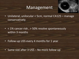
Perimenopausal Ovarian cysts.
Shobhana Mohandas.MD.DGO.FICOG.Dip Endoscopy.
Sun Medical centre, Thrissur, Kerala
10% of women will have some form of surgery
for the presence of an ovarian mass.
In premenopausal women almost all ovarian
masses and cysts are benign.
The overall incidence of a symptomatic ovarian
cyst in a premenopausal female being
malignant is approximately 1:1000 increasing to
3:1000 at the age of 50.
Green–top Guideline No. 62 RCOG/BSGE Joint Guideline I November 2011
Management
● Conservative management
● Use of laparoscopic techniques
● Referral to a gynaecological oncologist
Management
• Unilateral, unilocular < 5cm, normal CA125 – manage
conservatively
• < 1% cancer risk , > 50% resolve spontaneously
within 3 months
• Follow-up USS every 4 months for 1 year
• Same size after 3 USS – No more follow up
Post Menopausal Ovarian cysts.
Thin-walled, unilocular, sonolucent cysts less than 10 cm in
diameter with smooth, regular borders are usually benign
(malignancy rate = 0 to 1 percent, regardless of menopausal
status).
In one study, 2,763 postmenopausal women with this type of
cyst
were followed for a mean of 6.3 years and evaluated with
ultrasonography every six months. Almost 70 percent of the cysts
resolved spontaneously, and none of these simple cysts developed
into ovarian cancer.
Serial ultrasonography is sufficient to document the resolution of
cysts with these features.
Recommended intervals for ultrasonography
4-6 weeks initially,
3-6 months ,
Every 6 months American family physician Volume 84, Number 3 ◆
August 1, 2011
65 year old woman
Year: 1980
Courtesy:
Dr.Jose, General
Surgeon
Tumor: Pseudo
Mucinous
Cystadenoma
Of ovary
Olden days …
Earlier palpable ovary in the post menopausal
age was an indication for surgery
Where are we today ?
No role for cystectomy after menopause
Cyst Aspiration and oral contraceptive pill useless
DePriest ultrasound morphology index
1 point 2 points 3 points 4 points
Tumour volume <10 cm3 10 to 50
cm3
>50 to 200
cm3
>200 cm3
Cyst wall
Structure and thickness
smooth <3
mm
smooth >3
mm
papillary <3
mm
papillary >3
mm
Septal structure No septa Thin septa
<3 mm
Thick septa
3 to 10 mm
solid area
>10 mm
An ultrasound morphology index score <5 in a
pre-menopausal woman is in keeping with a
benign aetiology.
Post menopausal woman: Malignancy Index
>5
Positive predictive value of malignancy:
0.45.
Thickened wall structure, Volume >10cm:
Malignancy likely
CA- 125
• Raised in > 80 % ovarian cancers
greater than 200 U per mL [200 kU per L] in premenopausal
women and
greater than 35 U per mL [35 kU per L]
in postmenopausal women
positive predictive value
49 % in premenopausal women
98 % in postmenopausal women.
• But raised in only 50 % Stage 1
• Also raised in benign conditions and other malignancies
65 year old woman with 15 cm ovarian
cyst with some solid components
CA 125- 2 IU !!!!!!!!!!!!
Stage IV cystadenocarcinoma
Patient died 2 years later.
Risk of malignancy index (RMI)
RMI = U x M x CA-125
Ultrasound scans are scored 1 point for each of the following characteristics:
Multi-locular cyst
Evidence of solid areas
Evidence of metastases
Presence of ascites
Bi-lateral lesions.
U = 0 (for ultrasound score of 0);
U = 1 (for ultrasound score of 1);
U = 3 (for ultrasound score of 2 to 5).
M = 3 for all post-menopausal women dealt with by this guideline.
CA-125 is serum CA-125 measurement in U/mL.
Low-risk RMI = <25 (40% of women; risk of cancer is <3%).
Moderate-risk RMI = 25 to 250 (30% of women; risk of
cancer is 20%).
High-risk RMI = >250 (30% of women; risk of cancer is 75%).
8 cm ovarian cyst with solid
components
Normal CA-125
Loss of weight- 6 months:
Kruckenberg tumour,
primary not found
• complete blood count,
• cervical cultures, and measurement of HCG
• low-density lipoprotein cholesterol,
• And α-fetoprotein.
• CA 125 levels
Pelvic examination
Primary
infertility
NulliparityOlder age
F H/O breast or ovarian
cancer
Non polyposis
colorectal cancer
Lynch syndrome
Endometriosis
History taking
Identification of metastasis.
Ultrasound Exam
Lab testing
Types of adnexal masses
Benign ovarian Benign non-ovarian
• Functional cysts Paratubal cyst
• Endometriomas Hydrosalpinges
• Serous cystadenoma Tubo-ovarian abscess
• Mucinous cystadenoma Peritoneal pseudocysts
• Mature teratoma Appendiceal abscess
Diverticular abscess
Pelvic kidney
Primary malignant Secondary malignant
Ovarian Germ cell tumour Predominantly breast and
gastrointestinal carcinoma.
Epithelial carcinoma
Sex-cord tumour
Post hysterectomy patient with
excruciating pain in abdomen
with 7cm ovarian cyst
• Resistance index Risk of malignancy
• Colour Doppler - to study the vascularity
Role of MRI/CT/PET not yet established
Post Hysterectomy
Ovarian
Cysts
40Year old woman with history of hysterectomy comes
With abdominal pain and a simple ovarian cyst of 5 cms.
There is a vertical scar on the abdomen
22 year old Unmarried girl comes with uniilateral
6cm Ovarian cyst with solid components.
CA 125- 65 IU
Minimal ascites
Staging Laparotomy?
Staging Laparoscopy?
60 year old lady with 6 cm ovarian
cyst with
few solid components
CA – 125- 45 IU.
Laparoscopy?
Laparotomy?
Laparoscopic hysterectomy with BSO done.
Specimen removed through
endobag
Transitional cell tumour of the ovary
Post op CT: “recurrence “ of tumour 7cm!!!!
Chemotherapy
Alive and kicking 3 years later
ACOG and SGO guidelines
for referral to gynaecological oncology
Post-menopausal women with suspicious pelvic mass and:
Elevated CA-125 level (>35 U/mL)
Ascites
A nodular or fixed pelvic mass
Evidence of abdominal or distant metastasis
A family history of 1 or more first-degree relatives with
ovarian or breast cancer.
Pre-menopausal women with a suspicious pelvic mass
and:
Greatly elevated CA-125 level (> 200 U/mL)
Ascites
Evidence of abdominal or distant metastasis
A family history of 1 or more first-degree relatives with
ovarian or breast cancer
Thank you

Ovarian cyst in perimenopause

  • 1.
    Perimenopausal Ovarian cysts. ShobhanaMohandas.MD.DGO.FICOG.Dip Endoscopy. Sun Medical centre, Thrissur, Kerala
  • 2.
    10% of womenwill have some form of surgery for the presence of an ovarian mass. In premenopausal women almost all ovarian masses and cysts are benign. The overall incidence of a symptomatic ovarian cyst in a premenopausal female being malignant is approximately 1:1000 increasing to 3:1000 at the age of 50. Green–top Guideline No. 62 RCOG/BSGE Joint Guideline I November 2011
  • 3.
    Management ● Conservative management ●Use of laparoscopic techniques ● Referral to a gynaecological oncologist
  • 4.
    Management • Unilateral, unilocular< 5cm, normal CA125 – manage conservatively • < 1% cancer risk , > 50% resolve spontaneously within 3 months • Follow-up USS every 4 months for 1 year • Same size after 3 USS – No more follow up
  • 5.
    Post Menopausal Ovariancysts. Thin-walled, unilocular, sonolucent cysts less than 10 cm in diameter with smooth, regular borders are usually benign (malignancy rate = 0 to 1 percent, regardless of menopausal status). In one study, 2,763 postmenopausal women with this type of cyst were followed for a mean of 6.3 years and evaluated with ultrasonography every six months. Almost 70 percent of the cysts resolved spontaneously, and none of these simple cysts developed into ovarian cancer. Serial ultrasonography is sufficient to document the resolution of cysts with these features. Recommended intervals for ultrasonography 4-6 weeks initially, 3-6 months , Every 6 months American family physician Volume 84, Number 3 ◆ August 1, 2011
  • 6.
    65 year oldwoman Year: 1980 Courtesy: Dr.Jose, General Surgeon Tumor: Pseudo Mucinous Cystadenoma Of ovary
  • 7.
    Olden days … Earlierpalpable ovary in the post menopausal age was an indication for surgery Where are we today ?
  • 8.
    No role forcystectomy after menopause
  • 9.
    Cyst Aspiration andoral contraceptive pill useless
  • 10.
    DePriest ultrasound morphologyindex 1 point 2 points 3 points 4 points Tumour volume <10 cm3 10 to 50 cm3 >50 to 200 cm3 >200 cm3 Cyst wall Structure and thickness smooth <3 mm smooth >3 mm papillary <3 mm papillary >3 mm Septal structure No septa Thin septa <3 mm Thick septa 3 to 10 mm solid area >10 mm
  • 11.
    An ultrasound morphologyindex score <5 in a pre-menopausal woman is in keeping with a benign aetiology. Post menopausal woman: Malignancy Index >5 Positive predictive value of malignancy: 0.45. Thickened wall structure, Volume >10cm: Malignancy likely
  • 12.
    CA- 125 • Raisedin > 80 % ovarian cancers greater than 200 U per mL [200 kU per L] in premenopausal women and greater than 35 U per mL [35 kU per L] in postmenopausal women positive predictive value 49 % in premenopausal women 98 % in postmenopausal women. • But raised in only 50 % Stage 1 • Also raised in benign conditions and other malignancies
  • 13.
    65 year oldwoman with 15 cm ovarian cyst with some solid components CA 125- 2 IU !!!!!!!!!!!! Stage IV cystadenocarcinoma Patient died 2 years later.
  • 14.
    Risk of malignancyindex (RMI) RMI = U x M x CA-125 Ultrasound scans are scored 1 point for each of the following characteristics: Multi-locular cyst Evidence of solid areas Evidence of metastases Presence of ascites Bi-lateral lesions. U = 0 (for ultrasound score of 0); U = 1 (for ultrasound score of 1); U = 3 (for ultrasound score of 2 to 5). M = 3 for all post-menopausal women dealt with by this guideline. CA-125 is serum CA-125 measurement in U/mL.
  • 15.
    Low-risk RMI =<25 (40% of women; risk of cancer is <3%). Moderate-risk RMI = 25 to 250 (30% of women; risk of cancer is 20%). High-risk RMI = >250 (30% of women; risk of cancer is 75%).
  • 16.
    8 cm ovariancyst with solid components Normal CA-125 Loss of weight- 6 months: Kruckenberg tumour, primary not found
  • 17.
    • complete bloodcount, • cervical cultures, and measurement of HCG • low-density lipoprotein cholesterol, • And α-fetoprotein. • CA 125 levels Pelvic examination Primary infertility NulliparityOlder age F H/O breast or ovarian cancer Non polyposis colorectal cancer Lynch syndrome Endometriosis History taking Identification of metastasis. Ultrasound Exam Lab testing
  • 18.
    Types of adnexalmasses Benign ovarian Benign non-ovarian • Functional cysts Paratubal cyst • Endometriomas Hydrosalpinges • Serous cystadenoma Tubo-ovarian abscess • Mucinous cystadenoma Peritoneal pseudocysts • Mature teratoma Appendiceal abscess Diverticular abscess Pelvic kidney Primary malignant Secondary malignant Ovarian Germ cell tumour Predominantly breast and gastrointestinal carcinoma. Epithelial carcinoma Sex-cord tumour
  • 19.
    Post hysterectomy patientwith excruciating pain in abdomen with 7cm ovarian cyst
  • 25.
    • Resistance indexRisk of malignancy • Colour Doppler - to study the vascularity
  • 26.
    Role of MRI/CT/PETnot yet established
  • 27.
  • 28.
    40Year old womanwith history of hysterectomy comes With abdominal pain and a simple ovarian cyst of 5 cms. There is a vertical scar on the abdomen
  • 30.
    22 year oldUnmarried girl comes with uniilateral 6cm Ovarian cyst with solid components. CA 125- 65 IU Minimal ascites Staging Laparotomy? Staging Laparoscopy?
  • 32.
    60 year oldlady with 6 cm ovarian cyst with few solid components CA – 125- 45 IU. Laparoscopy? Laparotomy?
  • 33.
    Laparoscopic hysterectomy withBSO done. Specimen removed through endobag Transitional cell tumour of the ovary Post op CT: “recurrence “ of tumour 7cm!!!! Chemotherapy Alive and kicking 3 years later
  • 34.
    ACOG and SGOguidelines for referral to gynaecological oncology Post-menopausal women with suspicious pelvic mass and: Elevated CA-125 level (>35 U/mL) Ascites A nodular or fixed pelvic mass Evidence of abdominal or distant metastasis A family history of 1 or more first-degree relatives with ovarian or breast cancer.
  • 35.
    Pre-menopausal women witha suspicious pelvic mass and: Greatly elevated CA-125 level (> 200 U/mL) Ascites Evidence of abdominal or distant metastasis A family history of 1 or more first-degree relatives with ovarian or breast cancer
  • 36.