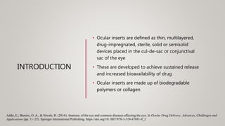
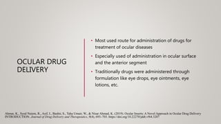
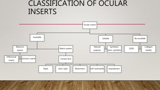
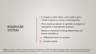
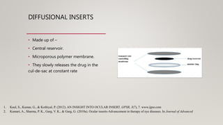
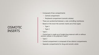
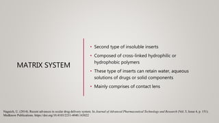
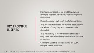
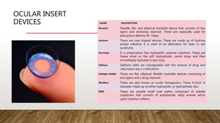
OCULAR INSERTS: ADVANCEMENTS IN
THERAPY USED FOR OCULAR DISEASES
SUBMITTED TO:
MISS DEEP SHIKHA SHARMA
(ASSISTANT PROFESSOR)
INTRODUCTION
• Ocular inserts are defined as thin, multilayered,
drug-impregnated, sterile, solid or semisolid
devices placed in the cul-de-sac or conjunctival
sac of the eye
• These are developed to achieve sustained release
and increased bioavailability of drug
• Ocular inserts are made up of biodegradable
polymers or collagen
Addo, E., Bamiro, O. A., & Siwale, R. (2016). Anatomy of the eye and common diseases affecting the eye. In Ocular Drug Delivery: Advances, Challenges and
Applications (pp. 11–25). Springer International Publishing. https://doi.org/10.1007/978-3-319-47691-9_2
OCULAR DRUG
DELIVERY
• Most used route for administration of drugs for
treatment of ocular diseases
• Especially used of administration in ocular surface
and the anterior segment
• Traditionally drugs were administered through
formulation like eye drops, eye ointments, eye
lotions, etc.
Ahmar, K., Syed Naiem, R., Asif, I., Bashir, S., Taha Umair, W., & Nisar Ahmad, K. (2019). Ocular Inserts: A Novel Approach in Ocular Drug Delivery
INTRODUCTION. Journal of Drug Delivery and Therapeutics, 9(4), 693–703. https://doi.org/10.22270/jddt.v9i4.3207
STRUCTURE OF EYE
• Most complex organ of human
eye
• It provides vision
• The eyeballs occupy 1/5th area of
the orbital space
• Functioning of eye:
• The light rays are converted into
electrochemical signals and
impulse are generated in the
neurons which provides signals to
brain
Buffault, J., Labbé, A., Hamard, P., Brignole-Baudouin, F., & Baudouin, C. (2020). The trabecular meshwork: Structure, function and clinical implications. A
review of the literature. In Journal francais d’ophtalmologie (Vol. 43, Issue 7, pp. e217–e230). NLM (Medline). https://doi.org/10.1016/j.jfo.2020.05.002
STRUCTURE OF EYE
The eyeball has three major layers:
• The outer fibrous layer: sclera and cornea
• The uveal vascular layer: choroid, ciliary
body and iris
• The inner nervous layer: retina
The eye is basically divided into two segments:
1. Anterior segment
2. Posterior segment
The anterior segment consists of:
• Cornea
• Iris
• Pupil
• Conjunctiva
• Ciliary body
• Aqueous humor
• Trabecular meshwork
• Lens
The posterior segment consists of :
• Vitreous humor
• Retina
• Sclera
• Choroid
• Optic nerve
CLASSIFICATION OF OCULAR
INSERTS
Ocular inserts
Insoluble
Reservoir
system
Diffusional
inserts
Osmotic inserts
Matrix system
Contact lens
Rigid Semi-rigid Elastomeric Soft hydrophilic biopolymeric
Soluble
Natural
polymer
Synthetic/
Semi- synthetic
Bio-erodible
SODI
Collagen
sheilds
INSOLUBLE
INSERTS
• These inserts are made up of insoluble polymers
• After drug delivery these are needed to be
removed
• The insoluble inserts are further classified as:
1. Reservoir system
2. Matrix system
Buffault, J., Labbé, A., Hamard, P., Brignole-Baudouin, F., & Baudouin, C. (2020). The trabecular meshwork: Structure, function and clinical implications. A
review of the literature. In Journal francais d’ophtalmologie (Vol. 43, Issue 7, pp. e217–e230). NLM (Medline). https://doi.org/10.1016/j.jfo.2020.05.002
RESERVOIR
SYSTEM
• It contains a solid matrix, a semi solid, a gel, a
colloid, a liquid or a carrier containing drug.
• The is made up natural or synthetic or organic or
hydrophilic or hydrophobic polymers
• Based on mechanism of drug release these are
further classified as:
(a) Diffusional inserts or ocuserts
(b) Osmotic inserts
Buffault, J., Labbé, A., Hamard, P., Brignole-Baudouin, F., & Baudouin, C. (2020). The trabecular meshwork: Structure, function and clinical implications. A
review of the literature. In Journal francais d’ophtalmologie (Vol. 43, Issue 7, pp. e217–e230). NLM (Medline). https://doi.org/10.1016/j.jfo.2020.05.002
DIFFUSIONAL INSERTS
• Made up of –
• Central reservoir.
• Microporous polymer membrane.
• They slowly releases the drug in the
cul-de-sac at constant rate
1. Kaul, S., Kumar, G., & Kothiyal, P. (2012). AN INSIGHT INTO OCULAR INSERT. IJPSR, 3(7), 7. www.ijpsr.com
2. Kumari, A., Sharma, P. K., Garg, V. K., & Garg, G. (2010a). Ocular inserts-Advancement in therapy of eye diseases. In Journal of Advanced
ELEMENTS OF DIFFUSIONAL INSERTS
Central reservoir Glycerin, water, ethylene glycol,
propylene glycol, ethyl cellulose mixed
with water, sodium alginate, ethylene,
or poly stearate.
Microporous membrane Polysulfones, polyvinyl chloride,
polycarbonates, cellulose esters,
crosslinked poly(ethyl oxide), crosslinked
polyvinyl pyrrolidine, crosslinked polyvinyl
alcohol.
OSMOTIC
INSERTS
• Composed of two compartments
• Central compartment
• Peripheral compartment (osmotic solutes)
• These are sandwiched between a rate controlling membrane.
• Based on the revoir the osmotic inserts are of two types:
• Type 1
• Type 2
• Type 1
• Central part is made up of single drug reservoir with or without
osmotic solutes in the polymeric matrix
• Type 2
• Central compartment is composed of two distinct compartments.
• Separate compartments for drug and osmotic solute
MATRIX SYSTEM
• Second type of insoluble inserts
• Composed of cross-linked hydrophilic or
hydrophobic polymers
• These type of inserts can retain water, aqueous
solutions of drugs or solid components
• Mainly comprises of contact lens
Nagaich, U. (2014). Recent advances in ocular drug delivery system. In Journal of Advanced Pharmaceutical Technology and Research (Vol. 5, Issue 4, p. 151).
Medknow Publications. https://doi.org/10.4103/2231-4040.143022
SOLUBLE INSERTS
• As the name indicates these inserts are soluble in
nature i.e., there is no need for removal after drug
release
• These are made up of hydrophilic polymers
• Based on the polymers used these are further
classified as:
1. Natural polymers
2. Synthetic polymers
Gukasyan, H. J., Hailu, S., & Karami, T. K. (2019). Ophthalmic Drug Discovery and Development. In Pharmaceutical Research (Vol. 36, Issue 5). Springer
New York LLC. https://doi.org/10.1007/s11095-019-2606-7
NATURAL POLYMERS
• Made up of natural polymers
• Manufacturing is not easy
• Best known polymers are collagen,
chitosan and gelatin
• Drug releases as the polymer dissolves
SYNTHETIC POLYMERS
• Man made
• These are preferred over natural
polymers because of the ease of
manufacturing.
• Generally used synthetic polymers are
methyl cellulose, hydroxy ethyl
cellulose, hydroxy propyl cellulose
• Drug is released by tear penetration
BIO ERODIBLE
INSERTS
• Inserts are composed of bio-erodible polymers
(example: polyester derivatives, crosslinked gelatin
derivatives)
• Dissolution occurs by hydrolysis of chemical bonds
• They are specifically used for implants because after
the release of drug, they are not needed to be
eliminated
• They have ability to modify the rate of release of
drug by erosion after altering the chemical structure
of polymers
• Commonly used bio-erodible inserts are SODI,
collagen shields, minidiscs
S, R. K., & K, N. R. (n.d.). Review on Ocular Inserts. In International Journal of PharmTech Research CODEN (Vol. 1, Issue 2).
OCULAR INSERT
DEVICES NAME DESCRIPTION
Ocuserts Flexible, flat, and elliptical insoluble device that consists of two
layers and enclosing reservoir. These are especially used for
pilocarpine delivery for 7days.
Laciserts These are rose shaped devices. These are made up of hydroxy
propyl cellulose. It is used as an alternative for tears in eye
syndrome
Dry drops It is preservative free hydrophilic polymer solutions. These are
freeze dried on the soft hydrophobic carrier strips and then
immediately hydrated in tear strip.
Gelfoam Gelfoam slabs are impregnated with the mixture of drug and
cetyl esters wax in chloroform.
Collagen shields These are flat, elliptical, flexible insoluble devices consisting of
two layers and a drug reservoir.
Minidiscs These are also known as ocular therapeutics. These 4-5mm in
diameter made up of either hydrophilic or hydrophobic disc.
SODI These are soluble small oval wafers composed of soluble
copolymer that consists of acetylamide, ethyl acetate which
upon insertion softens.
METHODS USED
FOR
PREPARATION OF
OCULAR INSERTS
• By electrospinning method
• Ionic gelation method
• Solvent cast technique
• Melt cast technique
• Film hydration technique
• Lyophilization
ADVANTAGES OF
OCULAR INSERTS
1. Systemic absorption is reduced
2. Precise dosing
3. Less systemic and visual side effects (because of less dose
frequency)
4. The intern ocular tissue is directly targeted through conjunctival
sacs
5. Improved patient compliance
6. Shelf life is increased
7. The rate of drug release is constant and slow
8. Increased bioavailability
9. Prolonged drug release
10. Less preservatives
CONCLUSION
The main aim to design ocular inserts is to overcome the
drawbacks of traditional ocular drug delivery systems i.e., eye
drops, eye gels etc. the ophthalmic inserts provides prolonged
residence time and sustained release of drugs in the cul-de-
sac.
For development of these insert variable techniques of
production are used according to drug stability and
compatibility with the polymers used.
The major advantage of ocular inserts is that these inserts are
palatable into unit dosage forms. Thus, accurate amount of
drug is delivered with prolonged release.
REFERENCES
1. Bhagav, P., Trivedi, V., Shah, D., & Chandran, S. (2011). Sustained release ocular inserts of brimonidine tartrate
for better treatment in open-angle glaucoma. Drug Delivery and Translational Research, 1(2), 161–174.
https://doi.org/10.1007/s13346-011-0018-2
2. Bhowmik, D., Sampath Kumar, K. P., Harish3, G., Duraivel, S., & Pragathi Kumar, B. (2012). Ocular Inserts: A
Novel Controlled Drug Delivery System Charecterization of Ayurvedic nanomedicine Abhrak Bhasma View
project Ocular Inserts: A Novel Controlled Drug Delivery System. THE PHARMA INNOVATION-JOURNAL,
1(12). https://doi.org/10.4018/978-1-61350-353-9.ch001
3. Farmaceutische, F., & Wetenschappen, D. F. (2017). Evaluation of newly developed HPMC ophthalmic inserts
with sustained release properties as a carrier for thermolabile therapeutics Evaluatie van nieuw ontwikkelde
HPMC oculaire inserten met verlengde afgifte.
4. Galloway, N. R., Amoaku, W. M. K., Galloway, P. H., & Browning, A. C. (2016). Basic Anatomy and Physiology
of the Eye. In Common Eye Diseases and their Management (pp. 7–16). Springer International Publishing.
https://doi.org/10.1007/978-3-319-32869-0_2
5. Hornof, M., Weyenberg, W., Ludwig, A., & Bernkop-Schnürch, A. (2003). Mucoadhesive ocular insert based on
thiolated poly(acrylic acid): Development and in vivo evaluation in humans. Journal of Controlled Release, 89(3),
419–428. https://doi.org/10.1016/S0168-3659(03)00135-4
6. Kumari, A., Sharma, P. K., Garg, V. K., & Garg, G. (2010b). Ocular inserts-Advancement in therapy of eye
diseases. In Journal of Advanced Pharmaceutical Technology and Research (Vol. 1, Issue 3, pp. 291–296).
https://doi.org/10.4103/0110-5558.72419
7. Shukr, M. H. (2016). Novel in situ gelling ocular inserts for voriconazole-loaded niosomes: Design, in vitro
characterisation and in vivo evaluation of the ocular irritation and drug pharmacokinetics. Journal of
Microencapsulation, 33(1), 71–79. https://doi.org/10.3109/02652048.2015.1128489
PUBLICATION
THANK YOU

Ocular inserts ppt

  • 1.
    OCULAR INSERTS: ADVANCEMENTSIN THERAPY USED FOR OCULAR DISEASES SUBMITTED TO: MISS DEEP SHIKHA SHARMA (ASSISTANT PROFESSOR)
  • 2.
    INTRODUCTION • Ocular insertsare defined as thin, multilayered, drug-impregnated, sterile, solid or semisolid devices placed in the cul-de-sac or conjunctival sac of the eye • These are developed to achieve sustained release and increased bioavailability of drug • Ocular inserts are made up of biodegradable polymers or collagen Addo, E., Bamiro, O. A., & Siwale, R. (2016). Anatomy of the eye and common diseases affecting the eye. In Ocular Drug Delivery: Advances, Challenges and Applications (pp. 11–25). Springer International Publishing. https://doi.org/10.1007/978-3-319-47691-9_2
  • 3.
    OCULAR DRUG DELIVERY • Mostused route for administration of drugs for treatment of ocular diseases • Especially used of administration in ocular surface and the anterior segment • Traditionally drugs were administered through formulation like eye drops, eye ointments, eye lotions, etc. Ahmar, K., Syed Naiem, R., Asif, I., Bashir, S., Taha Umair, W., & Nisar Ahmad, K. (2019). Ocular Inserts: A Novel Approach in Ocular Drug Delivery INTRODUCTION. Journal of Drug Delivery and Therapeutics, 9(4), 693–703. https://doi.org/10.22270/jddt.v9i4.3207
  • 4.
    STRUCTURE OF EYE •Most complex organ of human eye • It provides vision • The eyeballs occupy 1/5th area of the orbital space • Functioning of eye: • The light rays are converted into electrochemical signals and impulse are generated in the neurons which provides signals to brain Buffault, J., Labbé, A., Hamard, P., Brignole-Baudouin, F., & Baudouin, C. (2020). The trabecular meshwork: Structure, function and clinical implications. A review of the literature. In Journal francais d’ophtalmologie (Vol. 43, Issue 7, pp. e217–e230). NLM (Medline). https://doi.org/10.1016/j.jfo.2020.05.002
  • 5.
    STRUCTURE OF EYE Theeyeball has three major layers: • The outer fibrous layer: sclera and cornea • The uveal vascular layer: choroid, ciliary body and iris • The inner nervous layer: retina The eye is basically divided into two segments: 1. Anterior segment 2. Posterior segment The anterior segment consists of: • Cornea • Iris • Pupil • Conjunctiva • Ciliary body • Aqueous humor • Trabecular meshwork • Lens The posterior segment consists of : • Vitreous humor • Retina • Sclera • Choroid • Optic nerve
  • 6.
    CLASSIFICATION OF OCULAR INSERTS Ocularinserts Insoluble Reservoir system Diffusional inserts Osmotic inserts Matrix system Contact lens Rigid Semi-rigid Elastomeric Soft hydrophilic biopolymeric Soluble Natural polymer Synthetic/ Semi- synthetic Bio-erodible SODI Collagen sheilds
  • 7.
    INSOLUBLE INSERTS • These insertsare made up of insoluble polymers • After drug delivery these are needed to be removed • The insoluble inserts are further classified as: 1. Reservoir system 2. Matrix system Buffault, J., Labbé, A., Hamard, P., Brignole-Baudouin, F., & Baudouin, C. (2020). The trabecular meshwork: Structure, function and clinical implications. A review of the literature. In Journal francais d’ophtalmologie (Vol. 43, Issue 7, pp. e217–e230). NLM (Medline). https://doi.org/10.1016/j.jfo.2020.05.002
  • 8.
    RESERVOIR SYSTEM • It containsa solid matrix, a semi solid, a gel, a colloid, a liquid or a carrier containing drug. • The is made up natural or synthetic or organic or hydrophilic or hydrophobic polymers • Based on mechanism of drug release these are further classified as: (a) Diffusional inserts or ocuserts (b) Osmotic inserts Buffault, J., Labbé, A., Hamard, P., Brignole-Baudouin, F., & Baudouin, C. (2020). The trabecular meshwork: Structure, function and clinical implications. A review of the literature. In Journal francais d’ophtalmologie (Vol. 43, Issue 7, pp. e217–e230). NLM (Medline). https://doi.org/10.1016/j.jfo.2020.05.002
  • 9.
    DIFFUSIONAL INSERTS • Madeup of – • Central reservoir. • Microporous polymer membrane. • They slowly releases the drug in the cul-de-sac at constant rate 1. Kaul, S., Kumar, G., & Kothiyal, P. (2012). AN INSIGHT INTO OCULAR INSERT. IJPSR, 3(7), 7. www.ijpsr.com 2. Kumari, A., Sharma, P. K., Garg, V. K., & Garg, G. (2010a). Ocular inserts-Advancement in therapy of eye diseases. In Journal of Advanced
  • 10.
    ELEMENTS OF DIFFUSIONALINSERTS Central reservoir Glycerin, water, ethylene glycol, propylene glycol, ethyl cellulose mixed with water, sodium alginate, ethylene, or poly stearate. Microporous membrane Polysulfones, polyvinyl chloride, polycarbonates, cellulose esters, crosslinked poly(ethyl oxide), crosslinked polyvinyl pyrrolidine, crosslinked polyvinyl alcohol.
  • 11.
    OSMOTIC INSERTS • Composed oftwo compartments • Central compartment • Peripheral compartment (osmotic solutes) • These are sandwiched between a rate controlling membrane. • Based on the revoir the osmotic inserts are of two types: • Type 1 • Type 2 • Type 1 • Central part is made up of single drug reservoir with or without osmotic solutes in the polymeric matrix • Type 2 • Central compartment is composed of two distinct compartments. • Separate compartments for drug and osmotic solute
  • 12.
    MATRIX SYSTEM • Secondtype of insoluble inserts • Composed of cross-linked hydrophilic or hydrophobic polymers • These type of inserts can retain water, aqueous solutions of drugs or solid components • Mainly comprises of contact lens Nagaich, U. (2014). Recent advances in ocular drug delivery system. In Journal of Advanced Pharmaceutical Technology and Research (Vol. 5, Issue 4, p. 151). Medknow Publications. https://doi.org/10.4103/2231-4040.143022
  • 13.
    SOLUBLE INSERTS • Asthe name indicates these inserts are soluble in nature i.e., there is no need for removal after drug release • These are made up of hydrophilic polymers • Based on the polymers used these are further classified as: 1. Natural polymers 2. Synthetic polymers Gukasyan, H. J., Hailu, S., & Karami, T. K. (2019). Ophthalmic Drug Discovery and Development. In Pharmaceutical Research (Vol. 36, Issue 5). Springer New York LLC. https://doi.org/10.1007/s11095-019-2606-7
  • 14.
    NATURAL POLYMERS • Madeup of natural polymers • Manufacturing is not easy • Best known polymers are collagen, chitosan and gelatin • Drug releases as the polymer dissolves SYNTHETIC POLYMERS • Man made • These are preferred over natural polymers because of the ease of manufacturing. • Generally used synthetic polymers are methyl cellulose, hydroxy ethyl cellulose, hydroxy propyl cellulose • Drug is released by tear penetration
  • 15.
    BIO ERODIBLE INSERTS • Insertsare composed of bio-erodible polymers (example: polyester derivatives, crosslinked gelatin derivatives) • Dissolution occurs by hydrolysis of chemical bonds • They are specifically used for implants because after the release of drug, they are not needed to be eliminated • They have ability to modify the rate of release of drug by erosion after altering the chemical structure of polymers • Commonly used bio-erodible inserts are SODI, collagen shields, minidiscs S, R. K., & K, N. R. (n.d.). Review on Ocular Inserts. In International Journal of PharmTech Research CODEN (Vol. 1, Issue 2).
  • 16.
    OCULAR INSERT DEVICES NAMEDESCRIPTION Ocuserts Flexible, flat, and elliptical insoluble device that consists of two layers and enclosing reservoir. These are especially used for pilocarpine delivery for 7days. Laciserts These are rose shaped devices. These are made up of hydroxy propyl cellulose. It is used as an alternative for tears in eye syndrome Dry drops It is preservative free hydrophilic polymer solutions. These are freeze dried on the soft hydrophobic carrier strips and then immediately hydrated in tear strip. Gelfoam Gelfoam slabs are impregnated with the mixture of drug and cetyl esters wax in chloroform. Collagen shields These are flat, elliptical, flexible insoluble devices consisting of two layers and a drug reservoir. Minidiscs These are also known as ocular therapeutics. These 4-5mm in diameter made up of either hydrophilic or hydrophobic disc. SODI These are soluble small oval wafers composed of soluble copolymer that consists of acetylamide, ethyl acetate which upon insertion softens.
  • 17.
    METHODS USED FOR PREPARATION OF OCULARINSERTS • By electrospinning method • Ionic gelation method • Solvent cast technique • Melt cast technique • Film hydration technique • Lyophilization
  • 18.
    ADVANTAGES OF OCULAR INSERTS 1.Systemic absorption is reduced 2. Precise dosing 3. Less systemic and visual side effects (because of less dose frequency) 4. The intern ocular tissue is directly targeted through conjunctival sacs 5. Improved patient compliance 6. Shelf life is increased 7. The rate of drug release is constant and slow 8. Increased bioavailability 9. Prolonged drug release 10. Less preservatives
  • 19.
    CONCLUSION The main aimto design ocular inserts is to overcome the drawbacks of traditional ocular drug delivery systems i.e., eye drops, eye gels etc. the ophthalmic inserts provides prolonged residence time and sustained release of drugs in the cul-de- sac. For development of these insert variable techniques of production are used according to drug stability and compatibility with the polymers used. The major advantage of ocular inserts is that these inserts are palatable into unit dosage forms. Thus, accurate amount of drug is delivered with prolonged release.
  • 20.
    REFERENCES 1. Bhagav, P.,Trivedi, V., Shah, D., & Chandran, S. (2011). Sustained release ocular inserts of brimonidine tartrate for better treatment in open-angle glaucoma. Drug Delivery and Translational Research, 1(2), 161–174. https://doi.org/10.1007/s13346-011-0018-2 2. Bhowmik, D., Sampath Kumar, K. P., Harish3, G., Duraivel, S., & Pragathi Kumar, B. (2012). Ocular Inserts: A Novel Controlled Drug Delivery System Charecterization of Ayurvedic nanomedicine Abhrak Bhasma View project Ocular Inserts: A Novel Controlled Drug Delivery System. THE PHARMA INNOVATION-JOURNAL, 1(12). https://doi.org/10.4018/978-1-61350-353-9.ch001 3. Farmaceutische, F., & Wetenschappen, D. F. (2017). Evaluation of newly developed HPMC ophthalmic inserts with sustained release properties as a carrier for thermolabile therapeutics Evaluatie van nieuw ontwikkelde HPMC oculaire inserten met verlengde afgifte. 4. Galloway, N. R., Amoaku, W. M. K., Galloway, P. H., & Browning, A. C. (2016). Basic Anatomy and Physiology of the Eye. In Common Eye Diseases and their Management (pp. 7–16). Springer International Publishing. https://doi.org/10.1007/978-3-319-32869-0_2 5. Hornof, M., Weyenberg, W., Ludwig, A., & Bernkop-Schnürch, A. (2003). Mucoadhesive ocular insert based on thiolated poly(acrylic acid): Development and in vivo evaluation in humans. Journal of Controlled Release, 89(3), 419–428. https://doi.org/10.1016/S0168-3659(03)00135-4 6. Kumari, A., Sharma, P. K., Garg, V. K., & Garg, G. (2010b). Ocular inserts-Advancement in therapy of eye diseases. In Journal of Advanced Pharmaceutical Technology and Research (Vol. 1, Issue 3, pp. 291–296). https://doi.org/10.4103/0110-5558.72419 7. Shukr, M. H. (2016). Novel in situ gelling ocular inserts for voriconazole-loaded niosomes: Design, in vitro characterisation and in vivo evaluation of the ocular irritation and drug pharmacokinetics. Journal of Microencapsulation, 33(1), 71–79. https://doi.org/10.3109/02652048.2015.1128489
  • 21.
  • 22.