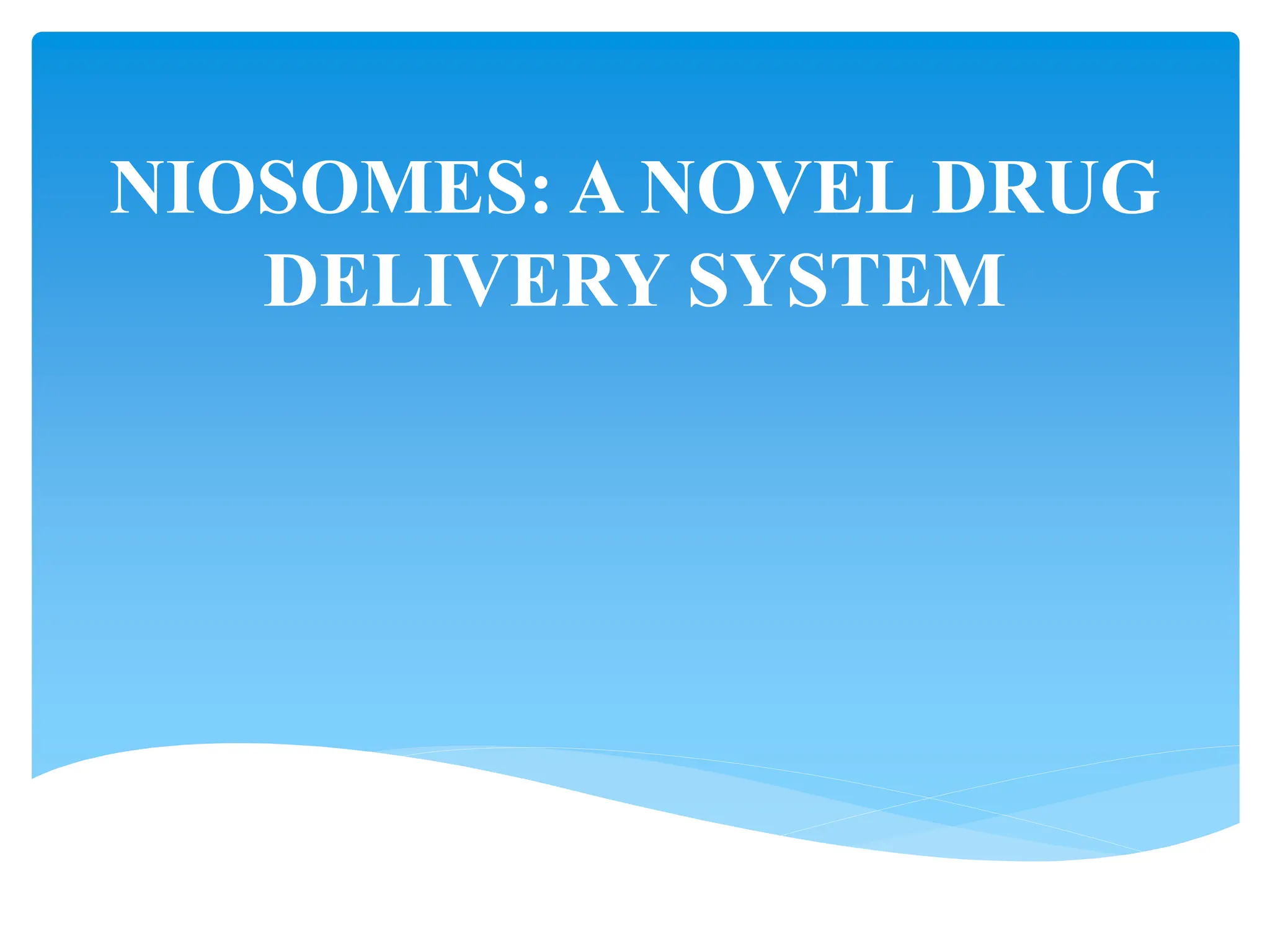
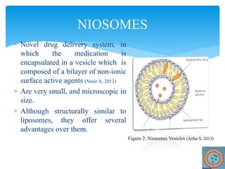
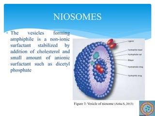
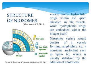
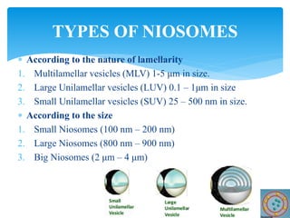
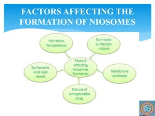
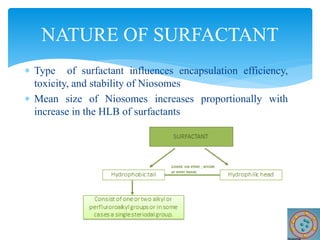
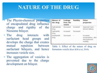
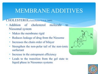
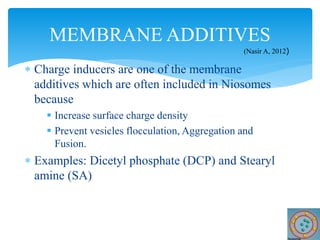
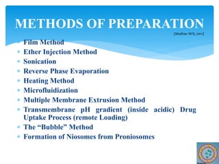
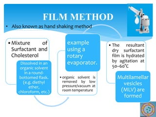
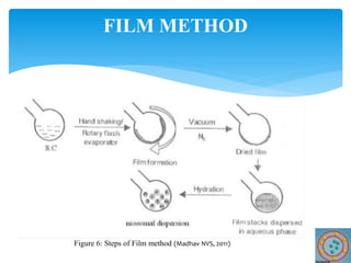
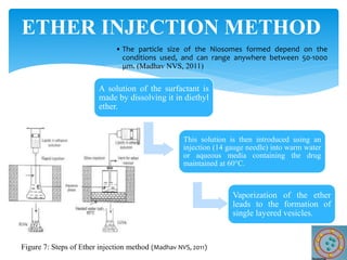
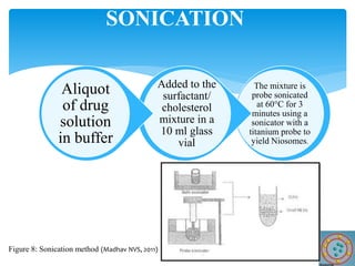
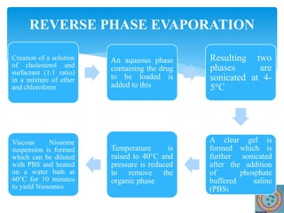
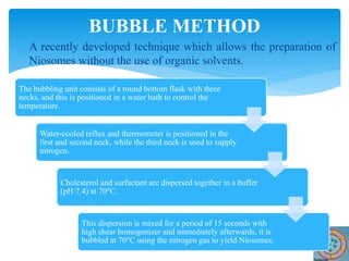
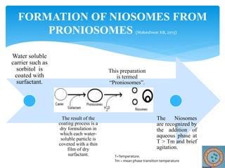
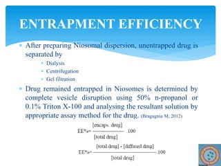
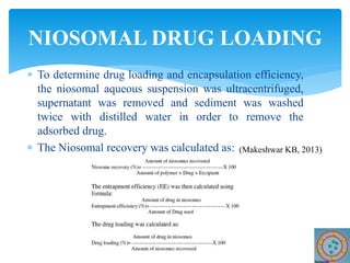
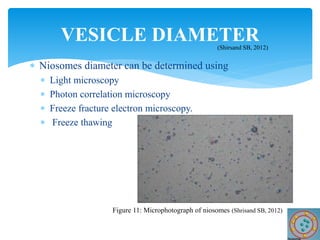
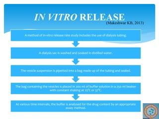
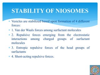
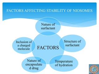
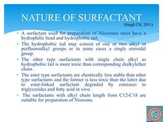
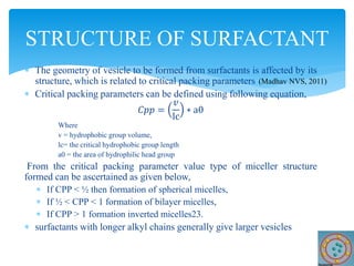
NIOSOMES: A NOVEL DRUG
DELIVERY SYSTEM
 Novel drug delivery system, in
which the medication is
encapsulated in a vesicle which is
composed of a bilayer of non-ionic
surface active agents (Nasir A, 2012)
 Are very small, and microscopic in
size.
 Although structurally similar to
liposomes, they offer several
advantages over them.
NIOSOMES
Figure 2: Niosomes Vesicles (Aitha S, 2013)
 The vesicles forming
amphiphile is a non-ionic
surfactant stabilized by
addition of cholesterol and
small amount of anionic
surfactant such as dicetyl
phosphate
NIOSOMES
Figure 3: Vesicle of niosome (Aitha S, 2013)
Figure 4: Structure of Niosomes
STRUCTURE
OF NIOSOMES
 similar to liposomes, in that they are also
made up of a bilayer.
 However, the bilayer in the case of
Niosomes is made up of non-ionic
surface active agents rather than
phospholipids.
 Made of a surfactant bilayer with its
hydrophilic ends exposed on the outside
and inside of the vesicle, while the
hydrophobic chains face each other
within the bilayer.
(Patel SM et al, 2012)
(Makeshwar KB, 2013)
STRUCTURE
OF NIOSOMES
 vesicle holds hydrophilic
drugs within the space
enclosed in the vesicle,
while hydrophobic drugs
are embedded within the
bilayer itself.
 Niosomes vesicle would
consist of a vesicle
forming amphiphile i.e. a
non-ionic surfactant such
as Span- 60, which is
usually stabilized by the
addition of cholesterol
(Makeshwar KB, 2013)
Figure 5: Structure of niosome (Makeshwar KB, 2013)
 Entrap solutes in a manner analogous to liposomes.
 Osmotically active and stable.
 Accommodate the drug molecules with a wide range of
solubility.
 Exhibits flexibility in their structural characteristics
(composition, fluidity and size)
 Performance of the drug molecules is increased.
 Better availability to the particular site by protecting the
drug from biological environment.
 Surfactants used in preparation are biodegradable,
biocompatible and non-immunogenic
SALIENT FEATURES OF
NIOSOMES (Makeshwar KB, 2013)
 Improve the therapeutic performance of the drug molecules by
 Delayed clearance from the circulation
 Protecting the drug from biological environment
 Restricting effects to target cells
 Niosomal dispersion in an aqueous phase can be emulsified in a
nonaqueous phase to
 Regulate the delivery rate of drug
 Administer normal vesicle in external non-aqueous phase.
 Handling and storage of surfactants requires no special conditions.
 Bioavailability of poorly absorbed drugs is increased.
 Targeted to the site of action by oral, parenteral as well as topical
routes.
ADVANTAGES OF NIOSOMES
DELIVERY SYSTEM
(Makeshwar KB, 2013)
 According to the nature of lamellarity
1. Multilamellar vesicles (MLV) 1-5 μm in size.
2. Large Unilamellar vesicles (LUV) 0.1 – 1μm in size
3. Small Unilamellar vesicles (SUV) 25 – 500 nm in size.
 According to the size
1. Small Niosomes (100 nm – 200 nm)
2. Large Niosomes (800 nm – 900 nm)
3. Big Niosomes (2 μm – 4 μm)
TYPES OF NIOSOMES
FACTORS AFFECTING THE
FORMATION OF NIOSOMES
 Type of surfactant influences encapsulation efficiency,
toxicity, and stability of Niosomes
 Mean size of Niosomes increases proportionally with
increase in the HLB of surfactants
NATURE OF SURFACTANT
 The surfactant/lipid ratio is generally 10-30 mM (1-2.5%
w/w)
 Increasing the surfactant/lipid level increases the total
amount of drug encapsulated
SURFACTANT AND LIPID LEVELS
NATURE OF THE DRUG
 The Physio-chemical properties
of encapsulated drug influence
charge and rigidity of the
Niosome bilayer.
 The drug interacts with
surfactant head groups and
develops the charge that creates
mutual repulsion between
surfactant bilayers, and hence
increases vesicle size.
 The aggregation of vesicles is
prevented due to the charge
development on bilayer.
Table 1: Effect of the nature of drug on
formation vesicle (Kazi KM et al, 2010)
CHOLESTEROL(Tamizharas S et al, 2009)
 Addition of cholesterol molecule to
Niosomal system
• Makes the membrane rigid
• Reduces leakage of drug from the Niosome
• Increases the chain order of bilayer
• Strengthen the non-polar tail of the non-ionic
surfactant
• Increase in the entrapment efficiency
• Leads to the transition from the gel state to
liquid phase in Niosomes systems
MEMBRANE ADDITIVES
Cholesterol
 Charge inducers are one of the membrane
additives which are often included in Niosomes
because
 Increase surface charge density
 Prevent vesicles flocculation, Aggregation and
Fusion.
 Examples: Dicetyl phosphate (DCP) and Stearyl
amine (SA)
MEMBRANE ADDITIVES
(Nasir A, 2012)
 Film Method
 Ether Injection Method
 Sonication
 Reverse Phase Evaporation
 Heating Method
 Microfluidization
 Multiple Membrane Extrusion Method
 Transmembrane pH gradient (inside acidic) Drug
Uptake Process (remote Loading)
 The “Bubble” Method
 Formation of Niosomes from Proniosomes
METHODS OF PREPARATION
(Madhav NVS, 2011)
•Mixture of
Surfactant and
Cholesterol
Dissolved in an
organic solvent
in a round-
bottomed flask.
(e.g. diethyl
ether,
chloroform, etc.)
•organic solvent is
removed by low
pressure/vacuum at
room temperature
example
using a
rotary
evaporator.
• The resultant
dry surfactant
film is hydrated
by agitation at
50–60°C
Multilamellar
vesicles
(MLV) are
formed
FILM METHOD
• Also known as hand shaking method
FILM METHOD
Figure 6: Steps of Film method (Madhav NVS, 2011)
A solution of the surfactant is
made by dissolving it in diethyl
ether.
This solution is then introduced using an
injection (14 gauge needle) into warm water
or aqueous media containing the drug
maintained at 60°C.
Vaporization of the ether
leads to the formation of
single layered vesicles.
• The particle size of the Niosomes formed depend on the
conditions used, and can range anywhere between 50-1000
μm. (Madhav NVS, 2011)
ETHER INJECTION METHOD
Figure 7: Steps of Ether injection method (Madhav NVS, 2011)
The mixture is
probe sonicated
at 60°C for 3
minutes using a
sonicator with a
titanium probe to
yield Niosomes.
Added to the
surfactant/
cholesterol
mixture in a
10 ml glass
vial
Aliquot
of drug
solution
in buffer
SONICATION
Figure 8: Sonication method (Madhav NVS, 2011)
Creation of a solution
of cholesterol and
surfactant (1:1 ratio)
in a mixture of ether
and chloroform
An aqueous phase
containing the drug
to be loaded is
added to this
Resulting two
phases are
sonicated at 4-
5°C
A clear gel is
formed which is
further sonicated
after the addition
of phosphate
buffered saline
(PBS)
Temperature is
raised to 40°C and
pressure is reduced
to remove the
organic phase
Viscous Niosome
suspension is formed
which can be diluted
with PBS and heated
on a water bath at
60°C for 10 minutes
to yield Niosomes
REVERSE PHASE EVAPORATION
 Non-toxic, Scalable and one-step method.
HEATING METHOD
Mixtures of non-ionic
surfactant, cholesterol
and/or charge inducing
molecules are added to an
aqueous medium e.g.
buffer, distilled H2O, etc
• In the presence of a
Polyol such as glycerol.
The mixture is
heated while
stirring at low
shear forces
• Until vesicles are
formed
 Recent technique used to prepare Unilamellar vesicles of
defined size distribution.
 based on submerged jet principle
MICROFLUIDIZATION
Two fluidized
streams interact at
ultra high velocities,
in precisely defined
micro channels
within the interaction
chamber
The impingement of thin
liquid sheet along a
common front is arranged
such that the energy
supplied to the system
remains within the area of
Niosomes formation
The result is a greater
uniformity, smaller
size and better
reproducibility of
Niosome are formed
MICROFLUIDIZATION
Figure 9: Steps of microfludization method (Madhav NVS, 2011)
 Good method for controlling Niosomes size.
MULTIPLE MEMBRANE EXTRUSION
METHOD
Mixture of surfactant, cholesterol and
dicetyl phosphate in chloroform is made
into thin film by evaporation
The film is hydrated with aqueous drug
solution
Resultant suspension is extruded through
polycarbonate membranes which are
placed in series for upto 8 passages
Figure 10: Multiple membrane
extrusion method (Madhav NVS, 2011)
Solution of surfactant
and cholesterol is made
in chloroform
Solvent is then evaporated
under reduced pressure to get
a thin film on the wall of the
round bottom flask, similar to
the hand shaking method
This film is then
hydrated using citric acid
solution by vortex
mixing
Resulting Multilamellar
vesicles are then treated
to three freeze thaw
cycles and sonicated
To the Niosomal
suspension, aqueous
solution containing
10mg/ml of drug is
added and vortexed
pH of the sample is
then raised to 7.0-7.2
using 1M disodium
phosphate
Mixture is heated at
60°C for 10 minutes to
give Niosomes
TRANSMEMBRANE pH GRADIENT DRUG
UPTAKE PROCESS
 A recently developed technique which allows the preparation of
Niosomes without the use of organic solvents.
BUBBLE METHOD
The bubbling unit consists of a round bottom flask with three
necks, and this is positioned in a water bath to control the
temperature.
Water-cooled reflux and thermometer is positioned in the
first and second neck, while the third neck is used to supply
nitrogen.
Cholesterol and surfactant are dispersed together in a buffer
(pH 7.4) at 70°C.
This dispersion is mixed for a period of 15 seconds with
high shear homogenizer and immediately afterwards, it is
bubbled at 70°C using the nitrogen gas to yield Niosomes.
FORMATION OF NIOSOMES FROM
PRONIOSOMES (Makeshwar KB, 2013)
Water soluble
carrier such as
sorbitol is
coated with
surfactant.
The result of the
coating process is a
dry formulation in
which each water-
soluble particle is
covered with a thin
film of dry
surfactant.
This preparation
is termed
“Proniosomes”.
The Niosomes
are recognized by
the addition of
aqueous phase at
T > Tm and brief
agitation.
T=Temperature.
Tm = mean phase transition temperature
a) Size, Shape and Morphology
b) Entrapment efficiency
c) Vesicle diameter
d) In vitro release
e) Vesicle charge
f) Bilayer rigidity and Homogeneity
g) Osmotic Shrinkage
h) Physical stability of vesicles at different temperature
i) Turbidity Measurement
CHARACTERIZATION OF NIOSOMES
 Structure of surfactant based vesicles has been visualized
and established using freeze fracture microscopy
 Photon correlation spectroscopy used to determine mean
diameter of the vesicles.
 Electron microscopy used for morphological studies of
vesicles
 Laser beam is generally used to determine size distribution,
mean surface diameter and mass distribution of Niosomes.
SIZE, SHAPE AND MORPHOLOGY
 After preparing Niosomal dispersion, unentrapped drug is
separated by
 Dialysis
 Centrifugation
 Gel filtration
 Drug remained entrapped in Niosomes is determined by
complete vesicle disruption using 50% n-propanol or
0.1% Triton X-100 and analysing the resultant solution by
appropriate assay method for the drug. (Bragagnia M, 2012)
ENTRAPMENT EFFICIENCY
 To determine drug loading and encapsulation efficiency,
the niosomal aqueous suspension was ultracentrifuged,
supernatant was removed and sediment was washed
twice with distilled water in order to remove the
adsorbed drug.
 The Niosomal recovery was calculated as:
NIOSOMAL DRUG LOADING
(Makeshwar KB, 2013)
 Niosomes diameter can be determined using
 Light microscopy
 Photon correlation microscopy
 Freeze fracture electron microscopy.
 Freeze thawing
VESICLE DIAMETER
(Shirsand SB, 2012)
Figure 11: Microphotograph of niosomes (Shrisand SB, 2012)
At various time intervals, the buffer is analysed for the drug content by an appropriate
assay method.
The bag containing the vesicles is placed in 200 ml of buffer solution in a 250 ml beaker
with constant shaking at 25°C or 37°C.
The vesicle suspension is pipetted into a bag made up of the tubing and sealed.
A dialysis sac is washed and soaked in distilled water.
A method of in-vitro release rate study includes the use of dialysis tubing.
IN VITRO RELEASE
(Makeshwar KB, 2013)
 The vesicle surface charge can play an important role in the
behaviour of Niosomes in vitro and in vivo.
 Charged Niosomes are more stable against aggregation and
fusion than uncharged vesicles.
 In order to obtain an estimate of the surface potential, the zeta
potential of individual Niosomes can be measured by
Microelectrophoresis, Fluorophores, and Dynamic light
scattering.
 Zeta potential is calculated by using Henry equation (S P Vyas, 2011)
ζ =
µ𝐸4πη
Σ
Where ζ is Zeta potential, µ𝐸 is electrophoretic mobility, η is
viscosity of the medium and Σ is dielectric constant
VESICLE CHARGE
(Makeshwar KB, 2013)
 The biodistribution and biodegradation of Niosomes are
influenced by rigidity of the bilayer.
 Homogeneity can occur both within Niosomes structures
themselves and between Niosomes in dispersion and
could be identified via. NMR, Differential Scanning
Calorimetry (DSC) and Fourier transform-infra red
spectroscopy (FT-IR) techniques.
 Membrane rigidity can be measured by means of
mobility of fluorescence probe as a function of
temperature. (Patel SM et al, 2012)
BILAYER RIGIDITY AND HOMOGENEITY
 Osmotic shrinkage of vesicles can be determined by
monitoring reductions in vesicle diameter, initiated by
addition of hypertonic salt solution to suspension of
Niosomes.
 Niosomes prepared from pure surfactant are osmotically
more sensitive in contrast to vesicles containing cholesterol.
OSMOTIC SHRINKAGE
 Aggregation or fusion of vesicles as a function of
temperature was determined as the changes in vesicle
diameter by laser light scattering method.
 The vesicles were stored in glass vials at room
temperature or kept in refrigerator (4oC) for 3 months.
 The changes in morphology of Multilamellar vesicles
(MLVs) and also the constituent separation were assessed
by an optical microscope.
 The retention of entrapped drug were measured 72 hours
after preparation and after 1, 2 or 3 months in same
formulations
PHYSICAL STABILITY OF VESICLES
AT DIFFERENT TEMPERATURE
 Niosomes were diluted with bidistilled water to give a total
lipid concentration of 0.312 mM
 After rapid mixing by sonication for 5 min
 Turbidity was measured as the absorbance with an
ultraviolet-visible diode array spectrophotometer.
TURBIDITY MEASUREMENT
 Vesicles are stabilized based upon formation of 4 different
forces:
 1. Van der Waals forces among surfactant molecules
 2. Repulsive forces emerging from the electrostatic
interactions among charged groups of surfactant
molecules
 3. Entropic repulsive forces of the head groups of
surfactants
 4. Short-acting repulsive forces.
STABILITY OF NIOSOMES
FACTORS
Nature of
surfactant
Structure of
surfactant
Temperature
of hydration
Nature of
encapsulate
d drug
Inclusion of
a charged
molecule
FACTORS AFFECTING STABILITY OF NIOSOMES
 A surfactant used for preparation of Niosomes must have a
hydrophilic head and hydrophobic tail.
 The hydrophobic tail may consist of one or two alkyl or
perfluoroalkyl groups or in some cases a single steroidal
group.
 The ether type surfactants with single chain alkyl as
hydrophobic tail is more toxic than corresponding dialkylether
chain.
 The ester type surfactants are chemically less stable than ether
type surfactants and the former is less toxic than the latter due
to ester-linked surfactant degraded by esterases to
triglycerides and fatty acid in vivo.
 The surfactants with alkyl chain length from C12-C18 are
suitable for preparation of Niosome.
NATURE OF SURFACTANT
(Singh CH, 2011)
 The geometry of vesicle to be formed from surfactants is affected by its
structure, which is related to critical packing parameters
 Critical packing parameters can be defined using following equation,
𝐶𝑝𝑝 =
𝑣
lc
∗ a0
Where
v = hydrophobic group volume,
lc= the critical hydrophobic group length
a0 = the area of hydrophilic head group
From the critical packing parameter value type of miceller structure
formed can be ascertained as given below,
 If CPP < ½ then formation of spherical micelles,
 If ½ < CPP < 1 formation of bilayer micelles,
 If CPP > 1 formation inverted micelles23.
 surfactants with longer alkyl chains generally give larger vesicles
STRUCTURE OF SURFACTANT
(Madhav NVS, 2011)
 The physico-chemical properties of encapsulated drug
influence charge and rigidity of the Niosome bilayer.
 The drug interacts with surfactant head groups and
develops the charge that creates mutual repulsion between
surfactant bilayers and hence increases vesicle size.
NATURE OF ENCAPSULATED DRUG
(Singh CH, 2011)
 Hydration temperature influences the shape and size of
the Niosome.
 For ideal condition it should be above the gel to liquid
phase transition temperature of system.
 Temperature change of Niosomal system affects
assembly of surfactants into vesicles and also induces
vesicle shape transformation
TEMPERATURE OF HYDRATION
(Madhav NVS, 2011)
 Niosomes as Drug Carriers
 Diagnostic imaging with Niosomes
 Drug Targeting
 Delivery to the brain
 Anti cancer drugs
 Anti infectives
 Targeting of bioactive agents
 To Reticulo-endothelial system (RES)
 To organs other than RES
NIOSOME DELIVERY APPLICATIONS
(Malhotra M et al, 1994)
 Ophthalmic drug delivery
 Delivery of peptide drugs
 Immunological application of Niosomes
 Transdermal delivery of drugs by Niosomes
 Delivery system for the vasoactive intestinal peptide
(VIP)
 Niosomes as carriers for Hemoglobin
 Niosomal vaccines
NIOSOME DELIVERY APPLICATIONS
 Sustained Release
 Localized Drug Action
OTHER APPLICATIONS
 Unfortunately, there is not enough research conducted to
investigate toxicity of Niosomes.
 It was determined that the ester type surfactants are less
toxic than ether type surfactants.
 In general, the physical form of Niosomes did not
influence their toxicity as evident in a study comparing
the formulations prepared in the form of liquid crystals
and gels.
 Nasal applications of these formulations caused toxicity in
the case of liquid crystal type Niosomes.
TOXICITY OF NIOSOMES
1. Aitha, S. (2013). slideshare. Retrieved 2014, from http://www.slideshare.net/swethaaitha/liposomes-
and-niosomes-15259272
2. Bragagnia M, Natascia M. (2012). Development and Characterization of Niosomal Formulations of
Doxorubicin Aimed at Brain Targeting. Journal of Pharmaceutical science, 184-196.
3. Kazi KM, M. A. (2010). Niosome: A future of targeted drug delivery systems. Journal of Advanced
Pharmaceutical technology and Research, 4, 374-380.
4. Madhav NVS, S. A. (2011). Niosomes: A Novel Drug Delivery System. International Journal of
Research in Pharmacy and Chemistry, 1(3), 498-511.
5. Makeshwar KB, W. S. (2013). Niosome: a Novel Drug Delivery System. Asian Journal of
Pharmapress, 3(1), 16-20.
6. Malhotra M, J. N. (1994). Niosomes as drug carriers (Vol. 31). Newdelhi: Indian Drugs.
7. Nasir A, H. S., A, K. (2012). Niosomes: An Excellent Tool for Drug Delivery. International Journal
of Research in Pharmacy and Chemistry, 2(2), 479-487.
8. Patel SM, R. D. (2012). Niosome as an Effective Drug Delivery: A Review. International Journal for
Pharmaceutical Research Scholars, 46-53.
9. Shirsand SB, P. M. (2012). Formulation and evaluation of Ketoconazole niosomal gel drug delivery
system. Int J Pharma investig, 201-207.
10. Singh CH, J. C. (2011). Formulation, characterization, stability and invitro evaluation of nimesulide
niosomes. Pharmacophore, 2, 168-185.
11. Tamizharas S, D. A. (2009). Development and characterization of niosomal drug delivery of
gliclazide. J Young Pharmacists, 1, 205-209.
12. Vyas SP, K. R. (2011). Targeted and Controlled Drug Delivery Novel carrier systems. New delhi:
CBS Publisher and Distributors.
REFERENCE
niosomes. Non-Ionic Surfactant Vesicles (Niosomes)

niosomes. Non-Ionic Surfactant Vesicles (Niosomes)

  • 1.
    NIOSOMES: A NOVELDRUG DELIVERY SYSTEM
  • 2.
     Novel drugdelivery system, in which the medication is encapsulated in a vesicle which is composed of a bilayer of non-ionic surface active agents (Nasir A, 2012)  Are very small, and microscopic in size.  Although structurally similar to liposomes, they offer several advantages over them. NIOSOMES Figure 2: Niosomes Vesicles (Aitha S, 2013)
  • 3.
     The vesiclesforming amphiphile is a non-ionic surfactant stabilized by addition of cholesterol and small amount of anionic surfactant such as dicetyl phosphate NIOSOMES Figure 3: Vesicle of niosome (Aitha S, 2013)
  • 4.
    Figure 4: Structureof Niosomes STRUCTURE OF NIOSOMES  similar to liposomes, in that they are also made up of a bilayer.  However, the bilayer in the case of Niosomes is made up of non-ionic surface active agents rather than phospholipids.  Made of a surfactant bilayer with its hydrophilic ends exposed on the outside and inside of the vesicle, while the hydrophobic chains face each other within the bilayer. (Patel SM et al, 2012) (Makeshwar KB, 2013)
  • 5.
    STRUCTURE OF NIOSOMES  vesicleholds hydrophilic drugs within the space enclosed in the vesicle, while hydrophobic drugs are embedded within the bilayer itself.  Niosomes vesicle would consist of a vesicle forming amphiphile i.e. a non-ionic surfactant such as Span- 60, which is usually stabilized by the addition of cholesterol (Makeshwar KB, 2013) Figure 5: Structure of niosome (Makeshwar KB, 2013)
  • 6.
     Entrap solutesin a manner analogous to liposomes.  Osmotically active and stable.  Accommodate the drug molecules with a wide range of solubility.  Exhibits flexibility in their structural characteristics (composition, fluidity and size)  Performance of the drug molecules is increased.  Better availability to the particular site by protecting the drug from biological environment.  Surfactants used in preparation are biodegradable, biocompatible and non-immunogenic SALIENT FEATURES OF NIOSOMES (Makeshwar KB, 2013)
  • 7.
     Improve thetherapeutic performance of the drug molecules by  Delayed clearance from the circulation  Protecting the drug from biological environment  Restricting effects to target cells  Niosomal dispersion in an aqueous phase can be emulsified in a nonaqueous phase to  Regulate the delivery rate of drug  Administer normal vesicle in external non-aqueous phase.  Handling and storage of surfactants requires no special conditions.  Bioavailability of poorly absorbed drugs is increased.  Targeted to the site of action by oral, parenteral as well as topical routes. ADVANTAGES OF NIOSOMES DELIVERY SYSTEM (Makeshwar KB, 2013)
  • 8.
     According tothe nature of lamellarity 1. Multilamellar vesicles (MLV) 1-5 μm in size. 2. Large Unilamellar vesicles (LUV) 0.1 – 1μm in size 3. Small Unilamellar vesicles (SUV) 25 – 500 nm in size.  According to the size 1. Small Niosomes (100 nm – 200 nm) 2. Large Niosomes (800 nm – 900 nm) 3. Big Niosomes (2 μm – 4 μm) TYPES OF NIOSOMES
  • 9.
  • 10.
     Type ofsurfactant influences encapsulation efficiency, toxicity, and stability of Niosomes  Mean size of Niosomes increases proportionally with increase in the HLB of surfactants NATURE OF SURFACTANT
  • 11.
     The surfactant/lipidratio is generally 10-30 mM (1-2.5% w/w)  Increasing the surfactant/lipid level increases the total amount of drug encapsulated SURFACTANT AND LIPID LEVELS
  • 12.
    NATURE OF THEDRUG  The Physio-chemical properties of encapsulated drug influence charge and rigidity of the Niosome bilayer.  The drug interacts with surfactant head groups and develops the charge that creates mutual repulsion between surfactant bilayers, and hence increases vesicle size.  The aggregation of vesicles is prevented due to the charge development on bilayer. Table 1: Effect of the nature of drug on formation vesicle (Kazi KM et al, 2010)
  • 13.
    CHOLESTEROL(Tamizharas S etal, 2009)  Addition of cholesterol molecule to Niosomal system • Makes the membrane rigid • Reduces leakage of drug from the Niosome • Increases the chain order of bilayer • Strengthen the non-polar tail of the non-ionic surfactant • Increase in the entrapment efficiency • Leads to the transition from the gel state to liquid phase in Niosomes systems MEMBRANE ADDITIVES Cholesterol
  • 14.
     Charge inducersare one of the membrane additives which are often included in Niosomes because  Increase surface charge density  Prevent vesicles flocculation, Aggregation and Fusion.  Examples: Dicetyl phosphate (DCP) and Stearyl amine (SA) MEMBRANE ADDITIVES (Nasir A, 2012)
  • 15.
     Film Method Ether Injection Method  Sonication  Reverse Phase Evaporation  Heating Method  Microfluidization  Multiple Membrane Extrusion Method  Transmembrane pH gradient (inside acidic) Drug Uptake Process (remote Loading)  The “Bubble” Method  Formation of Niosomes from Proniosomes METHODS OF PREPARATION (Madhav NVS, 2011)
  • 16.
    •Mixture of Surfactant and Cholesterol Dissolvedin an organic solvent in a round- bottomed flask. (e.g. diethyl ether, chloroform, etc.) •organic solvent is removed by low pressure/vacuum at room temperature example using a rotary evaporator. • The resultant dry surfactant film is hydrated by agitation at 50–60°C Multilamellar vesicles (MLV) are formed FILM METHOD • Also known as hand shaking method
  • 17.
    FILM METHOD Figure 6:Steps of Film method (Madhav NVS, 2011)
  • 18.
    A solution ofthe surfactant is made by dissolving it in diethyl ether. This solution is then introduced using an injection (14 gauge needle) into warm water or aqueous media containing the drug maintained at 60°C. Vaporization of the ether leads to the formation of single layered vesicles. • The particle size of the Niosomes formed depend on the conditions used, and can range anywhere between 50-1000 μm. (Madhav NVS, 2011) ETHER INJECTION METHOD Figure 7: Steps of Ether injection method (Madhav NVS, 2011)
  • 19.
    The mixture is probesonicated at 60°C for 3 minutes using a sonicator with a titanium probe to yield Niosomes. Added to the surfactant/ cholesterol mixture in a 10 ml glass vial Aliquot of drug solution in buffer SONICATION Figure 8: Sonication method (Madhav NVS, 2011)
  • 20.
    Creation of asolution of cholesterol and surfactant (1:1 ratio) in a mixture of ether and chloroform An aqueous phase containing the drug to be loaded is added to this Resulting two phases are sonicated at 4- 5°C A clear gel is formed which is further sonicated after the addition of phosphate buffered saline (PBS) Temperature is raised to 40°C and pressure is reduced to remove the organic phase Viscous Niosome suspension is formed which can be diluted with PBS and heated on a water bath at 60°C for 10 minutes to yield Niosomes REVERSE PHASE EVAPORATION
  • 21.
     Non-toxic, Scalableand one-step method. HEATING METHOD Mixtures of non-ionic surfactant, cholesterol and/or charge inducing molecules are added to an aqueous medium e.g. buffer, distilled H2O, etc • In the presence of a Polyol such as glycerol. The mixture is heated while stirring at low shear forces • Until vesicles are formed
  • 22.
     Recent techniqueused to prepare Unilamellar vesicles of defined size distribution.  based on submerged jet principle MICROFLUIDIZATION Two fluidized streams interact at ultra high velocities, in precisely defined micro channels within the interaction chamber The impingement of thin liquid sheet along a common front is arranged such that the energy supplied to the system remains within the area of Niosomes formation The result is a greater uniformity, smaller size and better reproducibility of Niosome are formed
  • 23.
    MICROFLUIDIZATION Figure 9: Stepsof microfludization method (Madhav NVS, 2011)
  • 24.
     Good methodfor controlling Niosomes size. MULTIPLE MEMBRANE EXTRUSION METHOD Mixture of surfactant, cholesterol and dicetyl phosphate in chloroform is made into thin film by evaporation The film is hydrated with aqueous drug solution Resultant suspension is extruded through polycarbonate membranes which are placed in series for upto 8 passages Figure 10: Multiple membrane extrusion method (Madhav NVS, 2011)
  • 25.
    Solution of surfactant andcholesterol is made in chloroform Solvent is then evaporated under reduced pressure to get a thin film on the wall of the round bottom flask, similar to the hand shaking method This film is then hydrated using citric acid solution by vortex mixing Resulting Multilamellar vesicles are then treated to three freeze thaw cycles and sonicated To the Niosomal suspension, aqueous solution containing 10mg/ml of drug is added and vortexed pH of the sample is then raised to 7.0-7.2 using 1M disodium phosphate Mixture is heated at 60°C for 10 minutes to give Niosomes TRANSMEMBRANE pH GRADIENT DRUG UPTAKE PROCESS
  • 26.
     A recentlydeveloped technique which allows the preparation of Niosomes without the use of organic solvents. BUBBLE METHOD The bubbling unit consists of a round bottom flask with three necks, and this is positioned in a water bath to control the temperature. Water-cooled reflux and thermometer is positioned in the first and second neck, while the third neck is used to supply nitrogen. Cholesterol and surfactant are dispersed together in a buffer (pH 7.4) at 70°C. This dispersion is mixed for a period of 15 seconds with high shear homogenizer and immediately afterwards, it is bubbled at 70°C using the nitrogen gas to yield Niosomes.
  • 27.
    FORMATION OF NIOSOMESFROM PRONIOSOMES (Makeshwar KB, 2013) Water soluble carrier such as sorbitol is coated with surfactant. The result of the coating process is a dry formulation in which each water- soluble particle is covered with a thin film of dry surfactant. This preparation is termed “Proniosomes”. The Niosomes are recognized by the addition of aqueous phase at T > Tm and brief agitation. T=Temperature. Tm = mean phase transition temperature
  • 28.
    a) Size, Shapeand Morphology b) Entrapment efficiency c) Vesicle diameter d) In vitro release e) Vesicle charge f) Bilayer rigidity and Homogeneity g) Osmotic Shrinkage h) Physical stability of vesicles at different temperature i) Turbidity Measurement CHARACTERIZATION OF NIOSOMES
  • 29.
     Structure ofsurfactant based vesicles has been visualized and established using freeze fracture microscopy  Photon correlation spectroscopy used to determine mean diameter of the vesicles.  Electron microscopy used for morphological studies of vesicles  Laser beam is generally used to determine size distribution, mean surface diameter and mass distribution of Niosomes. SIZE, SHAPE AND MORPHOLOGY
  • 30.
     After preparingNiosomal dispersion, unentrapped drug is separated by  Dialysis  Centrifugation  Gel filtration  Drug remained entrapped in Niosomes is determined by complete vesicle disruption using 50% n-propanol or 0.1% Triton X-100 and analysing the resultant solution by appropriate assay method for the drug. (Bragagnia M, 2012) ENTRAPMENT EFFICIENCY
  • 31.
     To determinedrug loading and encapsulation efficiency, the niosomal aqueous suspension was ultracentrifuged, supernatant was removed and sediment was washed twice with distilled water in order to remove the adsorbed drug.  The Niosomal recovery was calculated as: NIOSOMAL DRUG LOADING (Makeshwar KB, 2013)
  • 32.
     Niosomes diametercan be determined using  Light microscopy  Photon correlation microscopy  Freeze fracture electron microscopy.  Freeze thawing VESICLE DIAMETER (Shirsand SB, 2012) Figure 11: Microphotograph of niosomes (Shrisand SB, 2012)
  • 33.
    At various timeintervals, the buffer is analysed for the drug content by an appropriate assay method. The bag containing the vesicles is placed in 200 ml of buffer solution in a 250 ml beaker with constant shaking at 25°C or 37°C. The vesicle suspension is pipetted into a bag made up of the tubing and sealed. A dialysis sac is washed and soaked in distilled water. A method of in-vitro release rate study includes the use of dialysis tubing. IN VITRO RELEASE (Makeshwar KB, 2013)
  • 34.
     The vesiclesurface charge can play an important role in the behaviour of Niosomes in vitro and in vivo.  Charged Niosomes are more stable against aggregation and fusion than uncharged vesicles.  In order to obtain an estimate of the surface potential, the zeta potential of individual Niosomes can be measured by Microelectrophoresis, Fluorophores, and Dynamic light scattering.  Zeta potential is calculated by using Henry equation (S P Vyas, 2011) ζ = µ𝐸4πη Σ Where ζ is Zeta potential, µ𝐸 is electrophoretic mobility, η is viscosity of the medium and Σ is dielectric constant VESICLE CHARGE (Makeshwar KB, 2013)
  • 35.
     The biodistributionand biodegradation of Niosomes are influenced by rigidity of the bilayer.  Homogeneity can occur both within Niosomes structures themselves and between Niosomes in dispersion and could be identified via. NMR, Differential Scanning Calorimetry (DSC) and Fourier transform-infra red spectroscopy (FT-IR) techniques.  Membrane rigidity can be measured by means of mobility of fluorescence probe as a function of temperature. (Patel SM et al, 2012) BILAYER RIGIDITY AND HOMOGENEITY
  • 36.
     Osmotic shrinkageof vesicles can be determined by monitoring reductions in vesicle diameter, initiated by addition of hypertonic salt solution to suspension of Niosomes.  Niosomes prepared from pure surfactant are osmotically more sensitive in contrast to vesicles containing cholesterol. OSMOTIC SHRINKAGE
  • 37.
     Aggregation orfusion of vesicles as a function of temperature was determined as the changes in vesicle diameter by laser light scattering method.  The vesicles were stored in glass vials at room temperature or kept in refrigerator (4oC) for 3 months.  The changes in morphology of Multilamellar vesicles (MLVs) and also the constituent separation were assessed by an optical microscope.  The retention of entrapped drug were measured 72 hours after preparation and after 1, 2 or 3 months in same formulations PHYSICAL STABILITY OF VESICLES AT DIFFERENT TEMPERATURE
  • 38.
     Niosomes werediluted with bidistilled water to give a total lipid concentration of 0.312 mM  After rapid mixing by sonication for 5 min  Turbidity was measured as the absorbance with an ultraviolet-visible diode array spectrophotometer. TURBIDITY MEASUREMENT
  • 39.
     Vesicles arestabilized based upon formation of 4 different forces:  1. Van der Waals forces among surfactant molecules  2. Repulsive forces emerging from the electrostatic interactions among charged groups of surfactant molecules  3. Entropic repulsive forces of the head groups of surfactants  4. Short-acting repulsive forces. STABILITY OF NIOSOMES
  • 40.
    FACTORS Nature of surfactant Structure of surfactant Temperature ofhydration Nature of encapsulate d drug Inclusion of a charged molecule FACTORS AFFECTING STABILITY OF NIOSOMES
  • 41.
     A surfactantused for preparation of Niosomes must have a hydrophilic head and hydrophobic tail.  The hydrophobic tail may consist of one or two alkyl or perfluoroalkyl groups or in some cases a single steroidal group.  The ether type surfactants with single chain alkyl as hydrophobic tail is more toxic than corresponding dialkylether chain.  The ester type surfactants are chemically less stable than ether type surfactants and the former is less toxic than the latter due to ester-linked surfactant degraded by esterases to triglycerides and fatty acid in vivo.  The surfactants with alkyl chain length from C12-C18 are suitable for preparation of Niosome. NATURE OF SURFACTANT (Singh CH, 2011)
  • 42.
     The geometryof vesicle to be formed from surfactants is affected by its structure, which is related to critical packing parameters  Critical packing parameters can be defined using following equation, 𝐶𝑝𝑝 = 𝑣 lc ∗ a0 Where v = hydrophobic group volume, lc= the critical hydrophobic group length a0 = the area of hydrophilic head group From the critical packing parameter value type of miceller structure formed can be ascertained as given below,  If CPP < ½ then formation of spherical micelles,  If ½ < CPP < 1 formation of bilayer micelles,  If CPP > 1 formation inverted micelles23.  surfactants with longer alkyl chains generally give larger vesicles STRUCTURE OF SURFACTANT (Madhav NVS, 2011)
  • 43.
     The physico-chemicalproperties of encapsulated drug influence charge and rigidity of the Niosome bilayer.  The drug interacts with surfactant head groups and develops the charge that creates mutual repulsion between surfactant bilayers and hence increases vesicle size. NATURE OF ENCAPSULATED DRUG (Singh CH, 2011)
  • 44.
     Hydration temperatureinfluences the shape and size of the Niosome.  For ideal condition it should be above the gel to liquid phase transition temperature of system.  Temperature change of Niosomal system affects assembly of surfactants into vesicles and also induces vesicle shape transformation TEMPERATURE OF HYDRATION (Madhav NVS, 2011)
  • 45.
     Niosomes asDrug Carriers  Diagnostic imaging with Niosomes  Drug Targeting  Delivery to the brain  Anti cancer drugs  Anti infectives  Targeting of bioactive agents  To Reticulo-endothelial system (RES)  To organs other than RES NIOSOME DELIVERY APPLICATIONS (Malhotra M et al, 1994)
  • 46.
     Ophthalmic drugdelivery  Delivery of peptide drugs  Immunological application of Niosomes  Transdermal delivery of drugs by Niosomes  Delivery system for the vasoactive intestinal peptide (VIP)  Niosomes as carriers for Hemoglobin  Niosomal vaccines NIOSOME DELIVERY APPLICATIONS
  • 47.
     Sustained Release Localized Drug Action OTHER APPLICATIONS
  • 48.
     Unfortunately, thereis not enough research conducted to investigate toxicity of Niosomes.  It was determined that the ester type surfactants are less toxic than ether type surfactants.  In general, the physical form of Niosomes did not influence their toxicity as evident in a study comparing the formulations prepared in the form of liquid crystals and gels.  Nasal applications of these formulations caused toxicity in the case of liquid crystal type Niosomes. TOXICITY OF NIOSOMES
  • 49.
    1. Aitha, S.(2013). slideshare. Retrieved 2014, from http://www.slideshare.net/swethaaitha/liposomes- and-niosomes-15259272 2. Bragagnia M, Natascia M. (2012). Development and Characterization of Niosomal Formulations of Doxorubicin Aimed at Brain Targeting. Journal of Pharmaceutical science, 184-196. 3. Kazi KM, M. A. (2010). Niosome: A future of targeted drug delivery systems. Journal of Advanced Pharmaceutical technology and Research, 4, 374-380. 4. Madhav NVS, S. A. (2011). Niosomes: A Novel Drug Delivery System. International Journal of Research in Pharmacy and Chemistry, 1(3), 498-511. 5. Makeshwar KB, W. S. (2013). Niosome: a Novel Drug Delivery System. Asian Journal of Pharmapress, 3(1), 16-20. 6. Malhotra M, J. N. (1994). Niosomes as drug carriers (Vol. 31). Newdelhi: Indian Drugs. 7. Nasir A, H. S., A, K. (2012). Niosomes: An Excellent Tool for Drug Delivery. International Journal of Research in Pharmacy and Chemistry, 2(2), 479-487. 8. Patel SM, R. D. (2012). Niosome as an Effective Drug Delivery: A Review. International Journal for Pharmaceutical Research Scholars, 46-53. 9. Shirsand SB, P. M. (2012). Formulation and evaluation of Ketoconazole niosomal gel drug delivery system. Int J Pharma investig, 201-207. 10. Singh CH, J. C. (2011). Formulation, characterization, stability and invitro evaluation of nimesulide niosomes. Pharmacophore, 2, 168-185. 11. Tamizharas S, D. A. (2009). Development and characterization of niosomal drug delivery of gliclazide. J Young Pharmacists, 1, 205-209. 12. Vyas SP, K. R. (2011). Targeted and Controlled Drug Delivery Novel carrier systems. New delhi: CBS Publisher and Distributors. REFERENCE