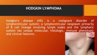
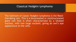
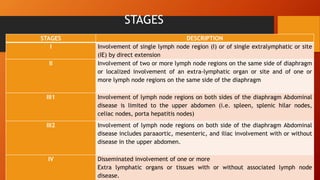
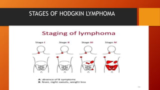
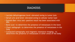
HODGKIN LYMPHOMA &
NON HODGKIN LYMPHOMA
HARSHITA
M.Sc. NURSING
CHILD HEALTH NURSING
HODGKIN LYMPHOMA
Hodgkin’s disease (HD) is a malignant disorder of
lymphoreticular system; a lymphoreticular neoplasm primarily
of B cell lineage involving lymph nodes and the lymphatic
system has unique molecular, histologic, immune phenotypic
and clinical features.
WHO CLASSIFICATION
Nodular lymphocytic-predominant Hodgkin
lymphoma (NLPHL)
Classical Hodgkin lymphoma
NLPHL
(NODULAR LYMPHOCYTIC PREDOMINANT HL)
Patients with NLPHL generally present with localized, non-bulky
disease.
The only symptom for most people with NLPHL is one or more
lumps. These are enlarged lymph nodes (swollen glands). They
are often in only one place in the body.
A few people have other general symptoms of lymphoma, like
night sweats, weight loss and fevers.
Classical Hodgkin lymphoma
The hallmark of classic Hodgkin lymphoma is the Reed-
Sternberg cell. This is a binucleated or multinucleated
giant cell that is often characterized by a bilobed
nucleus, with two large nucleoli, giving an owl’s eye
appearance to the cells.
CAUSES
• Variation in the incidence of HD in different ethnic
groups and association with human leukocyte antigen
suggests that inherited susceptibility plays an important
role in the pathogenesis.
• Environmental factors such as Epstein-Barr virus
infection,
• Familial clustering of cases and
• Higher incidence in twins may be some of the other
contributing factors.
SIGN AND SYMPTOMS
Lymphadenopathy, usually in the cervical, supraclavicular, and
mediastinal areas; mediastinal presentation common in
adolescents and young adults; (significant mediastinal adenopathy
may cause cough, dyspnea, or superior vena cava syndrome)
Painless, movable lymph nodes in tissues surrounding involved
area
Unexplained fever
Weight loss
Drenching night sweats
Malaise
Painless cervical or supraclavicular lymphadenopathy
STAGES
STAGES DESCRIPTION
I Involvement of single lymph node region (I) or of single extralymphatic or site
(IE) by direct extension
II Involvement of two or more lymph node regions on the same side of diaphragm
or localized involvement of an extra-lymphatic organ or site and of one or
more lymph node regions on the same side of the diaphragm
III1 Involvement of lymph node regions on both sides of the diaphragm Abdominal
disease is limited to the upper abdomen (i.e. spleen, splenic hilar nodes,
celiac nodes, porta hepatitis nodes)
III2 Involvement of lymph node regions on both side of the diaphragm Abdominal
disease includes paraaortic, mesenteric, and iliac involvement with or without
disease in the upper abdomen.
IV Disseminated involvement of one or more
Extra lymphatic organs or tissues with or without associated lymph node
disease.
STAGES OF HODGKIN LYMPHOMA
DIAGNOSIS
Complete blood count—diagnostic (anemia may indicate
advanced disease)
Erythrocyte sedimentation rate (ESR)—may be elevated
at diagnosis
Serum copper, iron, calcium, and alkaline phosphatase
levels—also may be elevated at diagnosis
Liver and renal function tests—to assess organ
involvement
Urinalysis—to determine renal involvement
DIAGNOSIS
Chest radiographic study—to determine mediastinal or
hilar node involvement
Computed tomography—to evaluate mediastinal,
pulmonary, and abdominal disease
Gallium and/or positron emission tomography (PET) scan
to determine the extent of involvement
Excisional lymph node biopsy—essential to diagnosis and
staging
Bone marrow biopsy if patient has stage 3 or 4 disease
according to imaging studies
TREATMENT
Treatment modalities have varied from total nodal
radiation therapy to chemotherapy to combination of
chemotherapy and radiotherapy with significant
improvement in survival rate throughout the last three
decades.
All children generally receive combination
chemotherapy as initial treatment.
TREATMENT
Radiation therapy can cause profound musculoskeletal
growth retardation and increase the risk for cardiovascular
disease and secondary solid malignancies in children.
The volume of radiation and the intensity/duration of
chemotherapy are determined by prognostic factors at
presentation, including presence of constitutional
symptoms, disease stage, and bulk.
NON HODGKIN LYMPHOMA
NON-HODGKIN LYMPHOMA
Non-Hodgkin’s Lymphoma (NHL) is neoplasm of a wide
range of cell types that comprise the immune system.
NHL most commonly occurs during the second decade
of life and occurs less frequently in children less than
three years of age. Pediatric NHL are high grade,
diffuse and aggressive with propensity for
dissemination
Three Therapeutically Categories
 B-cell NHL (Burkitt lymphoma/leukemia and diffuse
large B-cell lymphoma);
Lymphoblastic lymphoma (primarily precursor T-cell
lymphoma and, less frequently, precursor B-cell
lymphoma);
Anaplastic large cell lymphoma (T cell or null-cell
lymphomas).
CAUSES
1. B cells
B cells fight infection by producing antibodies that
neutralize foreign invaders. Most non-Hodgkin's
lymphoma arises from B cells. Subtypes of non-
Hodgkin's lymphoma that involve B cells include
diffuse large B-cell lymphoma, follicular lymphoma,
mantle cell lymphoma and Burkitt lymphoma.
CAUSES
2. T cells
T cells are involved in killing foreign invaders directly.
Non-Hodgkin's lymphoma occurs less often in T cells.
Subtypes of non-Hodgkin's lymphoma that involve T
cells include peripheral T-cell lymphoma and
cutaneous T-cell lymphoma.
SIGN AND SYMPTOMS
1. Intraabdominal Involvement
• Possible symptoms mimicking appendicitis (pain, right lower
quadrant tenderness)
• Ovarian, pelvic, retroperitoneal masses
• Ascites
• Vomiting
• Diarrhea
• Weight loss
SIGN AND SYMPTOMS
2. Mediastinal Involvement
• Pleural effusion
• Tracheal compression
• Superior vena cava syndrome
• Coughing, wheezing, dyspnea, respiratory distress
• Edema of upper extremities
• Mental status changes
SIGN AND SYMPTOMS
3. Primary Nasal, Paranasal, Oral, and Pharyngeal Involvement
• Nasal congestion
• Rhinorrhea
• Epistaxis
• Headache
• Proptosis
• Irritability
STAGES
STAGE DESCRIPTION
I
Single tumor (extranodal)Single anatomic area (nodal) excluding mediastinum or abdomen
II Single tumor (extranodal) with regional node involvement. Primary gastro intenstinal
tumor with
or without involvement of mesentric node.or
On same side of diaphragm:
a. Two or more nodal areas
b. Two single extranodal tumors with or without
regional node involvement
III All primary intrathoracic tumors. All extensive
primary intra-abdominal disease. Two or more
nodal or extranodal areas on both sides of
diaphragm
IV Any of the above with CNS or bone marrow
STAGES
DIAGNOSIS
• Bone marrow biopsy—to identify malignant cells with bone marrow
involvement
• Lumbar puncture—to determine presence of malignant cells in CNS
• Complete blood count—diagnostic for bone marrow dysfunction;
may show elevated white blood cell count, decreased hemoglobin
level, hematocrit, and platelet count
• Liver and kidney function tests—liver function test values may be
elevated with liver involvement; kidney function test values may
be elevated with kidney involvement
DIAGNOSIS
• Lactate dehydrogenase level—elevated owing to tumor lysis
• Serum uric acid level—elevated owing to cellular tumor load
• Epstein-Barr virus test—positive result has been associated with
NHL
• Bone scan—to determine the presence of metastases in the bone
• Chest radiograph—to determine the presence of metastases in the
lung
• Computed tomography and magnetic resonance imaging— to
determine the presence of metastases in other areas of the body
TREATMENT
Childhood NHL is an extremely chemosensitive
disease. Surgery plays a very limited role, mainly for
arriving at a diagnosis. Radiation of primary sites is
used very rarely in emergency situations. Hence,
multi-agent chemotherapy directed to the histologic
subtype and stage of the disease remains the
cornerstone of therapy.

nhlhl-190521214936.pdf

  • 1.
    HODGKIN LYMPHOMA & NONHODGKIN LYMPHOMA HARSHITA M.Sc. NURSING CHILD HEALTH NURSING
  • 2.
    HODGKIN LYMPHOMA Hodgkin’s disease(HD) is a malignant disorder of lymphoreticular system; a lymphoreticular neoplasm primarily of B cell lineage involving lymph nodes and the lymphatic system has unique molecular, histologic, immune phenotypic and clinical features.
  • 3.
    WHO CLASSIFICATION Nodular lymphocytic-predominantHodgkin lymphoma (NLPHL) Classical Hodgkin lymphoma
  • 4.
    NLPHL (NODULAR LYMPHOCYTIC PREDOMINANTHL) Patients with NLPHL generally present with localized, non-bulky disease. The only symptom for most people with NLPHL is one or more lumps. These are enlarged lymph nodes (swollen glands). They are often in only one place in the body. A few people have other general symptoms of lymphoma, like night sweats, weight loss and fevers.
  • 5.
    Classical Hodgkin lymphoma Thehallmark of classic Hodgkin lymphoma is the Reed- Sternberg cell. This is a binucleated or multinucleated giant cell that is often characterized by a bilobed nucleus, with two large nucleoli, giving an owl’s eye appearance to the cells.
  • 6.
    CAUSES • Variation inthe incidence of HD in different ethnic groups and association with human leukocyte antigen suggests that inherited susceptibility plays an important role in the pathogenesis. • Environmental factors such as Epstein-Barr virus infection, • Familial clustering of cases and • Higher incidence in twins may be some of the other contributing factors.
  • 7.
    SIGN AND SYMPTOMS Lymphadenopathy,usually in the cervical, supraclavicular, and mediastinal areas; mediastinal presentation common in adolescents and young adults; (significant mediastinal adenopathy may cause cough, dyspnea, or superior vena cava syndrome) Painless, movable lymph nodes in tissues surrounding involved area Unexplained fever Weight loss Drenching night sweats Malaise Painless cervical or supraclavicular lymphadenopathy
  • 8.
    STAGES STAGES DESCRIPTION I Involvementof single lymph node region (I) or of single extralymphatic or site (IE) by direct extension II Involvement of two or more lymph node regions on the same side of diaphragm or localized involvement of an extra-lymphatic organ or site and of one or more lymph node regions on the same side of the diaphragm III1 Involvement of lymph node regions on both sides of the diaphragm Abdominal disease is limited to the upper abdomen (i.e. spleen, splenic hilar nodes, celiac nodes, porta hepatitis nodes) III2 Involvement of lymph node regions on both side of the diaphragm Abdominal disease includes paraaortic, mesenteric, and iliac involvement with or without disease in the upper abdomen. IV Disseminated involvement of one or more Extra lymphatic organs or tissues with or without associated lymph node disease.
  • 9.
  • 10.
    DIAGNOSIS Complete blood count—diagnostic(anemia may indicate advanced disease) Erythrocyte sedimentation rate (ESR)—may be elevated at diagnosis Serum copper, iron, calcium, and alkaline phosphatase levels—also may be elevated at diagnosis Liver and renal function tests—to assess organ involvement Urinalysis—to determine renal involvement
  • 11.
    DIAGNOSIS Chest radiographic study—todetermine mediastinal or hilar node involvement Computed tomography—to evaluate mediastinal, pulmonary, and abdominal disease Gallium and/or positron emission tomography (PET) scan to determine the extent of involvement Excisional lymph node biopsy—essential to diagnosis and staging Bone marrow biopsy if patient has stage 3 or 4 disease according to imaging studies
  • 12.
    TREATMENT Treatment modalities havevaried from total nodal radiation therapy to chemotherapy to combination of chemotherapy and radiotherapy with significant improvement in survival rate throughout the last three decades. All children generally receive combination chemotherapy as initial treatment.
  • 13.
    TREATMENT Radiation therapy cancause profound musculoskeletal growth retardation and increase the risk for cardiovascular disease and secondary solid malignancies in children. The volume of radiation and the intensity/duration of chemotherapy are determined by prognostic factors at presentation, including presence of constitutional symptoms, disease stage, and bulk.
  • 14.
  • 15.
    NON-HODGKIN LYMPHOMA Non-Hodgkin’s Lymphoma(NHL) is neoplasm of a wide range of cell types that comprise the immune system. NHL most commonly occurs during the second decade of life and occurs less frequently in children less than three years of age. Pediatric NHL are high grade, diffuse and aggressive with propensity for dissemination
  • 16.
    Three Therapeutically Categories B-cell NHL (Burkitt lymphoma/leukemia and diffuse large B-cell lymphoma); Lymphoblastic lymphoma (primarily precursor T-cell lymphoma and, less frequently, precursor B-cell lymphoma); Anaplastic large cell lymphoma (T cell or null-cell lymphomas).
  • 17.
    CAUSES 1. B cells Bcells fight infection by producing antibodies that neutralize foreign invaders. Most non-Hodgkin's lymphoma arises from B cells. Subtypes of non- Hodgkin's lymphoma that involve B cells include diffuse large B-cell lymphoma, follicular lymphoma, mantle cell lymphoma and Burkitt lymphoma.
  • 18.
    CAUSES 2. T cells Tcells are involved in killing foreign invaders directly. Non-Hodgkin's lymphoma occurs less often in T cells. Subtypes of non-Hodgkin's lymphoma that involve T cells include peripheral T-cell lymphoma and cutaneous T-cell lymphoma.
  • 19.
    SIGN AND SYMPTOMS 1.Intraabdominal Involvement • Possible symptoms mimicking appendicitis (pain, right lower quadrant tenderness) • Ovarian, pelvic, retroperitoneal masses • Ascites • Vomiting • Diarrhea • Weight loss
  • 20.
    SIGN AND SYMPTOMS 2.Mediastinal Involvement • Pleural effusion • Tracheal compression • Superior vena cava syndrome • Coughing, wheezing, dyspnea, respiratory distress • Edema of upper extremities • Mental status changes
  • 21.
    SIGN AND SYMPTOMS 3.Primary Nasal, Paranasal, Oral, and Pharyngeal Involvement • Nasal congestion • Rhinorrhea • Epistaxis • Headache • Proptosis • Irritability
  • 22.
    STAGES STAGE DESCRIPTION I Single tumor(extranodal)Single anatomic area (nodal) excluding mediastinum or abdomen II Single tumor (extranodal) with regional node involvement. Primary gastro intenstinal tumor with or without involvement of mesentric node.or On same side of diaphragm: a. Two or more nodal areas b. Two single extranodal tumors with or without regional node involvement III All primary intrathoracic tumors. All extensive primary intra-abdominal disease. Two or more nodal or extranodal areas on both sides of diaphragm IV Any of the above with CNS or bone marrow
  • 23.
  • 24.
    DIAGNOSIS • Bone marrowbiopsy—to identify malignant cells with bone marrow involvement • Lumbar puncture—to determine presence of malignant cells in CNS • Complete blood count—diagnostic for bone marrow dysfunction; may show elevated white blood cell count, decreased hemoglobin level, hematocrit, and platelet count • Liver and kidney function tests—liver function test values may be elevated with liver involvement; kidney function test values may be elevated with kidney involvement
  • 25.
    DIAGNOSIS • Lactate dehydrogenaselevel—elevated owing to tumor lysis • Serum uric acid level—elevated owing to cellular tumor load • Epstein-Barr virus test—positive result has been associated with NHL • Bone scan—to determine the presence of metastases in the bone • Chest radiograph—to determine the presence of metastases in the lung • Computed tomography and magnetic resonance imaging— to determine the presence of metastases in other areas of the body
  • 26.
    TREATMENT Childhood NHL isan extremely chemosensitive disease. Surgery plays a very limited role, mainly for arriving at a diagnosis. Radiation of primary sites is used very rarely in emergency situations. Hence, multi-agent chemotherapy directed to the histologic subtype and stage of the disease remains the cornerstone of therapy.