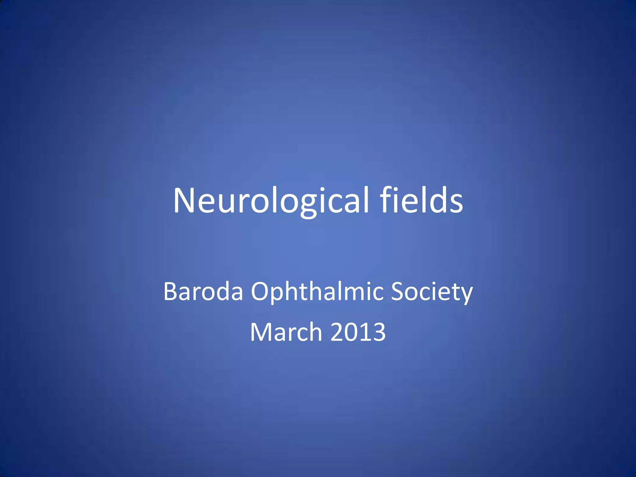
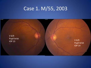
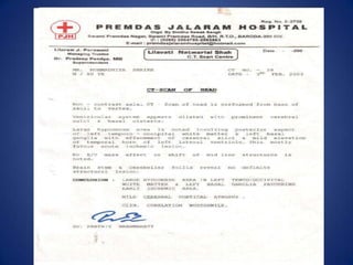
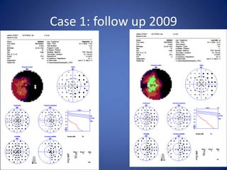
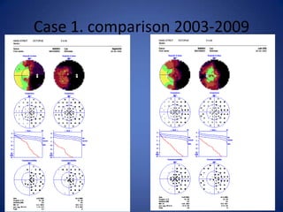
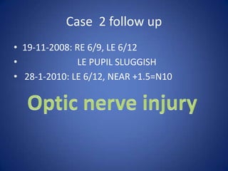
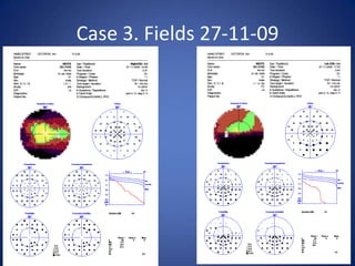
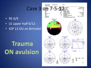
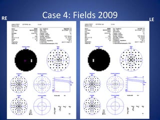
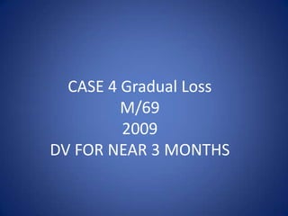
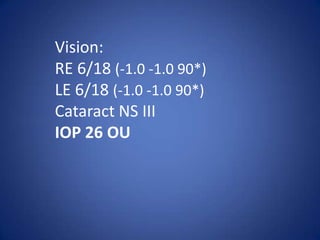
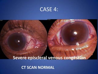
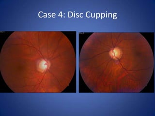
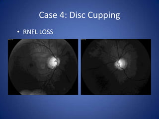
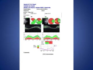
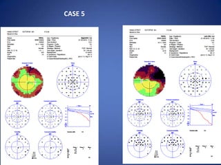
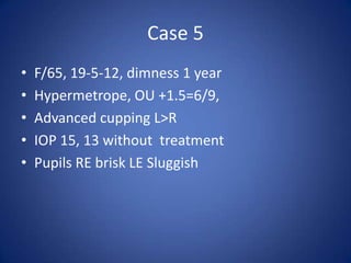
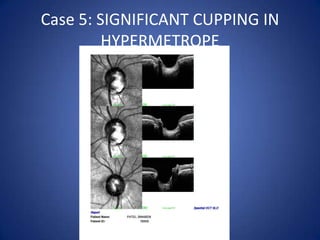
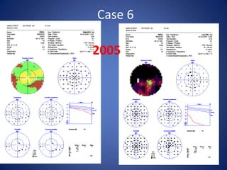
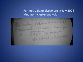
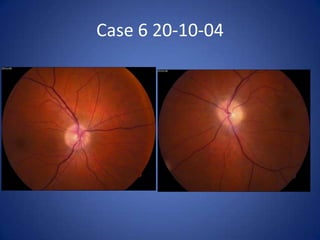
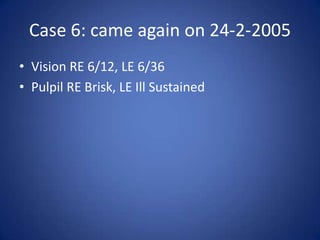
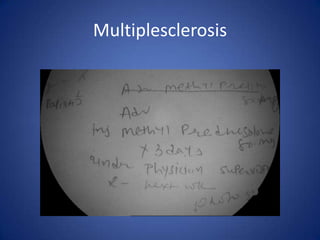
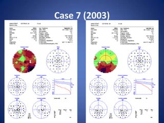
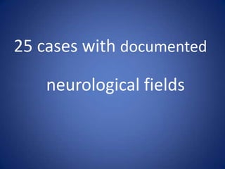
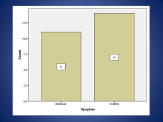
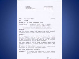
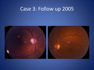
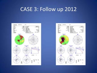
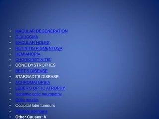
Neurological fields

Baroda Ophthalmic Society
       March 2013
Case 1.
Case 1. Gradual Loss

         M/55, 2003
DV 2 months, difficulty in reading

   Stroke 4months, Squint
Case 1. M/55, 2003


V 6/9
Pupil brisk
                                   V 6/9
IOP 13
                                   Pupil brisk
                                   IOP 14
Case 1: follow up 2009
Case 1. comparison 2003-2009
20-5-06 RE   Case 2.   20-5-06 LE
Case 2. Sudden Loss
       M/38
 ROAD ACCIDENT
Case 2.
Trauma 4/5/2006, UNCONCIOUS   DV LE NOTICED LATER


   6/9                         6/36, nasally
   Pupil brisk                 Pupil sluggish
   IOP 11                      IOP 14
2007   2008
Case 2 follow up
• 19-11-2008: RE 6/9, LE 6/12
•             LE PUPIL SLUGGISH
• 28-1-2010: LE 6/12, NEAR +1.5=N10
Case 3. Fields 27-11-09
Case 3.
     M/50
   22-8-2007
Dimness LE 6/24,
  NS grade III
  Hypertensive
Case 3
•   IOP RE 17, LE 13
•   Pupils reacting normally
•   LE Phaco 14-9-07
•   Post op 6/9 till 19-6-08
Case 3
•   Head Injury on 16-8-09, left temporal region
•   Reports loss of vision on 26-11-09.
•   MRI for left optic canal, nerve, reported NAD
•   Vision LE FC 2’
•   IOP 16 OU
Case 3 Fundus 2-12-09
Case 3 LE

              Naked Vessls


RNFL
Case 3. Sequence
Case 3 on 7-5-12
• RE 6/9
• LE upper half 6/12
• IOP 13 OU on Brimolol
RE   Case 4: Fields 2009   LE
CASE 4 Gradual Loss
         M/69
         2009
DV FOR NEAR 3 MONTHS
Vision:
RE 6/18 (-1.0 -1.0 90*)
LE 6/18 (-1.0 -1.0 90*)
Cataract NS III
IOP 26 OU
CASE 4:


Severe episcleral venous congestion
    CT SCAN NORMAL
Case 4: Disc Cupping
• RNFL LOSS
Case 4:
• RE Phaco + Trab on        • LE phaco + Trab on
• 8-3-2011                  • 4-2-2010
• IOP 12 with topical       • IOP 13 with topical
  drops                       drops
Case 4: Follow up
RE 6/9
IOP
RE 12
LE 13
Glaucoma +
?Cavernous
   sinus
Case 5
CASE 5
Case 5
•   F/65, 19-5-12, dimness 1 year
•   Hypermetrope, OU +1.5=6/9,
•   Advanced cupping L>R
•   IOP 15, 13 without treatment
•   Pupils RE brisk LE Sluggish
Optic Disc
Fields 25-5-12


    IOP
    RE 16
    LE 14
    TRAVACOM


     MRI
     FEW CH ISCHEMIC FOCCI
     BILAT. FRONTO-PARIETAL
     PARIETO-OCCIPT
Fields 12-12-12

     IOP
     RE 13
     LE 15

     TRAVACOM

     ADD BRIMO
Fields 22-3-13


     IOP
     RE 15
     LE 15


     CCT
     RE 539
     LE 544
Fields comparision
Case 5: SIGNIFICANT CUPPING IN
         HYPERMETROPE
Case 6: Sudden Loss
     20-10-04
       M/43
   Vision RE 6/9
      LE FC 6’
Case 6

2005
Perimetry done elsewhere in July 2004
Medmont cluster analysis
Case 6 20-10-04
Case 6: LE Field 20-10-04
Case 6: came again on 24-2-2005
• Vision RE 6/12, LE 6/36
• Pulpil RE Brisk, LE Ill Sustained
Multiplesclerosis
Case 6:
Last followup 4-3-2005
Case 7 (2003)
Case 7: Gradual loss
    24-6-1999
        F/42
Case 7: History
•   Dimness RE 8 months
•   Diagnosed Optic Atrophy elsewhere
•   NIDDM
•   Vision RE FC 4’, LE -2.5 90*= 6/18
•   Pupil RE Sluggish, LE Brisk
Case 7: Fundus Pictures
• Treated with Inj Methyl Pred IV.
Case 6: 22-07-2003
Types of Neurological defects
CENTRO-CAECAL
HEMI- QUADRANT ANOPIA
GLAUCOMA + NEUROLOGICAL FIELD LOSS
25 cases with documented

    neurological fields
•Case 3
Case 3: F/50 MAY 1993
DV GRADUAL 1WEEK   HEADACHE VOMITING 2DAYS
Case 3: Follow up 2005
CASE 3: Follow up 2012
•   MACULAR DEGENERATION
•   GLAUCOMA
•   MACULAR HOLES
•   RETINITIS PIGMENTOSA
•   HEMIANOPIA
•   CHORIORETINITIS
•   CONE DYSTROPHES
•   BEST'S DISEASE
•   STARGADT'S DISEASE
•   ACHROMATOPSIA
•   LEBER'S OPTIC ATROPHY
•   Ischemic optic neuropathy
•   Optic neuritis
•   Occipital lobe tumours
•   Pituitary adenoma
•   Other Causes: V

Neurological fields bos 2013