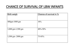
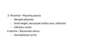
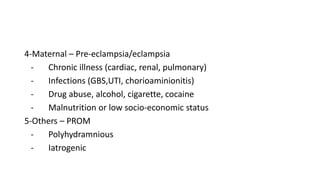
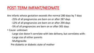
neonatology
Prepared Dr Jembere Belachew
Providing care a newborn baby
• Maintaining an open air way
• Immediately after birth, the birth attendant should assess the air way
of the new born
• If the baby does not breath with in 3 seconds or is blue (cyanosed)
after delivery, do not wait for a minute
• Clear the airway quickly and thoroughly, especially if the amniotic
fluid is meconium-stained
• Place the neonate on his/her back in horizontal position with head
down ward slightly extended, so that the mouth, pharynx and nose
clear of fluid, mucus, blood and meconium by gravity
- Gently suck out mucus with a bulb syringe or soft rubber or plastic mucus
extractor (1st mouth, then the nose)
- Ventilate the baby by Ambu bag or other available bag and mask:
- The mask should cover mouth and nose
- Push the lower jaw a little forward with your thumb and open the mouth
through slight pressure of a finger and the mask on the chin
-- Prevent air leak around the baby’s face
- Start 02 (100% at a rate of 40 LPM)
- Allow the baby to breath out
- Chest and abdomen must be moving with ventilation
- Proper breath sounds should be heard over the chest
Cleaning of the Eye, skin and Umblical cord of the newborn to prevent
infection
Eye
- Immediately after the head is born, the baby’s eyes should be cleaned
with a sterile guaze. (Eyes should be gently wiped with a swab)
-The eyes of all infants must be protected against gonococcal infection by
applying
-1% silver nitrate drops-the best proven therapy
-0.5% erythromycin or 1% tetracycline ointment (alternative measures)
• 2.5% povidone-iodine-may be effective at one time prophylactic agent
• Instil 2drops of 1% silver nitrate solution into each conjactiva sac shortly
after birth
- Infants born to mothers with active gonorrhea are at high risk for developing gonococcal
ophthalmitis and should be given:
- A single 125 mg/Im injection of ceftriaxone for prophylaxis
- For low birth weight infants, the dose is 25-50mg/kg
Skin and cord care
• To reduce the incidence of skin and peri-umbilical colonization with pathogenic bacteria
and infection (omphalitis), the entire skin and cord should be cleansed, once an infant’s
temperature has stabilized.
• Clean with sterile cotton soaked in warm water or mild, no medicated soap solution
• Use sterile cored clamp or autoclaved cord ties and tie well
• Rinse the neonate in warm water near or at body temperature
• Keep the child wrapped in a dry cloth at a warm place (Temperature of 30-350
C)
• Excess vernix and any blood on the baby’s body is removed using a
soft cloth soaked in lukewarm water
• Do not scrub the baby’s skin with a rough sponge immediately after
birth
• The Umblical stump is examined again for any bleeding and is re-tied
if necessary
• If necessary, paint the Umblical stump with an antibacterial
preparation
• Triple dye (a mixture of gentian violet, brillian green and proflavine)
• Antibiotic spray (a mixture of neomycin, bacitracin and polymyxine), in the
absence of triple dye
• A void bandaging around the abdomen, as it may interfere with
breathing and give rise to asphyxia
• There fore, care of the skin, eyes and cord of the new born is vital to
prevent infection
Feeding the neonate
• Successful infant feeding requires cooperation b/n the mother and her baby
• Promptly establishing comfortable, satisfying feeding practices contributes
greatly to the infant’s and mother’s emotional well-being
• Feeding should be initiated to maintain normal metabolism and growth during
the transition from fetal to extra uterine life
To promote maternal – infant bonding
To decrease risks of
- Hypoglycemia - Hyperbilirubinaemia and
- Hyperkalemia - Azotemia
-Neonates should start breast-feeding immediately after birth (with in 30 minutes)
- The neonate should feed the breast milk frequently and as much as he/she
wants (Every 3-4 hour/day= day and night)
Exclusive breast feeding
EBF- is giving breast milk alone for the first 6 months of life except medication
- WHO recommended that every infant should exclusively breast-fed until 6
months of age .
Advantages of EBF
- Breast-feeding is essential for the survival of the infant in most situations in
developing countries
- It has overwhelming advantages anywhere in the world
Colostrums - produced during the first few day of lactation is particularly beneficial
in preventing infections
- Human-milk is exactly right nutritionally for the young infant’s needs, and, even in
less than well-nourished women
• Advantages of EBF
• - Breast-feeding is essential for the survival of the infant in most
situations in developing countries
• - It has overwhelming advantages anywhere in the world
• Colostrums - produced during the first few day of lactation is
particularly beneficial in preventing infections
• - Human-milk is exactly right nutritionally for the young infant’s
needs, and, even in less than well-nourished women
Generally breast-feeding has the following advantages:-
Breast-milk is the natural food for full-term infants during the 1st months of
life
• Always readily available at proper temperature
• Needs no time for preparation
• Fresh and free of contaminating bacteria
• Allergy and intolerance to cow’s milk create significant disturbances and
feeding difficulties
• these are not seen in breast-fed infants
• Human-milk contains bacterial and viral antibodies
• Macrophages normally present in human colostrums and milk may be able
to synthesize complement, lysozyme and lactoferrine
• Supply the necessary nutrients to the infant
• It has psychological advantage for both the mother and infant
• Prematurely born baby usually thrive on breast milk
• NB- The low vitamin K content of human-milk may contribute to
hemorrhagic disease of the newborn, So, 1 mg of vit k administration
is recommend for all infants, especially for those who will be breast-
fed
-Formula- milk is used if the mother could never breast feed for
various reasons
- Animal milk and numerous “modified formulas” are widely
available, but are very expensive
-Whatever is fed to the non-breast-fed infant, is extremely dangerous
because of
- Likelihood of over dilution
- Bacterial contamination
- The feeding-bottle is especially risky as it is very difficult to clean
- Advise the mother to use cups or spoons for feeding her infant
• Signs of good attachment
- If an infant is not well attached, the result may be: pain and damage
to the nipples or: The infant may not take enough milk (breast-milk)
effectively ,may cause engorgement
- If the infant is not well attached
- He/she may be unsatisfied after breast feeds; want to feed very often
or for a very long time
- The infant may get too little milk and not gain weight
- The breast-milk may dry up
• All the above problems may improve if attachment can be improved
• The following four signs indicates good attachment
1- Chin touching breast (or very close)
2- Mouth wide open
3- Lower lip turned outward
4- More areola visible above than below the mouth
• If attachment is not good, you may see
• Chin not touching breast
• Mouth not wide open, lips pushed for word
• Lower lip turned in or
• More areola (or equal amount) visible below infants mouth than above it
Measuring APGAR score
Defn- It is a practical method of systematically assessing the newborn infant
immediately after birth to help identify infants requiring resuscitation for hypoxic-
acidosis
- Assessment at birth is by the APGAR score is done routinely at 1 and 5
minutes after delivery
- Purpose – To help identify infants requiring resuscitation for hypoxic-acidosis
(Asphyxia) or not
-The 1- minute APGAR score signals the need for immediate resuscitation
-The 5minute score indicates the probability of successfully resuscitating the
newborn
-APGAR scores of 0-3 at 20 minute predict high morbidity and mortality
APGAR Score system
Sign 0 1 2
Appearance (colour) Blue, pale grey Body pink blue extremities Completely pink
Pulse (HR) Absent Slow <100/min >100/min
Grimace (Response to
stimuli)
No response Grimace (slight) Cry, cough sneezing
Activity (Muscle
tone)
Limp Some flexion of extremities Active motion
Respiratory effort Absent Slow irregular gasping Good crying
Classification of APGAR score according total score (interpretation)
1) Apgar 7-10 = Normal
2) Apgar 4-6 = Moderate Asphyxia
3) Apgar 0-3 = Sever Asphyxia
A- Apgar 7-10
- Dry the baby and keep him/her warm
- Using suction is not necessary if the baby cries vigorously
B- Apgar 4-6
- Most of these babies are breathing
- If so and have HR> 100/minute, quick and gentle clearing of the airway
and stimulation by slapping the buttocks, with 02 by mask
-If there is no improvement, or getting worse and the HR<100/minute
Move on immediately to vigorous Rx as follows below (see Apgar 0-3)
C- Apgar 0-3
- Proceed in the following order as quickly and as carefully as possible
- Note the time
- Dry and cover the baby
- Supply heat from above the baby
ANEONATE WITH NEONATAL PROBLEMS
• LBW-infant is a baby whose birth weight is less than 2,500 gm.
• LBW- infant is a baby whose birth weight is less than 2,500 gm.
• VLBW-infant is low birth weight baby whose birth weight is <1,500 gm
• ELBW-infant – is a LBW baby whose weight is less than 1,000 gm
(Immature neonate)
• Premature/preterm-infant – is live born infant delivered before 37 weeks
from the 1st day of LMP
• Post-term infant – An infant born after 42 weeks of gestation from LMP,
regardless of birth weight
• LBW infants are premature if they only have gestational age < 37 weeks.
CHANCE OF SURVIVAL OF LBW INFANTS
Birth weigh Chance of survival in %
400gm-1000 gm 10%
1,000 gm-1,500 gm 40%-50%
1,500 gm- 2000 gm 75-85%
• In developing world approximately 70% of LBW infant have IUGR
• VLBW infants account for > 50% of neonatal deaths
• Many LBW infants are also preterm
• The majority of infants above 2,000 gm do not need specialized care
and can be kept comfortably with their mothers
• Make sure they can feed and keep them warm
• Infants below 2,000gm, and especially those less than 1,750 gm, do
need specialized care, which in most cases can be provided only by
hospitals.
IDENTIFIABLE CAUSES OF PRETERM BIRTH AND/OR IUGR
• Generally the causes of LBW/Preterm/IUGR are classified into five
categories:
• 1- Fetal- Fetal distress due to lung immaturity, infection or aspiration
• Multiple gestation
• Erythroblastosis
• Non- immune hydrops- chromosomal disorders
• Chronic fetal infections
• Congenital malformations
• Radiation injury
2- Placental – Placenta praevia
- Abruptio placenta
- Small weight, decreased surface area, infarction
- Infection, tumor
3-Uterine – Bicorunate uterus
- Incompetence cervix
4-Maternal – Pre-eclampsia/eclampsia
- Chronic illness (cardiac, renal, pulmonary)
- Infections (GBS,UTI, chorioaminionitis)
- Drug abuse, alcohol, cigarette, cocaine
- Malnutrition or low socio-economic status
5-Others – PROM
- Polyhydramnious
- Iatrogenic
PROBLEMS OF THE LBW/PREMATURE INFANTS
• The problems of the LBW/Premature infants are mostly associated with
anatomical and physiological immaturity of the various organs of the infant
• 1- Respiratory system problem
• Respiratory distress is common in very immature babies, especially the VLBW
infants and those with a gestation of < 32 weeks.
• Mainly due to lung immaturity, but it may be caused by infection or aspiration
-Apnea/cyanosis - Poorly developed lungs
-Prone to Atelectasis - Lack of chest-wall stability
-Respiratory distress syndrome (HMD) - Poor/reduced surfactant production
-Poor gag and cough reflex -Poorly developed respiratory muscles
• 2-DIGESTIVE SYSTEM
• The GIT though physiologically mature has not had opportunity to function
till after birth.
• Small stomach (likely to vomit)
• Gastro esophageal regurgitation
• Decreased food tolerance or absorption, especially fat, vit D and all fat
soluble vitamins
• Do not express hunger by crying- not feed at regular intervals –
Hypoglycemia- Brain damage
• Abdominal distension due to poorly developed muscles
• Hypoglycemia, hypo albuminemia and hypoprothrombinaemia due to
hepatic immaturity
3- POOR THERMAL STABILITY (HYPOTHERMIA)
-Body temperature determines survival as well as postnatal growth
-Small infants are unable to maintain normal body temperature
unless the environment is warm enough.
-Cold stress
- Hypoglycemia
-Hypoxia-acidosis
• Little subcutaneous fat
• Poor vasomotor control of blood flow to the skin (poor shivering)
• Large surface area to body wt ratio
• Decreased sweat glands – can’t perspire
• Less active with decreased muscle and fat deposits
• 4- POORLY DEVELOPED RENAL AND HEPATIC FUNCTION
• Poor electrolyte and fluid regulation
• Liver unable handle or conjugate bilirubin- high rate of jaundice and
Hyperbilirubinaemia
• Liver Vit-k store or production not developed
• Liver do not store glucose (hypoglycaemia)
• A steady decrease in Hgb after birth and in production of blood-
anemia
• 5- CNS – Slow response to stimuli
- Uncoordinated sucking and swallowing which may lead to aspiration
- Poorly developed vital centres
• 6-Increased risk of infection
- LBW infants, particularly those born very prematurely, have a very low
resistance against all infections of the newborn
- Infection is a major cause for death
- Increased risk of infection in LBW/preterm infants is due to:
- Don’t receive enough antibodies from the mother
- No IGM present
- Decreasing, chemotaxis, opsonization and phagocytosis
POST-TERM IMFANT/NEONATE
Are infants whose gestation exceeds the normal 280 days by 7 days
-25% of all pregnancies are born on or after 287 days
-12% of all pregnancies are born on or after 294 days
-5% of all pregnancies are born on or after 301 days
• Cause- unknown
-Large size doesn’t correlate with late delivery, but correlates with:
-Large size of either parents
-Multigravida
Pre diabetic or diabetic state of mother
Characteristics of post-term/post maturity
• Looks 1-3 weeks of age (old)
• Large birth weight
• Absence of lanugos
• Decreased or absent vernix caseosa
• Long nails
• Abundant scalp hair
• White parchment like or desquamating skin, cracked and
wrinkled skin
• Increased alertness
• Only 20% of neonates with placental insufficiency are post term
• When delivery is delayed more than 3 weeks beyond term, there is
a significant increase in mortality – Three times riskier than term
The risks are:
- Placenta insufficiency (fetal hypoxia, and distress)
-Meconium aspiration syndrome
-Hypoxic encephalopathy
-Birth injury
-Congenital malformation
CHARACTERSTICS OF PREMATURE INFANTS
1) Premature infants are tiny, scrawny and red
2) Have thin extremities with very little muscles and subcutaneous fat
3) Have disproportionately large head and abdomen
4) Have thin, relatively translucent and usually wrinkled skin
5) Have more visible abdominal and scalp veins
6) Have plentiful lanugos aver the extremities, back and shoulder
7) Their ears are soft with minimal cartilages
8) Have soft bones of skull, which have
9) Ribs yield with each labored breath
10) Undescended testes in male, and quiet prominent labia and
clitoris in female
11) Soles of the feet and palms of the hands have few creases
12) Many of the typical newborn reflexes are weak or absent
-The above characteristics of pre-term infant helps for diagnoses
purpose, and to differentiate from term-infants.
Prevention of low birth weight
• The association b/n LBW and socio-economic status indicates that the
ultimate solution to this problem of LBW will result from
socioeconomic development including appropriate health care.
1) Improved socio-economic status
2) Appropriate Health care
3) Prevention of maternal malnutrition and illness during pregnancy
4) Prenatal care and effective practice of FP, leading to child
spacing, improved general health and nutritional status.
2- CARE OF ANEONATE WITH SEPSIS AND MENINGITIS
• Neonatal sepsis-
• is a clinical syndrome characterized by systemic signs and symptom,
and bacteraemia during the first month of life.
• The incidence is relatively low (1 to 8 cases 1,000 live births)
• The risk of mortality is approximately 25%
• Meningitis in the neonate is usually a sequel a of bacteraemia,
however, it is discussed with neonatal sepsis, because they commonly
share etiology and pathogenesis
• The incidence of meningitis is usually a fraction of number of infants
with sepsis, varying in different settings from 1/4th to 1/3rd
Neonatal sepsis
• There are two major forms (subtypes) of neonatal sepsis:
• 1-Early onset sepsis (disease)
• Presents as a fulminant, multi systemic illness during the first 72 hours or 5-
7 days of life.
• Mostly caused by organisms prevalent in the genital tract or in labour room
and maternity operation theatre.
• Common pathogens
Group B streptococci - Klebsiella
Escherichia coli - Pseudomonas
Staphylococci aurous - Entrobacter species
• C/M – Majority manifest with respiratory distress due to an intrauterine
pneumonia) fever, failure to feed etc…
- Route of infection:
-Ascending infection (PROM)
- Passage of baby through infected birth canal
- During resuscitation in labor room
• Common pathogens
- Group B streptococci - Klebsiella
- Escherichia coli - Pseudomonas
- Staphylococci aurous - Entrobacter species
• Risk factors of Early-onset of Neonatal sepsis
• Birth Asphyxia
• Unclean vaginal examination
• Foul smelling liquor
• Duration of labor > 24 hours
• Birth weight < 2000 gm (2500gm/&/or gestation <37 weeks
• Premature rupture of membrane > 24 hours
• Maternal pyrexia
 When at least two or more of the above high-risk factors are present, the body is
considered to be infected and should be investigated properly for sepsis and
treated with appropriate antibiotics
2- Late-onset of neonatal sepsis
Is more commonly recognized after the first week of life or after 72
hours of life (Delayed by a minimum of 4 days)
 Acquired as nosocommunal infection from the nursery or lying in ward
 In most cases symptoms appear by the end of first week of life
 Common pathogens are:
 Klebsiella pneumoniae
 Entrobacteria
 Eschercia coli 2/3rd
cases are caused by these bacteria
 Gm negative bacilli
 Pseudomonas
 Salmonella
The rest are caused by gram- positive organism:
 Staphylococci aurous
 Staphylococci albus/epidermitis
Source of infection includes
 Incubators
 Resuscitation equipments
 Feeding bottles
 Catheter, face masks, infusion sets.
Clinical features of neonatal sepsis
 The clinical presentation could be silent in a very small baby who
suddenly may die with out exhibiting any s/s of infection.
o Behavioural changes and feeding changes (poor or failed sucking)
o Gradually or suddenly becomes lethargic, inactive, unresponsive and may
refuse to suck, then become pale.
o Hypothermia/Fever, RD
o Episodes of apnoeic spells
o Hepato-splenomegally
o Cyanosis
o Failure to gain weight or unexplained wt. loss
 Localized features of organ and system involved
Diagnostic work up and interpretation of Neonatal sepsis
 ESR (Value more than 10mm/hr is suggestive of infection)
 Absolute Neutrophils count (ANC)
- Do WBC and differential count 1st
• ANC= TLC x percentage of PMN cells
• Abnormal value <1,500 or >7,500
 Ratio of immature to total Neutrophils if I/T > 0.89, it is suggestive of
sepsis
- Gastric Aspiration for polymorphs
- Collect with in 1hours of birth
- Mix with 1 drop of heparin
- Drop on slide and thick smear
-Leishman’s stain
- (If>5 PMN/HPF – Infected amniotic fluid)
- Blood culture and sensitivity
- CSF analysis
- CXR, U/A and urine culture
Management of Neonatal sepsis
-First-line drugs:
 Crystalline pencillin or Ampicilline + Gentamycine
 Crystalline pencillin 50,000-1000- IV/Kg/day 2 doses for <1 weeks of age and 4
doses if >1 weeks of age
 Gentamycine 5mg/kg/day (2 doses)
 Ampicilline 100 mg/kg/day (2 dose for <1 wk age and 3 dose if >1 wk age)
- If there is meningitis, double the dose of cry. Pencillin and Ampicilline
-Second – Line drugs
Hospital acquired infection or if the mother has gram negative infection,
give the following
 Cefotaxine or ceftriaxone + Amino glycoside
 Supportive blood transfusion in DIC, sclerema and
Hyperbilirubinaemia
 Increase peripheral and pulmonary perfusion
 Corrects coagulation abnormality
 Removes toxins
Preventions of infection
• Hand washing (2min before examination and 30 sec b/n examination)
 Nursery Attire (Gown, shoes, baby gown) should be clean
 Avoid health personnel, if they are sick
 Patient placement
 Feeding-preparing formula/feeding materials
 General maintenance of labour room or neonatal unit
 Care of equipment (baby cots, tubes, thermometer should be clean)
 Skin and cord care
• N.B Better over treat than under treat, but do not overdose.
Neonatal Meningitis
Meningitis:- is an inflammation of the meninges, the membrane that
covers the brain and spinal cord
• It may be caused by bacteria, less commonly by tubercle bacilli or
viruses, or especially in immuno suppressed children by fungi or
parasite
Bacterial meningitis
-Out of all neonatal sepsis cases, 1/3 may have co-existing neonatal meningitis
-Purulent meningitis is a serious disease, caused by meningococci or
pneumococci, or other bacteria entering the meninges.
-In neonates clinical signs of meningeal irritation are absent
- In a neonate with septicaemia, the following s/s should arouse the
suspicion of neonatal meningitis
 Presence of fever
 Onset of convulsion or twitching
 Staring look
 Bulging anterior fontanel
 Abnormal high pitched cry or excessive crying
Lumbar puncture should be performed in:
 All new borns with suspected septicaemia or
• Those with above features
• Abnormal CSF
 More than 30 WBC per cubic millimetre
 More than 60% of WBC being polymorphs
 CSF glucose to blood glucose ratios< 50%
 Protein concentration > 150mg /dl in term baby and > 175 mg/dl in
preterm baby
 Positive gram stain and /or presence of micro-organisms in CSF
culture
Management
 Crystalline pencillin 300mg(500,000 IV) /kg/day given IV or Im in 3-hourly
doses, until the temperature is stable and normal, there after in 6 hourly doses for
14days
• Plus
• CAF 100mg/kg/day Im in 6-hourly doses for 3-5 days and then change
to oral CAF for a total of 14 days
 Ampicillin 200-400mg/kg/day
 In neonatal meningitis, the three best combinations of antibiotic are:
• CAF 25-50 mg/kg/day for babies > 2 kg of birth weight
• May be ↑ed to 50-75 mg/kg/day p 14 days
• Plus
• Pencilline 200,000-300,000 IV/kg/day Im or IV slowly
• 2- Ampicillin 200mg/kg/day in two to three doses
• Plus
 Gentamycine 5mg/kg/day in two doses Im or Iv- slowly
3- Ampicilline 200mg/kg/day in two to three doses
• Plus
 Cefotaxim 200mg/kg/day initially Iv or Im in four divided dose for 3 to 5 days then
orally for 14 days after that the CSF has cleared.
 Give phenobarbitone 5mg/kg/day in 3 divided doses with a minimum of 10mg three
times a day for children who have had a convulsion.
 Fluid – If the child can’t drink, an NGT should be passed and fluid / ORS should be
gives
 150ml /kg/day in children up to 10kg and 100ml/kg/day in bigger children up to a maximum of
2 lit/day.
 For very sick child or if vomiting, give maintenance IV fluid
 100ml/kg/24hours up to 10kg, 50-75ml /kg/24 hours in bigger children.
• N.B. Provide appropriate nursing care
3- MANAGING AND PREVENTING NEONATAL TETANUS
• Tetanus- Is an acute, often fatal, disease caused by an exotoxin
produced by the bacterium clostridium tetani
• Neonatal tetanus- is an acute, spastic paralytic illness in the
newborn (neonatal) period caused by tetanospasmin (neurotoxin)
produced by clostridium tetani.
 It is characterized by generalized rigidity and convulsive spasms of skeletal
muscles.
• The muscle stiffness usually involves the jaw (lockjaw) and neck and ten becomes
generalized.
Epidemiology:
 Occurs worldwide and endemic in 90 developing countries
 Neonatal tetanus is the most common form and kills about:
500,000 infants each year (un-immunized mothers)
Over 70% of these deaths occur just in 10 tropical Asian, and African
countries
 15,000-30,000 un-immunized mothers die world wide each year from
maternal tetanus (postpartum, postabortal, or post surgical wound
infection with c. tetani)
Etiology
 clostridium tetani
 A slender, gram-positive, and anaerobic rod
 May develop a terminal spore, giving it a drumstick appearance
 Resistance to heat and can’t survive in the presence of oxygen
 The spores are very resistance to heat and the usual antiseptics
Route of infection
 Umbilical stump
 Injection site (Drugs, vaccines)
 Circumcision
Pathogenesis
 C.tetani, usually enters the body through the wound (route of entry) In the
presence of anaerobic (low oxygen) conditions, the spores germinate and produce
toxins
 The toxins disseminated via blood and binds at the neuromuscular junction
Endocytosed by motor nerves Axonal transport and goes to spinal inhibitory
interneuron’s and prevents neurotransmitter release.
- Blocks normal inhibition of antagonistic muscles that is the basis of voluntary
coordinated movements.
 As a result, the affected muscles sustain maximal contraction
 The toxins act at several sites with in the CNS, including:
 Peripheral motor endplates
 Spinal cord
 Brain
 Sympathetic nervous system
Clinical manifestations of tetaus
• The typical clinical manifestation of tetanus are caused, when tetanus
toxins interferes with release of neurotransmitters, blocking inhibitor
impulses
 This leads to unopposed muscle contraction and spasm.
 Muscle spasm, Trismus or “lockjaw”
 Facial expression “Risus sardonicus”
 Sustained spasm of the back muscle “opisthotonus position”
 Tonic titanic seizure like activity
 Suddenly fails to suck, irritable
Paralysis or diminished movement
diagnosis
By clinical signs
 Un-immunized patient (and /or mother) born with in the preceding
• 2 wks with trismus, rigid muscles and clear sensorium.
Management
1- Remove source of tetanospasmin
Clean and debride wounds
Leave wounds open
Cleanse Umblical stump with antiseptics in newborns
Antibiotics
Pencillin G 100,000 IV/kg Im bid for 5 days
2- Neutralize circulating toxins by TAT 5000 IV Im
3- Provide supportive cares
A-Muscle relaxants and sedatives
- Diazepam 0.3 mg/kg Im or IV every 6 hourly, alternating with:
- Chlorpromazine 2mg/kg po or Im or
- Phenobarbitone 5mg/kg every 6 hourly po or Im
• B-Meticulous nursing care
- Quiet environment
- Avoid a auditory or visual stimuli
- Respiratory, oxygen, suction and Tracheostomy equipment should be available
- Meticulous care of mouth, skin, bladder and bowel.
• Prognosis
 Quality of supportive care determines the outcome
 Mortality is highest in:
-The very young and the very old
- short incubation period (<1wks)
- Short period of onset (< 3 days)
• Hypoxic insult to brain may result in:
-Cerebral palsy
-Mental retardation
-Behavioural disorders
• Prevention
-Maternal immunization (TT)
- Prompting clean delivery and nursery and cord cut
-TAT for home delivery
-Active immunization after that an episode of tetanus at should be
given
-Strengthening disease report
 RDS is also known as hyaline membrane disease (HMD), occurs almost
exclusively in premature infants
 The presence of two out of the following five features is defined as
respiratory distress
- Respiratory rate> 60/min
- Chest-indrawing
- Grunting
- Flaring of alaenasi
- Cyanosis
 The incidence and severity of RDS are related inversely to the gestational
age of the infant
 Common causes of RDS or HMD are classified into two:
MANAGING AND PREVENTING RESPIRATORY DISTRESS
SYNDROME (RDS)
• 1-Pulmonary causes:- They are again subdivided into three:
• A-Lung parenchymal disease:
o Hyaline membrane disease
o Meconium aspiration syndrome
o Congenital pneumonia
o Transient tachypnea of newborn
o Air leak
o Pulmonary haemorrhage
o Bronchopulmonary dysplesia
• B-Congenital Airway obstructions:
o Nasal or nasopharyngeal: choanal atresia, nasal edema
o Oral cavity: macroglosia, micrognatia and retrognathia
o Neck: congenital goitre, cystic hygroma
o Larynx: Web, subglottic, stenosis, haemangioma and laryngomalacia
o Trachea: Tracheomalacia, congenital tracheal stenosis
• C-Intrathoracic malformations
o Pulmonary hypoplesia or agenesis
o Diaphragmatic hernia
o Intra thoracic cysts
o Congenital lobar emphysema
• 2-Extrapulmonary causes of RDS: These are also further
divided into four:
A-Cardiac and circulatory causes
-Congenital heart diseases
-Congestive heart failure
-Hypervolemia
-Cardiac arrhythmia
• B-Metabolic causes
-Hypoglycemia
-Hypocalcaemia
-Hypothermia
-Metabolic acidosis
C-Neurological causes
-Neonatal meningitis
-Neonatal seizure
-Hypoxic-ischemic encephalopathy
-Extreme immaturity
-Intracranial haemorrhage
D- Haematological causes
-Anemia
-Polycythemia
PATHOPHYSIOLOGY OF RDS
 A relative deficiency of surfactant, which leads to decrease in lung
compliance and functional residual capacity with increased dead
space, causes RDS
 The resulting large ventilation-perfusion mismatch and right to left
shunt may involve as much as 80% of cardiac out put.
 Macroscopically, the lungs appear airless and ruddy (i.e. liver like).
Thus the lungs of these infants require a higher critical opening
pressure to inflate
 Microscopically, diffuse Atelectasis of distal airspaces along with
distension of some distal airways and per lymphatic areas are
observed
GENERAL APPROACH TO A NEONATE WITH RDS
 Proper history is most important; ANC and perinatal detailed Hx.
A-For Hyaline membrane disease
 Gestation
 Previous preterm babies
 Antenatal steroid prophylaxis
 APH and maternal DM
B-For congenital pneumonia: Ask for
 PROM
 Duration of labour
 Maternal fever
 Unclean vaginal examination
 Foul smelling liquor
C-For meconium aspiration syndrome
 Meconium stained liquor
 Hx of interapartum or postnatal suctioning
 Gestation
D-For congenital airway obstruction
 Polyhydramnious
 Excessive salivation since early neonatal period
 Difficulty of feeding
 Hx of traumatic delivery and birth asphyxia should be asked in all patients
 Do meticulous physical examination of the new born
Clinical manifestations of RDS
Progressive signs of respiratory distress are noted soon after birth and
include:
 Tachypnea
 Expiratory grunting (from partial closure of glottis)
 Sub costal and intercostals retractions
 Cyanosis
 Nasal flaring
• Dx- Hx, C/M and physical examination
 Analysis of blood gases (Respiratory and metabolic acidosis along with
hypoxia)
 Chest – Radiographs of an infant with RDS exhibits
o Bilateral diffuse reticular granular or ground glass appearance
o Air bronchograms
o Poor lung expansions
 Echocardiographic evaluation in selected infants
o For diagnosing PDA
o For determining the direction and degree of shunting
• Mg’t – Strategies to prevent premature birth and prudent use of
antenatal steroids to mature fetal lungs may decrease the incidence and
severity of RDS
 Give Vit-k, if not given at birth
 Give basic supportive cares
- Keep them in the thermo neutral zone
- Maintain adequate oxygenation
- Maintain adequate circulation
• (make sure that there is normal urine out put and normal capillary
filling time and normal weight)
 Delivery and resuscitation
 Surfactant replacement therapy
 Initiate specific treatment for specific problems
 Correction of metabolic abnormalities metabolic abnormalities
 Chest tube for air leak
• Mg’t -of cardiac failure
 Volume expander for hypovolemia
 Antibiotic for congenital pneumonia
 Surgery for congenital structural anomalies
• N.B- The outcome of RDS has improved with increased:
-Use of antenatal steroids to improve pulmonary maturity
-Early postnatal surfactant therapy to replace surfactant deficiency
-Gentle techniques of ventilation to minimize damage to the immature lungs
5-MANAGING PERINATAL ASPHEXIA
• Defn- It is an insult to the fetus or new born due to lack of oxygen
/hypoxia/and/or lack of perfusion/ischemia/to various organs
 It is a failure to establish efficient breathing at one-minute of
age/APGAR-score 0-6% .
 In most of the case it is associated with lactic acidosis
 The definition depending on APGAR-score , is not applicable on
preterm, birth trauma, and babies with congenital neurological
anomalies
• Incidence- 1-1.5% in developing country
 Directly related to GA and birth weight
 9% of newborns < 36 WKS of gestation are prone to have PNA
 PNA is responsible for 20% of perinatal death
Causes of PNA
 90% PNA are due to placental insufficiency (due to inability of the
placenta to provide adequate oxygen and remove co2 and hydrogen
from the fetus)
 10% of PNA are secondary to:
 Cardiovascular anomalies
 Pulmonary anomalies
 Neurological anomalies
PATHOPHYSIOLOGY OF PNA
 Fetus deprived of oxygen – Initial period of rapid breathing- primary
apnoea (responds to 02 and stimulation) – If asphyxia continues, it
leads to gasping respiration.
 ↓HR, ↓BP and ↓ed 02, apnoea develops – progressive brain damage
with central respiratory depression, which can only be improved by
cerebral oxygenation and circulation
 If asphyxia is not reversed on time, hypoxic damage leads to ischemic
challenge to the fetus.
Reflexes are initiated (diving reflex) causing shunting of blood to the most
vital organs (Heart, brain, adrenals) and away from lungs, GIT, liver, kidneys,
spleen, skeleton, muscle and skin
As asphyxia progresses with sever hypoxia and acidosis, there is a decrease in
the heart rate and decrease in COP which leads to decrease BP.
Anaerobic metabolic due to lack of 02 to the tissue leads to excessive
production of lactic acidosis and progressive hypoxia will lead to multi-organ
damage.
Target organs of PNA includes
 Kidneys ---------- 50% of PNAS
 CNS ---------- 28% of PNAS
 CVS ---------- 25% of PNAS
 RS- --------- 23%of PNAS
- Death due to multi-organ failure
- Brain injury due to PNA is termed as hypoxic ischemic Encephalopathy (HIE)
Predisposing /Risk/factors for PNA
 Hypoventilation during anesthesia
 Cyanotic heart diseases
 Respiratory failure or co-poisoning
 Spinal anesthesia – hypotension
 Administration of excessive oxytocin
 Compression or knotting of the cord
 Drug addiction of the mother (e.g. cocaine)
 Toxaemia and post maturity
 Severe anemia (severe haemorrhage or haemolytic disease)
 Severe shock (interfere 02 supply to vital organ)
 Cerebral damage, necrosis or injury
Clinical presentations
• Intrauterine growth restriction
 Slow heart rate during labour
 Scalp blood analysis may show a PH< 7.20.
 Presence of yellow, meconium-stained amniotic fluid at delivery (Fetal
distress)
 Depression and failure to breathe spontaneously at birth
 Pallor. Cyanosis, apnoea, a slow heart rate and unresponsiveness to
stimulation are also signs of Hypoxic-Ischemic encephalopathy – seizure
 Congestive heart failure and cardio-genic shock
 Persistent pulmonary hypertension
 Respiratory distress syndrome signs
 Gastrointestinal perforation with haemorrhage
 Hematuria and acute tubular necrosis
diagnosis
- Based on clinical manifestation, P/E
 Analysis blood for PH-value
• Mg’t Supportive and directed at the organ system manifestations
 Careful attention to ventilatory status and adequate oxygenation
 Blood volume
 Hemodynamic status
 Acid-base balance
 Possible infection
Phenobarbital, allopurinal, calcium channel blockers may be given
• Continuous electro encephalographic monitoring in case of seizure
Nursing care
 Monitor the condition of the fetus
 Maintaining patent airway, by suctioning or cleaning the mouth or
nose and positioning
 Provide ventilation and cardiac message (Resuscitation) – ABCs
 Place the asphyxiated infant immediately after that birth under radiant
heater (to avoid hypothermia)
 Administration 02 and other medications as ordered
 Monitor closely for signs of multi-organ hypoxic – ischemic tissue
injury.
 Classical hemorrhagic disease of the new born presents from the second to fifth day of the life or
by 48 – 72 hours after birth .
 It is due to failure of maturation of the coagulation mechanism of the newborn .
 The disease results from deficiency of the prothrombine complex, especially:
 Prothrombine
 Factor-Vll
 This is probably due to Vit.K deficiency and occurs as the child’s intestinal flora is not established
and hepatic function is immature .
 A moderate decrease in factors II, VII, IX and X normally occurs in all newborn infants by 48-72
hours after birth; with a gradual return to birth levels by 7-10 days of age .
 Bleeding tendency can be caused by a deficiency of factors normally involved in clotting of blood
and sealing of the injured vessels .
1) Lack of thrombocytes
2) Lack of clotting factors
3) Abnormal vessels
6-PROVIDING CARE FOR ANEWBORN WITH HEMORRHAGIC
DISORDERS
 Hemorrhagic disease of the newborn responsive to vitamin K therapy:
 Exclusively breast-fed infants
 Premature infants
 Hemorrhagic disease of the newborn which is unresponsive to
vitamin K therapy:
 Disseminated intravascular coagulopathy (DIC)
• Congenital deficiencies of one or more of the other clothing factors
• Classification
Phenobarbital Interferes Vit K function
Phenytoin
LATE-ONSET HEMORRHAGIC DISEASE OF THE NEWBORN
 Onset after one week (> 1wk)
 Is after associated with vitamin –K mal absorption
• E.g. Neonatal hepatitis or
 Biliary atresia
 Hemorrhagic disease of the newborn resulting from severe transient
deficiencies in Vit- K. dependent factors is characterized by bleeding
that tends to be:
 Gastrointestinal
 Nasal
 Subgaleal
 Due to circumcision
 Vitamin K facilitates post-transcriptional carboxylation of factors II,
VII, IX, and X
• Cause – Vitamin K deficiencies
• Deficiency of coagulating factors
• Risk-Factor- Exclusive Breast-fed infants
• prematurity (premature infants)
• Infants born to mothers receiving anticonvulsive medication
• Infants with severe hepatic disease
 The clinical course is frequently characterized by
 Hypoxia
 Acidosis
 Shock
 Haemangioma
 Infection
 Widespread subcutaneous ecchymosis in premature infants at or
immediately after birth are apparently a result of:
• Fragile superficial blood vessels rather than a coagulation defect
Clinical presentations
• The presenting clinical manifestation is haemorrhagic tendency, manifesting in
any system, but most marked in the:
 GIT
 Skin and
 Mucous membrane, nose
 Prolonged blood coagulation time
 Prolonged partial thromboplastin time
 Haematomas, Melena, post circumcision and Umblical cord bleeding
 Clinically apparent deficiency of factor viii and IX in the newborn period
 Injection site bleeding
 Intracranial bleeding
 Decreased factor II, VII,IX and X
 Subgaleal haemorrhage.
• N.B-Hemorrhagic disease of the newborn is mainly due to deficiency of vitamin K
• Dx- Based on C/M and PE
 History of taking anticonvulsive medication during pregnancy , Laboratory investigation
management
• Vitamin K1 1-5 mg IV (or VitK 2.5mg IV, 2.5mg Im) once – cessation of bleeding with in few
hours
 Old bloods may continue to come from the stomach or in the stool, but fresh bleeding should stop
with in 6 hours (if not, refer urgently)
 Children with a general bleeding tendency, or continuing bleeding from one place only should be
referred to hospital for diagnosis
 Meticulous nursing care for the newborn infant
• N.B- Infants with CNS or other bleeding posing an immediate threat to life should receive fresh
frozen plasma, Vit K and blood
 Administration of 1mg of Vit .K Im for all newborn infants at birth, especially:
 Exclusively breast-fed infants
 Premature infants
 All difficult deliveries
• All infants of diabetic mothers
7- Providing care for a neonate with Jaundice (Neonatal
Hyperbilirubinaemia)
• Jaundice is yellowish discolouration of skin, sclera and mucous
membranes due to high bilirubin level in the blood
• Jaundice is observed during the first week of life in approximately
60% of term infants and 80% of preterm infants
• The yellowish pigment responsible for jaundice is bilirubin
• Bilirubin is formed from haemoglobin when this is set free in the
plasma from red blood cells that have been broken down or
haemolysed
• Bilirubin normally is combined in the liver cells with other substances to
form “conjugated” or “direct” bilirubin, which is then excreted in the bile
and responsible for the brown or green colour of the stools.
• Before bilirubin passes through the liver cells, it is called “unconjugated” or
“Indirect” bilirubin
• Damages brain cells, if it rises above a certain level in the blood, especially
in the premature or newborn infants. (Neurologic damage)
• The skin pigmentation is due to accumulation of unconjugated (indirect,
lipid soluble) bilirubin.
• Indirect bilirubin rises when large numbers of red cells are broken down as,
• In haemolytic anaemia of the newborn
• When the liver cells are too immature to combine bilirubin into the
direct bilirubin that is harmless to brain cells.
• Direct bilirubin rises when its outflow from liver cells into the
intestine is obstructed, as in:
• Neonatal hepatitis
• Congenital obstruction (atresia) of the bile duct
Classification of Neonatal Jaundice based on their causes
 Based on their causes neonatal jaundice can be divided into two groups
• 1- Physiological Jaundice
 It is also known as Icterus Neonatorum
 Appears around the 2nd to 3rd days in an otherwise well baby
 Under normal circumstances, the level of indirect-reacting bilirubin in umblical cord
serum is 1-3 mg/dl, and rises at a rate of less than 5mg/dl per 24 hours
 Peak levels are reached b/n the 2nd and 4th days at 5-6 mg/dl and decreasing to below
2mg/dl b/n 5th and 7th days of life.
 Bilirubin levels are never higher than 12 mg/100 ml (most of it is indirect bilirubin)
 Physiologic jaundice is believed to be the result of increased bilirubin production after
the break down of fetal red blood cells combined with transient limitation in the
conjugation of bilirubin by the liver.
 In utero, the foetal bilirubin passes through the placenta to be dealt with in the mother’s
body
 after birth, the liver cells of the baby take a few days to take over this task
 ↑ed bilirubin production from the fetal red blood cell – Jaundice for a few days until the
liver cells are fully functional
 prediction of which neonatal infants are at risk for exaggerated physiologic jaundice can
be based on hour-specific bilirubin levels in the 1st 24-72 hours of life
 Persistent indirect Hyperbilirubinaemia beyond 2 weeks, suggests:
 Haemolysis
 Hereditary glucuronyl transferase deficiency
 Breast-milk jaundice
 Hypothyroidism or
 Intestinal obstruction
 Reasons
A) “Physiologic polycythemia” and short lifespan of neonatal RBC↑ Bilirubin
B) Hepatic uptake, conjugation and excretion of bilirubin is limited due to transient enzyme
deficiency (especially in preterm)
C) Due to paucity of bacterial flora in gut of the neonate, and also over activity of glucoronidase
enzyme↑ Unconjugation and ↑circulation of bilirubin
• for physiologic Hyperbilirubinaemia are:
2- Pathological Jaundice
• Jaundice in the new born noticed in the 1st 24 hours after birth
 Or
- Which does not start to decrease after 5 days should be taken seriously
 There are a number of excessive breakdown of red blood cells in the blood of the new born which
can lead to the production of more bilirubin than the baby’s liver cells can deal with
 Causes of pathological Hyperbilirubinaemia
A. Clinical Jaundice appearing with in 24 hours of birth Haemolytic disease of the new born due to
feto-maternal blood group incompatibility in the Rhesus, ABO, & minor blood group system.
1. Intra uterine infection
2. Deficiency of RBC enzymes
E.g. G6 phosphate dehydrogenase- Pyruvate kinase, hexokinase
3. Administration of large amount of drugs to the mother during pregnancy
E.g. Sulfonamides, Diazepam, oxytocin, Nitrofurantoin
4. Hereditary spherocytosis
B.Clinical Jaundice Appearing at 24-72 hours of age:
- This is the time for physiologic Jaundice, but can be aggravated and prolonged by:
- Prematurity - Hypoglycaemia
- Birth Asphyxia - Drugs
- Hypothermia - Cephalohematoma & bruising
- Polycythemia
C- Jaundice Appearing after 72 hours and with in first 2 weeks of life
• 1- Septicemia
• 2- Neonatal hepatitis
• 3- Extrahepatic biliary atresia
• 4- Breast-milk Jaundice
• 5- Metabolic disorders
• 6- Hypertrophic pyloric stenosis and intestinal obstruction
N.B- Jaundice in the first 24 hours after birth is serious.
Clinical manifestations
- Jaundice in new born progresses in Cephalo-caudal direction, and thus the extent
of yellowness of the skin can correspond to bilirubin level.
- Face ------------- 5.7 mg/dl
- Chest ------------ 10 mg/dl
- Lower Abdomen --- 12 mg/dl or thigh
- Sole/palm -------- 15 mg/dl
 However it is always mandatory to determine TSB by laboratory before one takes
action for hyperbilirubinemia
Diagnosis
• Based on C/M and lab. Investigation
• Mg.t- Principles of management of neonate with hyperbilirubinemia
• 1- Avoid any drug, which may interfere with bilirubin metabolism
• 2- Correct any factor which makes CNS more susceptible to bilirubin toxicity
E.g. Hypoxia, Hypoglycaemia , Acidosis
3- Adequate feeding should be confirmed
4- Drug therapy is required as a replacement therapy in hypothyroidism or to
stimulate liver enzymes
E.g. Phenobarbitone
5- Lowering serum bilirubin by exchange transfusion and phototherapy
Make sure the baby gets adequate feeds
Find the cause and treat infection if present
Phototherapy should be started at a bilirubin level of:
- 10mg/100ml in preterm or sick full-term babies
- 15-19 mg/100ml in full-term babies
Management
- Principles of management of neonate with hyperbilirubinemia
1- Avoid any drug, which may interfere with bilirubin metabolism
2- Correct any factor which makes CNS more susceptible to bilirubin toxicity
E.g. Hypoxia, Hypoglycemia, Acidosis
3- Adequate feeding should be confirmed
4- Drug therapy is required as a replacement therapy in hypothyrodism or to
stimulate liver enzymes
E.g. Phenobarbitone
5- Lowering serum bilirubin by exchange transfusion and phototherapy
Make sure the baby gets adequate feeds
Find the cause and treat infection if present
Phototherapy should be started at a bilirubin level of:
- 10mg/100ml in preterm or sick full-term babies
- 15-19 mg/100ml in full-term babies
Exchange transfusion is considered if
- Bilirubin is rising very fast
- Initial levels of bilirubin are very high
- The baby’s haemoglobin is lower than normal or falling  showing
haemolysis is severe
• Danger signs in new born Jaundice:
- Mothers who had a previously affected baby
- Jaundice appearing in the first 24hours
- Jaundice becoming rapidly more severe
- Increasing pallor and jaundice continuing to increase after 5 days
• N.B- Kernicterus or Bilirubin Encephalopathy is due to indirect bilirubin toxicity to the
brain
• Occurs if the bilirubin level > 20 mg/100dl
• - Usually occurs 2-5 days in term and as late as 7th day in preterm after birth
8- Managing Neonatal Hypothermia
- If baby’s temperature falls below 36.50c, this is called Hypothermia or cold
injury
- Due to certain characteristics, new borns, especially LBW babies are at
increase risk of heat loss
- Large body surface area in relation to weight
- Large head in proportion to body
- Little subcutaneous fat
• When heat loss exceeds the baby’s ability to produce heat, its body temperature
drops below the normal range and it becomes hypothermic
Classification of Hypothermia
• 1- Mild Hypothermia
• - The newborn with a temperature of 36-36.40c
• - The newborn is under cold stress. Which should give rise to concern
• 2- Moderate Hypothermia
- A baby with a temperature of 32.0-35.90c
3- Severe Hypothermia
- A temperature below 320c
 The newborn is most volunerable to hypothermia during the first few hours after
birth although the condition may occur later too.
Causes –
• Situations contributing to excessive heat loss
• 1- External factors
 Cold environment
 Wet or naked baby
 Cold linen
 During transport
 Various procedures (e.g.- bathing, surgery)
 Increased air flow current with low humidity
• 2-Poor ability to conserve heat in LBW infants
• 3-Poor metabolic heat production
 Several factors interfere with metabolic heat production, which includes
A) Deficiency of brown fat, eg. Preterm and SGAS
B) CNS damage due to anoxia and CNS malformation
C) Hypoxia
D) Hypoglycemia
 Hypothermia of the newborn is due to more of lack of knowledge than to lack of
equipment
 Incorrect car of the baby at birth is the most important factor influencing the
occurrence of hypothermia
 Cold delivery room
 Newborn is often left wet and uncovered after delivery until the placenta is
delivered
 Weighing the newborn naked and washed soon after delivery
 Delayed initiation of breast-feeding
 The baby is kept in a nursery, apart from the mother
 Hypothermic newborns have decreased sucking ability, impaired feeding,
will lead to decreased heat production and worsening hypothermia
Consequences of hypothermia
- Hypoxia
- Hypoglycemia due to consumed glucose during thermogenesis
- Metabolic acidosis
- Bradycardia and Hypotension
- Depressed immunity (sepsis)
 Mechanisms of Heat-Loss
 There are for four (4) principal mechanism of heat loss:
• 1- Convection: loss of heat energy to surrounding cool air (From skin to moving
• air)
 Loss of heat depends on:
- Temperature difference b/n skin and air
- speed of air movement
- the surface area exposed
 The cooler the air and the higher speed of velocity, the greater is the heat loss
2- Conduction: Loss of heat energy to cooler materials where neonate lies or
resting
- Conductive heat loss is less since it is unusual to keep a newborn on a cool
surface
3- Radiation: Transfer of heat b/n objects of higher temperature to next solid object
of lower temperature. (Body to cooler object)
4- Evaporation: A major source of heat loss from the newborn
 Greater surface area of contact, increased velocity and thinner skin layer, leads to
an increase evaporative loss of heat either from skin or lungs
Clinical manifestations
• Cold feet to touch
- Cold skin all over the body (If hypothermia continued)
- The baby is less active, suckles poorly and has weak cry (If hypothermia
allowed to continue)
- Face and extremities may develop a bright red colur (in severely hypothermic
babies)
- Sclerem (hardening of the skin associated with reddening and oedema) may
occur on:
 The back and limb or
 Over the whole baby
- Lethargic, slow, shallow and irregular breathing
- Slow heart rate (Bradycardia)
- Low blood sugar and metabolic acidosis
- Gereralized internal bleeding (especially in the lungs) and respiratory distress
• NB- It is important to realize, however, that all these signs (the above) are non-
specific and may indicate other sever diseases, such as bacterial infection in the
newborn boby.
-
Diagnosis -
• Based C/M and Hx, as well as P/E
Management –
• Newborns found to be hypothermic must be rewarmed as soon as possible.
- Keep infants and room warm
- Over head radiant heater
- 10% dextrose and oxygen
- treatment of infection
 The rewarming process should be continued until the baby’s temperature
reaches the normal range.
- The temperature should be checked every hour
- The temperature of the device being used or the room adjusted accordingly
- The baby should continue to be fed
 Once the baby’s temperature reaches 340c, the rewarming process should be slowed down to
avoid overheating
Prevention
-Watch out for a falling temperature, especially in a preterm and LBW baby
- Keep every newborn dry and properly clothed
- Keep the delivery and nursery room warm
- Feed the newborn properly
• NB- Due to poor heat regulating mechanism of the newborn, the infant or premature babies
tend to develop moderate to high fever (Hyperthermia) and is just as dangerous as
hypothermia of the newborn.
9- Managing Neonatal Hypoglycemia
• Hypoglycemia is defined as blood glucose level less than 40mg/dl irrespective of
gestation and day of life
 A plasma glucose level of less than 30mg/dl in the first 24hours of life and less than 45
mg/dl thereafter constivenss hypoglycemia in the newborn
 Patients with Hypoglycemia may be asymptomatic or may present with sever CNS and
cardiopulmonary disturbances
 Sustained or repetitive Hypoglycemia in infants and children has a major impact on
normal brain development and function.
 There is evidence that hypoxemia and ischemia potentiate hypoglycemia causing brain
damage that may permanently impair neurologic development
Cause
 Hyperinsulinism or persistent hyperinsulinemic Hypoglycemia of
infancy (PHHI)
 Limited glycogen stores (e.g.- Prematurity, IUGR)
 Depleted glycogen stores (Eg.- Asphexia –per inatal stress, starvation)
 Increased glucose utilization (e.g. hyperthermia, polycythemia, sepsis,
growth hormones deficiency)
 Decreased glycogenolysis, gluconeogenesis, or utilization of alternate
fuels
• e.g. Inborn errors of metabolism, adrenal insufficiency)
Pathophysology
 Normal blood glucose is regulated very narrowly, usually from 80-90
mg/dl
 Glucose is the major energy source for fetus and neonate
 The newborn brain depends upon glucose almost exclusively
 Up to 90% of total glucose used is consumed by the brain
 Alternate fuels (e.g.- ketones, lactate) are produced in very low
quantities
 The usual rate of glucose utilizaiton is 4-8 mg/kg/min
 Glucose regulatory mechanisms are sluggish at birth. Thus the infant
is susceptible to hypoglycemia when
- Glucose demands are increased, or
- Exogenous or endogenous glucose supply is limited
 Severe or prolonged hypoglycemia may result in long term neurologic damage
 In the newborn serum glucose levels decline after birth until age 1-3 hours, then
they spontaneously increase
 Liver glycogen stores become rapidly depleted with in hours of birth; and
gluconeogenes is primarily from alanine can account for 10% of glucose turnover
in the newborn infant by several hours of age.
 Generally Neonatal Hypoglycemia is due to:
- Inappropriate change in hormone secretion
- Inadequate substrate reserve in the form of hepatic glycogen
• Inadequate lipid stores for the release of fatty acids
Clinical presentations
- Are broad and can be from a combination of adrenergic stimulation or from
decreased availability of glucose for the CNS
A)Infants of the 1st or 2nd day of life may be asymptomatic or have life-
threatening CNS and cardiopulmonary disturbances
- Hypotonia - CHF
- Lethargy and Apathy - Cyanosis
- Poor-feeding - Apnea
- Seizures - Hypothermia
B) C/Ms associated with activation of the autonomic nervous system
- Anxiety, tremulousness - Pallor
- Diaphoresis - Hunger, nausea, and vomiting
- Tachycardia
C) C/Ms of hypoglycorrhachia( low gluc.in CSF) or neuroglycopenia
- Headache
- Mental confusion, staring, behavioural changes difficulty concentrating
- Visual disturbances (e.g. decreased actvity, diplopia)
Dysarthria (imperfect articulation of speech)
- Seizures
- Ataxia, somnolence, coma
- Stroke (hemiplegia, aphasia), paraesthesia, dizziness, amnesia, decerebrate
and/or decorticate posturing
• Dx-
Based on C/M and PE
- Laboratory studies
- Serum or plasma glucose lever
- Serum insulin
• Urine for ketone and organic acid analysis
Management
- Manage both symptomatic and Asymptomatic cases similarly
- Treat associated problems, like polycythemia, sepsis etc
- Mini bolus: 200 mg/kg of dextrose (10%2 ml/kg) over 1 minute, then
followed by 10% dextrose, 6ml/kg/minute
- Check blood glucose after 15 minutes, if normal (>40mg/dl), continue the
same infusion, but if it is low, increase infusion rate by 2 mg/kg/min an dif
still low keep on increasing the glucose infusion rate by 2ml/kg/minute Q 15
minute
 To maximum of 12-14 ml/kg/min. while doing this, take care of fluid over load
- Once blood glucose is normal recheck Q 15 minute for two more times. If
normal check at 1 hourly interval twice, then 2 hourly twice and the 4 hourly
- Start tube-feeding with expressed breast milk or formula feed simultaneously.
-This avoids drastic fluctuation in blood glucose level
-If blood glucose level remains normal for 12 hrs or if it exceeds 100mg/dl,
decrease the infusion rate by 2mg/kg/minute every 6 hrly while checking blood glucose
-Once the infusion is down to 4 mg/kg/minute, increase the feeds and taper off IV fluids
Prevention
1- Early feeding of Infants of Diabetic mothers and small for Dates
- Start 1 hour of age
- 3 feeds at 1 hour interval, 5ml/kg/feed
- If normal blood sugar continue feed 2 hrly
- All feeds should be breast milk or formula feeds
2- Once the baby is transferred to the mother, Breast-Feeding should be supervised
for adequate intake (IDMs, SFDs)
3- In babies on IV- fluids, ensure that there is no discontinuation of drip.
Providing care for neonate with congenital abnormalities
• Congenital Anomalies- are abnormal malformations existing at, and
usually before birth.
- It Refer to conditions that are present at birth, regardless of their causation
• Congenital malformation – gross structural defect present at birth
 Congenital defects which are not detectable as gross structure, like microscopic defects,
inborn errors of metabolism, etc are included in the broader term congenital anomalies
 Congenital malformations are the causes of death especially in the intrauterine life, and
of considerable disability
 20% of death during 3rd trimester of pregnancy
 15% of death during the neonatal period
 The causes of congenital malformations in man are largely unknown, but the etiological
factors could be roughly divided into three
A.Genetic Factor
- Dominant traits, recessive traits, sex linked recessive traits, variable
and some mal formations (cleft lip, clubfoot, congenital heart
diseases etc) are some of congenital malformation associated with
genetic abnormalities B.
B. Chromosomal Aberrations
 Down’s syndrome - Gonadal dysgenesis
 D1-trisomy syndrome - Klienefelter’s syndrome
 E-18 trisomy
- These are Congenital malformations resulted from chromosomal
abnormalities
c. Environmental Factors
Maternal infection with Rubella during pregnancy can cause
congenital malformation of the
 Heart and eye - Mental retardation
 Deafness - Dental defect
Cytomegalovirus infection of the fetal brain in utero can causes
 Mental retardation - Cerebral calcification
 Chorioretinitis - Hydrocephalus
Toxoplasmosis involving the fetus in the utero can produce
 Hydrocephalus - Mental retardation
 Eye defects - Chorioretinitis
 Microcephaly
Maternal syphilis in pregnancy can cause
 Mental retardation
 Deafness
Irradiation of the fetus through irradiation of the maternal pelvis in utero
can produce
 Mutation leading to malformation
 Fetal and neonatal death
 Microcephaly with mental retardation
Thalidomide administration to the mother in pregnancy can cause
 Haemangioma - Facial paralysis
 Malformation of ear -Malformation of alimentary canal
Hepatitis has a relationship with increase incidence of congenital disorder
 Mongolism
 Hydrocephalus Surgical anesthesia – Hypoxia and hypercapina
• Advanced maternal and paternal ages
Providing care for a neonate with cleft palate and lip
- Cleft palate and lips are congenital deformities due to the failure of
various parts of the upper lip and palate to fuse in the normal manner
•most common facial deformations
- Both cleft palate and cleft lip may be present together
• Cleft – lip- It may be unilateral or bilateral
o May be extended up into the nostrils
• Etiology – Not entirely clear (unknown) but it appears to be
influenced by genetic
- and other environmental factors
- Cleft lip occurs in approximately 1 in every 1.000 births, most commonly in
boys.
- This defect results from failure of the maxillary and premaxillary process to fuse
during the 5th to 8th week of intrauterine life
• Dx- Physical examination
• Mg’t – The child should be admitted several days before the operation, and
• Observed for signs of cold- if present the operation should be delayed and
• throat swab should be taken
- The child should be trained in spoon feeding, putting the food well to the back of
the tongue
- Surgery is the best mg’t for cleft lip, usually performed by plastic surgeon
- Cleft lip is operated on a bout the age of three month
Cleft palate
- The child born with a cleft palate, but with an intact lip does not have the external
disfigurement that may be so distressing to the new mother
o But it is more serious
- Although a cleft lip and palate frequently appear together, or may appear alone
Etiology – unknown
with certainty
- o Relatives/heredity Plays a role
- o Environmental factor
- A cleft palate may involve the soft palate alone, or it may extend into the nose and into
the hard palate
- Cleft palate occurs in 1 in every 2,500 births;
- incidence in girls is double that in boys
- It may be bilateral or unilateral, an isolated defect, or in conjunction with the cleft lip
• Mg’t – The goal is to give the child a union of the cleft parts that would allow
intelligible and pleasant speech and to avoid injury to the maxillary
growth
• Surgery
• Timing of surgery individualized according to the size, the placement
and degree of deformity
 Optimal time b/n 6 months and 5 years
 Mg’t of cleft lip and palate needs members of the professional teams
which Include:
-Paediatrician - Speech therapist
- Plastic surgeon - Social worker
- Orthodontist - Public and clinical nurse
Providing care for a neonate with clubfoot
• Defn- Club foot is a congenitally deformed foot, which is twisted out of the shape
or position
- May be dersiflexion (Talipes calcareous)
- May be plantar flexion (Talipes equines)
- Abducted everted (Talipes valgus)
- Abducted inverted (Talipes varus)
• Types – There are many types of club foot (Talipes; these includes
 Talipes equinovarus
 Talipes calcanovalgus
 Talipes equinovarus is the most common type of clubfoot, in which the toes
pointing downward, the foot is turned in and the deformity can not be easily
corrected by hand
Club foot
Causes-
• Not clear; a hereditary factor is observed
- An arrested growth of germ plasma of the foot during the 1st trimester of pregnancy
 Congenital clubfoot is a deformity in which the entire foot inverted, the heel is drawn up
and the forefoot is adducted
- It may appear as a single anomaly or in combination/connection with other defects E.g.
Spina bifida
- Clubfoot may be unilateral or bilateral
• Dx- Easily detected in a newborn infant
- But must be differentiated form a persisting fetal “position of comfort” assumed in
utero
• N.B- Positional deformity can be easily corrected by the use of passive exercise, but the
true clubfoot deformity is fixed
• Non- surgical Mg’t
- Manipulation, bandaging or applying cast during early neonatal period
• Surgical mg’t
- Used for children who do not respond for non-surgical measures,
usually older children
- Involves several procedures depending on the age of the child and up
on the degree of deformity
neonate with Hernias /Diaphragmatic Umblical and Inguinal Hernias/
• 1-Diaphragmatic Hernia
Defn – Herniation of abdominal contents into the thoracic cavity
- May occur as a result of a congenital or traumatic defect in the
diaphragm
- Acquired/traumatic herniation is not common in children, but the
congenital one is common
• Cause – Not well understood
- Exposure to drugs
- Disrupted developing thoracic mesenchyme
Clinical manifestations
• Majority of infants with CDH( congenital diaphragmatic hernia) experiences sever
respiratory distress with in the 1st hours of life – with in 24 hours
- Dyspnoea
- Cyanosis
- Vomiting
- Intestinal sounds (peristaltic sounds especially on the left side) of the chest on
auscultation with evidence of Mediastinal shift
- Incarceration of the intestine will proceed to ischemia with sepsis and cardio
respiratory collapse
• N.B – Unrecognized CDH has been the cause of sudden death in infants and
toddlers
Dx-
Ultra Sonography is a common diagnostic procedure during prenatal period
- X-ray films and C/Ms
Mg’t Infants has to be nursed in the sitting position/posture/
- Provide small frequent feeds
- Intubation and gavages feeding may be given
- surgery
Umbilical hernia
• Defn – It is the protrusion of the abdominal content through the Umbilical ring
produced by its incomplete closure
- The swelling is covered by skin and is noticeable when the baby cries, coughs or
strains
- If the Umbilical ring does not admit the tip of the index finger, the hernia tends to
close spontaneously by one to two years of age
- In case of a wider ring the hernia may close in 5 to 6 years or may require surgery
• Cause – Not well known, but believed to be caused by incomplete closure of the
Umbilical ring due to congenital defects
• C/M- Protrusion at site of umbilicus, which is covered with skin
- The protrusion or swelling disappears when child lying on his/her
back
- The protrusion is visible when the child is in a sitting position, when
the baby cries, coughs or strains.
Dx- Based on P/E and C/M
• Mg’t If the ring of the umbilicus is small, it may be closed
spontaneously
- Psychological support of the family
- Surgical repair if the ring is wide
Inguinal Hernia
Defn- It is the protrusion of the intestine through the inguinal wall or canal
 An inguinal hernia is the most common condition requiring operation in
paediatric age group
- Incidence of inguinal hernia in children is estimated to be b/n 10 and
20/1,000 live births
- A bout 50% will present before 1 year of age (most are seen in the first 6
month of life)
- Indirect inguinal hernia is the most common type of inguinal hernia in
children
- 60% of inguinal hernia are on the right side
- 30% of inguinal hernia are on the left side
- 10% of inguinal-hernia are bilateral
• N.B – Premature infants have the higher incidence of inguinal hernia = 30%
• Cause – Failure of obliteration of the layers of the processes vaginalis (This
results in a persistent patency of the process vaginalis)
- Obliteration distally with patency proximally leads to An indirect inguinal hernia
obliteration proximally with patency distally leads to an isolated Hydrocele
(Hydrocele of the tunica vaginalis)
- Obliteration of the processus vaginalis proximally and distally but patency in the
mid portion of the spermatic cord leads to Hydrocele of the cord
- A complete failure of obliteration of the processus vaginalis leads to a complete
inguinal hernia
• C/M – An inguinal hernia usually appears as a bulge in the inguinal region and
extends towards the scrotum
- The bulge may be present only during crying or straining
- During sleep, rest or relaxed the hernia reduces spontaneously with a
noticeable bulge or enlargement of the scrotum
 The Hx. of intermittent groin, labial or scrotal swelling that
spontaneously reduces is classic for an indirect inguinal hernia
• Distinguish b/n hydrocele of the cord and incarcerated inguinal hernia
(in the later there is s/s of intestinal obstruction)
• Dx- Based on care full Hx, P/E and C/M
• Mg’t – The treatment of choice is operative repair
• Complications
1. Incarcerated Hernia – A condition which occurs when the contents of the hernia sac
can not be reduced back into the abdominal cavity
2. Postoperative Apnoea – A life – threatening complication of hernia repair in premature
infants
- The cause of this apnoea is unknown, but it may be due to immaturity of the brain stem
ventilator mechanism
congenital abnormalities of cardiovascular system
• Defn- Congenital heart diseases are conditions that affect the anatomical structure
of the heart at birth or shortly before birth
 It is an inborn abnormalities or malformation of the cardiovascular system
- Cardiovascular malformation varies b/n 1 to 3.2 per 1,000 and accounts for more
than 50% of deaths by all congenital defects in the first year of life
• Etiology- Most of the causes of congenital heart diseases are unknown
 Maternal infection, drugs, radiation
 Types of congenital heart disease
1- Acyanotic congenital heart disease (with left to right shunt)
-Atrial septal defect - Coarctation of Aorta
- Ventricular septal defect - Aortic stenosis
-Patent Ductus arteriosus
2- Cyanotic congenital heart Diseases with Reversed (Right to left) OR Bidirectional shunt
- Fallot’s Tetra logy
- Tricuspid Atresia
- Transposition of Great vessels
1- Acyanotic congenital heart disease (with left to right shunt)
-This is a condition in which there is no central cyanosis (cyanosis is absent)
- Abnormal communication in the heart or between the big vessels is one of the
defect.
This condition is seen in the following defects
A- Atrial septal defect
 At the end of the 1st month of fetal life, the posterior superior portion of the
common atrium a septum divide into left and right (Septum primum)
 A defect in the lower portion of septum primum is ostium primum
 An opening which also appears in the upper portion of ostium primum is ostium
secondum
- In ASD, the shunt is usually from left to right due to more pressure in the left
atrium even though it is only 3-5mm of Hg.
- In large ASD, the pressure in both atria may be equal and flow will depend up on
the relative resistance offered by pulmonary and systemic circulation
- The resistance offered by the pulmonary circulation is less than that of the left
ventricle and as such the shunt is from left to right.
- C/M – There may be no symptom during child hood; however, the following are
present
- Dyspnoea on exertion
- Palpitation and weakness
In infants
 Feeding difficulties
 Growth retardation
 Recurrent respiratory infection
- Skeletal abnormalities, ejection systolic and mid diastolic murmur
• Wide split 2nd heart sound, which is characteristically fixed in
respiration
• Dx. – X-ray- Shows cardiomegally (enlarged right ventricle and right
atrium)
- Enlarged pulmonary artery
• ECG
• Mg’t – Surgical treatment
- Repair of the defect by direct vision and by cardio pulmonary bypass is necessary
in most of the cases
Ventricular septal Defect (VSD)
 The ventricle is divided into two chambers by a septum, which grows upwards
along the anterior and posterior margin in the 2nd month of intrauterine life
• A bout 90% of cases of VSD is due to the failure of closure of the inter ventricular
foramen and is associated with abnormality in the division of truncus arteriosus
into aorta and pulmonary artery
 Because of the higher pressure in the left ventricle, the blood is shunted from left
to right ventricle
 As the systolic blood is not contaminated by venous blood there is no cyanosis
• C/M – Majority of cases with small shunts are asymptomatic
- In moderate left to right shunt
-Mild exercise intolerance
- Repeated respiratory infection
- In severe left to right shunt (large defect)
 Congestive heart failure
 Pan systolic murmur with maximum intensity in the left 3rd and 4th interspaces
 A thrill accompanies the murmur in 90% of cases
 Audible 3rd heart sounds
 Dx- X-ray – Shows enlargement of left ventricle with prominent pulmonary
artery
• Enlargement of left atrium
 ECG- Shows incomplete right bundle branch pattern in large defects
- Left ventricular hypertrophy
- Biventricular hypertrophy in sever cases
- Cardiac catheterization and Angiocardiography
• Mg’t-
• Small defects may close spontaneously
- Repair of other defects under direct vision (suture for bigger defects)
- Pulmonary banding as a palliative measure
- Pre and postoperative nursing care
Patent Ductus Arteriosus
 The Ductus arteriosus connects the main or the left pulmonary artery
near its origin with the descending aorta just beyond the origin of the
left subclavian artery
 R.A R.V  Pulmonary ArteryDuctus Arteriosus  Descending
Aorta  systemic circulation
 Immediately after birth, due to expansion of lungs and establishment
of pulmonary circulation very little blood goes through the Ductus
 Anatomically the Ductus closes by the 3rd month
 The patent Ductus is usually 1cm long and 1mm to 7.5 mm in width
• C/M – Repeated chest infections
- Dyspnea on exertion
- Tiredness common complaints
- CHF in large shunts
- Collapsing pulse due to Left to Right shunt
- Continuous or machinery murmur, which is best heard in 1st and
2nd interspaces
- In infants may be heard only during systole
diagnosis
• Dx- Chest X-ray findings shows
 Dilatation of pulmonary artery
 Pulmonary plethora
 Left a trial and Left ventricular enlargement
 Enlarged and pulsatile aorta
Electrocardiogram (ECG)
 Normal in mild cases
 Left ventricular hypertrophy
 Deep Q wave and inverted T-wave (Indicates diastolic over load)
Cardiac catheterization
 Reveals arise of 02 saturation a bout 5% in pulmonary artery over that of right ventricle
Mg’s – Surgical ligation and exclusion of the duct as early as possible, usually results in a
complete cure
• Restores the cardiac size to normal and abolishes the risk of complications
Fetal circulation
Coarctation of Aorta
- The stricture in the majority is just below the origin of left subclavion
artery (pre-ductal coarctation)
 The following congenital anomalies may be associated with coarctation of aorta
 Aortic stenosis
 Patent ductus arteriosus
 Bicuspid aortic valve
 The patent ductus arteriosus is the most commonly associated anomaly chiefly of
the pre-ductal type
- Due to resistance to the flow of blood, the left ventricle hypertrophies in all
cases
• C/M –
• May be asymptomatic until late in life
- Occasional headache or epistaxis
- There may be Left ventricular failure
- Small femoral pulse
• Dx- X-ray finding may show
- Left ventricular enlargement
- Prominence of ascending aorta
- Aortogram will reveal the site of coarctotion and collaterals
• Mg’t – In case of CHF- immediate and energetic Rx is indicated
- Surgical Rx consists of resection of coarcted segment with end to end
anatomises with or a graft as early as possible
Complications of coarctation of aorta includes:
 Congestive Heart Failure
 Bacterial Endocarditis
 Rupture of the Aorta
 Cerebral haemorrhage
Aortic stenosis
 Congenital aortic stenosis accounts for about 5% of all cardiac
malformations
 More common in males, which is 3:1 ratio
 In majority the stenosis is valvular
 As there is obstruction to the left ventricular emptying, there is increased pressure
and work load for the Left ventricle
• C/M – It may be asymptomatic
- Signs of CHF in sever stenosis
- Angina pectoris and syncope
- Ejection systolic murmur at the aortic area
• Dx. – Chest X-ray
- In valvular stenosis, there may be prominence of ascending aorta and valvular
calcification
- Evidence of Left ventricular enlargement
• Electrocardiogram
- May be normal in mild cases
- Show left ventricular hypertrophy
- Serial ECG may help in assessment for surgery
• Cardiac catheterization
- Show the gradient b/n the left ventricle and aorta
- Shows elevated systolic pressure in the left ventricle
• Mg’t – Surgery is indicated in sever cases of aortic stenosis
- Children should be left along in mild cases (But restrict them from strenuous
exercise)
- Proper preoperative and postoperative cares
Cyanotic congenital heart Diseases with Reversed (Right to left)
OR Bidirectional shunt
 Cyanotic congenital heart diseases are congenital anomalies in which
there is a central cyanosis, largely due to shunts of blood from the
right to the left side of heart.
A. Fallot’s Tetra logy
In this congenital anomaly, extensive hemodynamic studies showed
that there are two fundamental lesions
 Obstruction to Rt. Ventricular out flow
 A ventricular septal defect
Other less significant lesions includes
 Rt. Ventricular hypertrophy (20 lesion)
 Dextro position of Aorta
• C/M – Cyanosis- A prominent feature appear during infancy
- Clubbing - Retarded growth and development
- Dyspnea on exertion
Syncope which is characterized by:
- Irritability
- Crying
- Sudden Dyspnea to hypoxic attack, and occurs
- ↑ed cyanosis commonly in 2 month to 2years
- convulsion
• DX- -X-ray
- Normal size heart
- Narrow base and concavity of the left border of the heart
- Rounded a pex and tilted upwards= Boot shaped heart
•
 Electrocardiogram
- Right ventricular preponderance pattern with Rt. Axis deviation
- Prominent p-wave may be found
•
 Angiocardiography
- Shows simultaneous filling of the aorta and pulmonary artery after a Rt.
Ventricular dye injection
- May show stenosis in the region of outflow tract of the Rt. Ventricle and
pulmonary value
- Shows ventricular septa defect
Complication includes the following:
- Anoxia
- CVA(cerebro vascular Accidents)
- Brain abscess
- Bacterial endocarditis
- Pulmonary haemorrhage or infections
B- Tricuspid Atresia
In this congenital anomaly, there is no outlet from the right atrium to
the right ventricle
• C/M – Cyanosis is evident at birth
- Increased left ventricular impulse
- Holo systolic murmur, audible along the left sternal border
- Exertional dyspnea
- Easy fatigability
- Polycythemia
• Dx. – Roentgenographic studies
- Show either pulmonary under circulation or over circulation
 Electrocardiogram
- Show left axis deviation and left ventricular hypertrophy, except in those with
transposition of the great arteries
• Mg’t – Depends on the adequacy of pulmonary blood flow
- surgery
ANEONATE WITH HYPOSPADIASIS
• HYPOSPADIAS
• Refers to a urethral opening that is on the ventral surface of the penile
shaft
• Affects 1 in 250 male new born
- There is incomplete development of the prepuce, termed a dorsal
hood, in which the foreskin is on the sides and dorsal aspects of the
penile shaft and absent ventrally.
• Cause- uterine exposure to estrogenic or anti androgenic Endocrine
disrupting chemicals
• Eg- Polychlorbiphenyls
- Phytoestrogens
 Unknown in some cases
 Approximately 10% of boys with hypospadias have an undescended
testes
 Congenital inguinal hernias are also common
C/M – Urethral meatus or opening on the ventral surface of the penile shaft
DX - Based physical examination and HX
Mg’t Begins in the new born period
 Avoid circumcision, b/c the foreskin is used in the repair
 The ideal age for repair in a healthy infant is 6-12 month.
• Complications of untreated hypospadias include:
1. Deformity of the urinary stream, either ventral deflection or sever splaying
2. Sexual dysfunction 20 to penile curvature
3. Infertility if the urethral meatus is proximal
4. Meatal stenois (congenital) → Extremely rare
• Postoperative complications of hypospadias repair include:
1. Urethrocutaneous fistula
2. Hematoma
3. Wound infection
4. Meatal stenosis
5. Urethral diverticulum
6. Wound dehiscence
Epispadias
 A condition in which the opening (urethral meatus) is on the dorsal (top)
surface of the penis
In boys:
- The prepuce is distributed primarily on the ventral aspect of the penile shaft and
the urethral meatus is on the dorsum of the penis
In girls:
- The clitoris is bifid
- The urethra is split dorsally
- Should be repaired by 6-12 month of age
 In severe cases of epispadias, both in male and female
o The sphincter is incompletely formed
o There is total incontinence
• N.B- DX, Mg’t and nursing care of a child with epispadias is similar with
that of a child with hypospadias
Care of a child with Hydrocephalus
 Hydrocephalus is the term used for enlargement of the ventricular
system due to accumulation of CSF
- As a result of imbalance b/n production and absorption of CSF
 CSF from choroid plexus (lateral ventricles) → Aqueduct of sylvius
→ 4th ventricle → Foramina of magendie and luschka →
subarachnoid space→ venous circulation .
• N.B – Hydrocephalus is almost always due to interference in the above
pathways
o CSF production
o CSF circulation
o CSF absorption
• Cause- Abnormality of the aqueduct or a lesion in the 4 th ventricle
- Aqueductal steneosis
- Aqueductal glioosis due to the following condition
- Neonatal meningitis
• Subarachnoid hemorrhage
• Intrauterine viral infection
• Mumps & meningoencepnalitis
• Hydrocephalus can be distinguished or classified as:
1-Obstructive /non – communicating hydrocephalus
 Due to the obstruction to the flow of CSF with in the ventricular
system
 Is the commonest cause of congenial hydrocephalus.
 Dilatation of the ventricular system proximal to the sit of obstruction.
• 2- Communicating Hydrocephalus
 There is interference of absorption of CSF
• May be due to either acclusion of subarachnoid cisterns around the
brain stem or obliteration of the subarachnoid spaces over the
convexities of the brain
C/M – The clinical presentation of hydrocephalus is variable and depends on
many factors including
 Age of onset
 The nature of the lesion causing obstruction
 The duration and rate of increase of the ICP
 Enlargement of the head (HC> + 2 SD)
 Widely opened and bulged anterior fontanel
 Dilated scalp veins and soft skull
 Broad and prominent forehead
• Sun setting eye sign- Eyes may deviate down ward b/c of impingement
of the dilated suprapineal recess on the tectum
 Wide suture lines
 Babinski sign
 Irritability, lethargy, poor appetite and vomiting
• * “Cracked-pot” or macewen sing- A sign produced on
percussion of the skull, which indicate separation of the sutures.
• Mg.t – therapy for hydrocephalus depends on the causes
 Medical Mg’t includes the use of acetazolamide and furosemide
 Provide temporary relief by reducing the rate of CSF production
 Extra cranial shunts (Ventriculoperitoneal shunt)
 Ventricles a trial shunt (modified spitz Holter valve) connects the lateral ventricle
to the Rt. Atrium through the jugular veins
 Prognosis – prognosis of hydrocephalus is usually poor
 Complication- Bacterial infection (staphylococcus epidermidis), proteus
 Kinking, plugging, displacement of the shunt tubing
 Pulmonary emboli
 A cute shunt failure cause progressive increase in intracranial pressure
a neonate with spinabifida
Spina bifida and meningomyelocele
 is a condition due to the failure the arches which forms the brain and
the spinal cord to join together in early fetal life.
 Results if the closure of the spinal canal is incomplete
 Defect in the neural arch, in lumbosacral region
 Is a failure of posterior laminea of the vertebral to close
• Opening – Protrusion of the spinal meninges or spinal cord may occur
– meningocele or myelomeningocele
• Meningocele and Myelomeningoceles occur due to a midline defect in the
skin, vertebral column and the neural tube
• Occurance- The incidence of spina bifida is 1 in 1,000 births in USA
Types
A) Meningocele- The meninges can be seen from outside but the spinal cord
is developed normally
B) Myelomeningocele – A condition in which both the meninges and the
spinal cord tissue can be seen involved
C) Spina Bifida occulta
• Common anomaly consists of a midline defect of the vertebral bodies
without protrusion of the spinal cord or meninges.
• Causes * Unknown
- Neural tube defects due to genetic predisposition
- Nutritional and environmental factors
• E.g. – Folic acid supplementation decrease neural tube defect
- Trimethoprim and anticonvulsants (carbamazepine, phenytoin, Phenobarbital)
increases the risk of myelomeningocele.
• C/M – Asymptomatic and lack of neurologic signs
- Patches of hair, a lipoma, discoloration of the skin
- Dermal sinus in the midline of spine (lower back)
- A redish swelling at the back which is covered by transparent membrane
• depends on the extent and location of the lesion
- Bowel and bladder incontinence
- Saclike cystic structure covered by a thin layer of partially epithelialized tissue
- Flaccid paralysis of lower extremities.
- Absence of deep tendon reflexes
- Lack of response to touch and pain
- High incidence of postural abnormalities of the lower extremities
• DX- Based on C/m and PE
 Spine roentgenography, ultrasonography, CT-scan and MRI
• Mg’t Spinal bifida with only a membranous covering should always be covered
with a sterile dressing
• Generally, surgical correction of the defect as early as possible birth is advised
Exencephaly/Anencephaly
• Characterized by failure of the cephalic part of the neural tube to
close
• As a result, the vault of the skull does not form, leaving the
malformed brain exposed
• Later this tissue degenerates, leaving a mass of necrotic tissue
• Anencephaly is a common abnormality (1/1500) that occurs 4 times
more often in females than in males
A CHILD WITH IMPERFORATE ANUS
 Imperforate anus is a congenital malformation in which the rectal
pouch ends blindly at a distance above the anus and there is no anal
orifice .
• There may be a fistula b/n the rectum and the vagina in females or b/n the rectum
and the urinary tract in males
 Incidence varies from 1 in 3,000 to 1 in 5,000 live births
• C/M – No meconium passage (> 24 hours)
- Abdominal distension
- Rectal thermometer resistance
- Anal dimple only
- No flatus
• DX- Based C/M and PE
- X-ray
• Mg’t – Surgical correction colostomy
• Toilet training to a void faecal impaction

neontology.pptx

  • 1.
  • 2.
    Providing care anewborn baby • Maintaining an open air way • Immediately after birth, the birth attendant should assess the air way of the new born • If the baby does not breath with in 3 seconds or is blue (cyanosed) after delivery, do not wait for a minute • Clear the airway quickly and thoroughly, especially if the amniotic fluid is meconium-stained • Place the neonate on his/her back in horizontal position with head down ward slightly extended, so that the mouth, pharynx and nose clear of fluid, mucus, blood and meconium by gravity
  • 3.
    - Gently suckout mucus with a bulb syringe or soft rubber or plastic mucus extractor (1st mouth, then the nose) - Ventilate the baby by Ambu bag or other available bag and mask: - The mask should cover mouth and nose - Push the lower jaw a little forward with your thumb and open the mouth through slight pressure of a finger and the mask on the chin -- Prevent air leak around the baby’s face - Start 02 (100% at a rate of 40 LPM) - Allow the baby to breath out - Chest and abdomen must be moving with ventilation - Proper breath sounds should be heard over the chest
  • 4.
    Cleaning of theEye, skin and Umblical cord of the newborn to prevent infection Eye - Immediately after the head is born, the baby’s eyes should be cleaned with a sterile guaze. (Eyes should be gently wiped with a swab) -The eyes of all infants must be protected against gonococcal infection by applying -1% silver nitrate drops-the best proven therapy -0.5% erythromycin or 1% tetracycline ointment (alternative measures) • 2.5% povidone-iodine-may be effective at one time prophylactic agent • Instil 2drops of 1% silver nitrate solution into each conjactiva sac shortly after birth
  • 5.
    - Infants bornto mothers with active gonorrhea are at high risk for developing gonococcal ophthalmitis and should be given: - A single 125 mg/Im injection of ceftriaxone for prophylaxis - For low birth weight infants, the dose is 25-50mg/kg Skin and cord care • To reduce the incidence of skin and peri-umbilical colonization with pathogenic bacteria and infection (omphalitis), the entire skin and cord should be cleansed, once an infant’s temperature has stabilized. • Clean with sterile cotton soaked in warm water or mild, no medicated soap solution • Use sterile cored clamp or autoclaved cord ties and tie well • Rinse the neonate in warm water near or at body temperature • Keep the child wrapped in a dry cloth at a warm place (Temperature of 30-350 C)
  • 6.
    • Excess vernixand any blood on the baby’s body is removed using a soft cloth soaked in lukewarm water • Do not scrub the baby’s skin with a rough sponge immediately after birth • The Umblical stump is examined again for any bleeding and is re-tied if necessary • If necessary, paint the Umblical stump with an antibacterial preparation • Triple dye (a mixture of gentian violet, brillian green and proflavine) • Antibiotic spray (a mixture of neomycin, bacitracin and polymyxine), in the absence of triple dye
  • 7.
    • A voidbandaging around the abdomen, as it may interfere with breathing and give rise to asphyxia • There fore, care of the skin, eyes and cord of the new born is vital to prevent infection
  • 8.
    Feeding the neonate •Successful infant feeding requires cooperation b/n the mother and her baby • Promptly establishing comfortable, satisfying feeding practices contributes greatly to the infant’s and mother’s emotional well-being • Feeding should be initiated to maintain normal metabolism and growth during the transition from fetal to extra uterine life To promote maternal – infant bonding To decrease risks of - Hypoglycemia - Hyperbilirubinaemia and - Hyperkalemia - Azotemia -Neonates should start breast-feeding immediately after birth (with in 30 minutes) - The neonate should feed the breast milk frequently and as much as he/she wants (Every 3-4 hour/day= day and night)
  • 9.
    Exclusive breast feeding EBF-is giving breast milk alone for the first 6 months of life except medication - WHO recommended that every infant should exclusively breast-fed until 6 months of age . Advantages of EBF - Breast-feeding is essential for the survival of the infant in most situations in developing countries - It has overwhelming advantages anywhere in the world Colostrums - produced during the first few day of lactation is particularly beneficial in preventing infections - Human-milk is exactly right nutritionally for the young infant’s needs, and, even in less than well-nourished women
  • 10.
    • Advantages ofEBF • - Breast-feeding is essential for the survival of the infant in most situations in developing countries • - It has overwhelming advantages anywhere in the world • Colostrums - produced during the first few day of lactation is particularly beneficial in preventing infections • - Human-milk is exactly right nutritionally for the young infant’s needs, and, even in less than well-nourished women
  • 11.
    Generally breast-feeding hasthe following advantages:- Breast-milk is the natural food for full-term infants during the 1st months of life • Always readily available at proper temperature • Needs no time for preparation • Fresh and free of contaminating bacteria • Allergy and intolerance to cow’s milk create significant disturbances and feeding difficulties • these are not seen in breast-fed infants • Human-milk contains bacterial and viral antibodies • Macrophages normally present in human colostrums and milk may be able to synthesize complement, lysozyme and lactoferrine
  • 12.
    • Supply thenecessary nutrients to the infant • It has psychological advantage for both the mother and infant • Prematurely born baby usually thrive on breast milk
  • 13.
    • NB- Thelow vitamin K content of human-milk may contribute to hemorrhagic disease of the newborn, So, 1 mg of vit k administration is recommend for all infants, especially for those who will be breast- fed -Formula- milk is used if the mother could never breast feed for various reasons - Animal milk and numerous “modified formulas” are widely available, but are very expensive
  • 14.
    -Whatever is fedto the non-breast-fed infant, is extremely dangerous because of - Likelihood of over dilution - Bacterial contamination - The feeding-bottle is especially risky as it is very difficult to clean - Advise the mother to use cups or spoons for feeding her infant
  • 15.
    • Signs ofgood attachment - If an infant is not well attached, the result may be: pain and damage to the nipples or: The infant may not take enough milk (breast-milk) effectively ,may cause engorgement - If the infant is not well attached - He/she may be unsatisfied after breast feeds; want to feed very often or for a very long time - The infant may get too little milk and not gain weight - The breast-milk may dry up • All the above problems may improve if attachment can be improved
  • 16.
    • The followingfour signs indicates good attachment 1- Chin touching breast (or very close) 2- Mouth wide open 3- Lower lip turned outward 4- More areola visible above than below the mouth • If attachment is not good, you may see • Chin not touching breast • Mouth not wide open, lips pushed for word • Lower lip turned in or • More areola (or equal amount) visible below infants mouth than above it
  • 17.
    Measuring APGAR score Defn-It is a practical method of systematically assessing the newborn infant immediately after birth to help identify infants requiring resuscitation for hypoxic- acidosis - Assessment at birth is by the APGAR score is done routinely at 1 and 5 minutes after delivery - Purpose – To help identify infants requiring resuscitation for hypoxic-acidosis (Asphyxia) or not -The 1- minute APGAR score signals the need for immediate resuscitation -The 5minute score indicates the probability of successfully resuscitating the newborn -APGAR scores of 0-3 at 20 minute predict high morbidity and mortality
  • 18.
    APGAR Score system Sign0 1 2 Appearance (colour) Blue, pale grey Body pink blue extremities Completely pink Pulse (HR) Absent Slow <100/min >100/min Grimace (Response to stimuli) No response Grimace (slight) Cry, cough sneezing Activity (Muscle tone) Limp Some flexion of extremities Active motion Respiratory effort Absent Slow irregular gasping Good crying
  • 19.
    Classification of APGARscore according total score (interpretation) 1) Apgar 7-10 = Normal 2) Apgar 4-6 = Moderate Asphyxia 3) Apgar 0-3 = Sever Asphyxia A- Apgar 7-10 - Dry the baby and keep him/her warm - Using suction is not necessary if the baby cries vigorously
  • 20.
    B- Apgar 4-6 -Most of these babies are breathing - If so and have HR> 100/minute, quick and gentle clearing of the airway and stimulation by slapping the buttocks, with 02 by mask -If there is no improvement, or getting worse and the HR<100/minute Move on immediately to vigorous Rx as follows below (see Apgar 0-3) C- Apgar 0-3 - Proceed in the following order as quickly and as carefully as possible - Note the time - Dry and cover the baby - Supply heat from above the baby
  • 21.
    ANEONATE WITH NEONATALPROBLEMS • LBW-infant is a baby whose birth weight is less than 2,500 gm. • LBW- infant is a baby whose birth weight is less than 2,500 gm. • VLBW-infant is low birth weight baby whose birth weight is <1,500 gm • ELBW-infant – is a LBW baby whose weight is less than 1,000 gm (Immature neonate) • Premature/preterm-infant – is live born infant delivered before 37 weeks from the 1st day of LMP • Post-term infant – An infant born after 42 weeks of gestation from LMP, regardless of birth weight • LBW infants are premature if they only have gestational age < 37 weeks.
  • 22.
    CHANCE OF SURVIVALOF LBW INFANTS Birth weigh Chance of survival in % 400gm-1000 gm 10% 1,000 gm-1,500 gm 40%-50% 1,500 gm- 2000 gm 75-85%
  • 23.
    • In developingworld approximately 70% of LBW infant have IUGR • VLBW infants account for > 50% of neonatal deaths • Many LBW infants are also preterm • The majority of infants above 2,000 gm do not need specialized care and can be kept comfortably with their mothers • Make sure they can feed and keep them warm • Infants below 2,000gm, and especially those less than 1,750 gm, do need specialized care, which in most cases can be provided only by hospitals.
  • 24.
    IDENTIFIABLE CAUSES OFPRETERM BIRTH AND/OR IUGR • Generally the causes of LBW/Preterm/IUGR are classified into five categories: • 1- Fetal- Fetal distress due to lung immaturity, infection or aspiration • Multiple gestation • Erythroblastosis • Non- immune hydrops- chromosomal disorders • Chronic fetal infections • Congenital malformations • Radiation injury
  • 25.
    2- Placental –Placenta praevia - Abruptio placenta - Small weight, decreased surface area, infarction - Infection, tumor 3-Uterine – Bicorunate uterus - Incompetence cervix
  • 26.
    4-Maternal – Pre-eclampsia/eclampsia -Chronic illness (cardiac, renal, pulmonary) - Infections (GBS,UTI, chorioaminionitis) - Drug abuse, alcohol, cigarette, cocaine - Malnutrition or low socio-economic status 5-Others – PROM - Polyhydramnious - Iatrogenic
  • 27.
    PROBLEMS OF THELBW/PREMATURE INFANTS • The problems of the LBW/Premature infants are mostly associated with anatomical and physiological immaturity of the various organs of the infant • 1- Respiratory system problem • Respiratory distress is common in very immature babies, especially the VLBW infants and those with a gestation of < 32 weeks. • Mainly due to lung immaturity, but it may be caused by infection or aspiration -Apnea/cyanosis - Poorly developed lungs -Prone to Atelectasis - Lack of chest-wall stability -Respiratory distress syndrome (HMD) - Poor/reduced surfactant production -Poor gag and cough reflex -Poorly developed respiratory muscles
  • 28.
    • 2-DIGESTIVE SYSTEM •The GIT though physiologically mature has not had opportunity to function till after birth. • Small stomach (likely to vomit) • Gastro esophageal regurgitation • Decreased food tolerance or absorption, especially fat, vit D and all fat soluble vitamins • Do not express hunger by crying- not feed at regular intervals – Hypoglycemia- Brain damage • Abdominal distension due to poorly developed muscles • Hypoglycemia, hypo albuminemia and hypoprothrombinaemia due to hepatic immaturity
  • 29.
    3- POOR THERMALSTABILITY (HYPOTHERMIA) -Body temperature determines survival as well as postnatal growth -Small infants are unable to maintain normal body temperature unless the environment is warm enough. -Cold stress - Hypoglycemia -Hypoxia-acidosis
  • 30.
    • Little subcutaneousfat • Poor vasomotor control of blood flow to the skin (poor shivering) • Large surface area to body wt ratio • Decreased sweat glands – can’t perspire • Less active with decreased muscle and fat deposits
  • 31.
    • 4- POORLYDEVELOPED RENAL AND HEPATIC FUNCTION • Poor electrolyte and fluid regulation • Liver unable handle or conjugate bilirubin- high rate of jaundice and Hyperbilirubinaemia • Liver Vit-k store or production not developed • Liver do not store glucose (hypoglycaemia) • A steady decrease in Hgb after birth and in production of blood- anemia
  • 32.
    • 5- CNS– Slow response to stimuli - Uncoordinated sucking and swallowing which may lead to aspiration - Poorly developed vital centres • 6-Increased risk of infection - LBW infants, particularly those born very prematurely, have a very low resistance against all infections of the newborn - Infection is a major cause for death - Increased risk of infection in LBW/preterm infants is due to: - Don’t receive enough antibodies from the mother - No IGM present - Decreasing, chemotaxis, opsonization and phagocytosis
  • 33.
    POST-TERM IMFANT/NEONATE Are infantswhose gestation exceeds the normal 280 days by 7 days -25% of all pregnancies are born on or after 287 days -12% of all pregnancies are born on or after 294 days -5% of all pregnancies are born on or after 301 days • Cause- unknown -Large size doesn’t correlate with late delivery, but correlates with: -Large size of either parents -Multigravida Pre diabetic or diabetic state of mother
  • 34.
    Characteristics of post-term/postmaturity • Looks 1-3 weeks of age (old) • Large birth weight • Absence of lanugos • Decreased or absent vernix caseosa • Long nails • Abundant scalp hair • White parchment like or desquamating skin, cracked and wrinkled skin • Increased alertness
  • 35.
    • Only 20%of neonates with placental insufficiency are post term • When delivery is delayed more than 3 weeks beyond term, there is a significant increase in mortality – Three times riskier than term The risks are: - Placenta insufficiency (fetal hypoxia, and distress) -Meconium aspiration syndrome -Hypoxic encephalopathy -Birth injury -Congenital malformation
  • 36.
    CHARACTERSTICS OF PREMATUREINFANTS 1) Premature infants are tiny, scrawny and red 2) Have thin extremities with very little muscles and subcutaneous fat 3) Have disproportionately large head and abdomen 4) Have thin, relatively translucent and usually wrinkled skin 5) Have more visible abdominal and scalp veins 6) Have plentiful lanugos aver the extremities, back and shoulder 7) Their ears are soft with minimal cartilages 8) Have soft bones of skull, which have
  • 37.
    9) Ribs yieldwith each labored breath 10) Undescended testes in male, and quiet prominent labia and clitoris in female 11) Soles of the feet and palms of the hands have few creases 12) Many of the typical newborn reflexes are weak or absent -The above characteristics of pre-term infant helps for diagnoses purpose, and to differentiate from term-infants.
  • 38.
    Prevention of lowbirth weight • The association b/n LBW and socio-economic status indicates that the ultimate solution to this problem of LBW will result from socioeconomic development including appropriate health care. 1) Improved socio-economic status 2) Appropriate Health care 3) Prevention of maternal malnutrition and illness during pregnancy 4) Prenatal care and effective practice of FP, leading to child spacing, improved general health and nutritional status.
  • 39.
    2- CARE OFANEONATE WITH SEPSIS AND MENINGITIS • Neonatal sepsis- • is a clinical syndrome characterized by systemic signs and symptom, and bacteraemia during the first month of life. • The incidence is relatively low (1 to 8 cases 1,000 live births) • The risk of mortality is approximately 25% • Meningitis in the neonate is usually a sequel a of bacteraemia, however, it is discussed with neonatal sepsis, because they commonly share etiology and pathogenesis • The incidence of meningitis is usually a fraction of number of infants with sepsis, varying in different settings from 1/4th to 1/3rd
  • 40.
    Neonatal sepsis • Thereare two major forms (subtypes) of neonatal sepsis: • 1-Early onset sepsis (disease) • Presents as a fulminant, multi systemic illness during the first 72 hours or 5- 7 days of life. • Mostly caused by organisms prevalent in the genital tract or in labour room and maternity operation theatre. • Common pathogens Group B streptococci - Klebsiella Escherichia coli - Pseudomonas Staphylococci aurous - Entrobacter species
  • 41.
    • C/M –Majority manifest with respiratory distress due to an intrauterine pneumonia) fever, failure to feed etc… - Route of infection: -Ascending infection (PROM) - Passage of baby through infected birth canal - During resuscitation in labor room • Common pathogens - Group B streptococci - Klebsiella - Escherichia coli - Pseudomonas - Staphylococci aurous - Entrobacter species
  • 42.
    • Risk factorsof Early-onset of Neonatal sepsis • Birth Asphyxia • Unclean vaginal examination • Foul smelling liquor • Duration of labor > 24 hours • Birth weight < 2000 gm (2500gm/&/or gestation <37 weeks • Premature rupture of membrane > 24 hours • Maternal pyrexia  When at least two or more of the above high-risk factors are present, the body is considered to be infected and should be investigated properly for sepsis and treated with appropriate antibiotics
  • 43.
    2- Late-onset ofneonatal sepsis Is more commonly recognized after the first week of life or after 72 hours of life (Delayed by a minimum of 4 days)  Acquired as nosocommunal infection from the nursery or lying in ward  In most cases symptoms appear by the end of first week of life  Common pathogens are:  Klebsiella pneumoniae  Entrobacteria  Eschercia coli 2/3rd cases are caused by these bacteria  Gm negative bacilli  Pseudomonas  Salmonella
  • 44.
    The rest arecaused by gram- positive organism:  Staphylococci aurous  Staphylococci albus/epidermitis Source of infection includes  Incubators  Resuscitation equipments  Feeding bottles  Catheter, face masks, infusion sets.
  • 45.
    Clinical features ofneonatal sepsis  The clinical presentation could be silent in a very small baby who suddenly may die with out exhibiting any s/s of infection. o Behavioural changes and feeding changes (poor or failed sucking) o Gradually or suddenly becomes lethargic, inactive, unresponsive and may refuse to suck, then become pale. o Hypothermia/Fever, RD o Episodes of apnoeic spells o Hepato-splenomegally o Cyanosis o Failure to gain weight or unexplained wt. loss  Localized features of organ and system involved
  • 46.
    Diagnostic work upand interpretation of Neonatal sepsis  ESR (Value more than 10mm/hr is suggestive of infection)  Absolute Neutrophils count (ANC) - Do WBC and differential count 1st • ANC= TLC x percentage of PMN cells • Abnormal value <1,500 or >7,500  Ratio of immature to total Neutrophils if I/T > 0.89, it is suggestive of sepsis - Gastric Aspiration for polymorphs - Collect with in 1hours of birth - Mix with 1 drop of heparin - Drop on slide and thick smear -Leishman’s stain - (If>5 PMN/HPF – Infected amniotic fluid) - Blood culture and sensitivity - CSF analysis - CXR, U/A and urine culture
  • 47.
    Management of Neonatalsepsis -First-line drugs:  Crystalline pencillin or Ampicilline + Gentamycine  Crystalline pencillin 50,000-1000- IV/Kg/day 2 doses for <1 weeks of age and 4 doses if >1 weeks of age  Gentamycine 5mg/kg/day (2 doses)  Ampicilline 100 mg/kg/day (2 dose for <1 wk age and 3 dose if >1 wk age) - If there is meningitis, double the dose of cry. Pencillin and Ampicilline
  • 48.
    -Second – Linedrugs Hospital acquired infection or if the mother has gram negative infection, give the following  Cefotaxine or ceftriaxone + Amino glycoside  Supportive blood transfusion in DIC, sclerema and Hyperbilirubinaemia  Increase peripheral and pulmonary perfusion  Corrects coagulation abnormality  Removes toxins
  • 49.
    Preventions of infection •Hand washing (2min before examination and 30 sec b/n examination)  Nursery Attire (Gown, shoes, baby gown) should be clean  Avoid health personnel, if they are sick  Patient placement  Feeding-preparing formula/feeding materials  General maintenance of labour room or neonatal unit  Care of equipment (baby cots, tubes, thermometer should be clean)  Skin and cord care • N.B Better over treat than under treat, but do not overdose.
  • 50.
    Neonatal Meningitis Meningitis:- isan inflammation of the meninges, the membrane that covers the brain and spinal cord • It may be caused by bacteria, less commonly by tubercle bacilli or viruses, or especially in immuno suppressed children by fungi or parasite Bacterial meningitis -Out of all neonatal sepsis cases, 1/3 may have co-existing neonatal meningitis -Purulent meningitis is a serious disease, caused by meningococci or pneumococci, or other bacteria entering the meninges. -In neonates clinical signs of meningeal irritation are absent
  • 51.
    - In aneonate with septicaemia, the following s/s should arouse the suspicion of neonatal meningitis  Presence of fever  Onset of convulsion or twitching  Staring look  Bulging anterior fontanel  Abnormal high pitched cry or excessive crying Lumbar puncture should be performed in:  All new borns with suspected septicaemia or • Those with above features
  • 52.
    • Abnormal CSF More than 30 WBC per cubic millimetre  More than 60% of WBC being polymorphs  CSF glucose to blood glucose ratios< 50%  Protein concentration > 150mg /dl in term baby and > 175 mg/dl in preterm baby  Positive gram stain and /or presence of micro-organisms in CSF culture Management  Crystalline pencillin 300mg(500,000 IV) /kg/day given IV or Im in 3-hourly doses, until the temperature is stable and normal, there after in 6 hourly doses for 14days
  • 53.
    • Plus • CAF100mg/kg/day Im in 6-hourly doses for 3-5 days and then change to oral CAF for a total of 14 days  Ampicillin 200-400mg/kg/day  In neonatal meningitis, the three best combinations of antibiotic are: • CAF 25-50 mg/kg/day for babies > 2 kg of birth weight • May be ↑ed to 50-75 mg/kg/day p 14 days • Plus • Pencilline 200,000-300,000 IV/kg/day Im or IV slowly • 2- Ampicillin 200mg/kg/day in two to three doses
  • 54.
    • Plus  Gentamycine5mg/kg/day in two doses Im or Iv- slowly 3- Ampicilline 200mg/kg/day in two to three doses • Plus  Cefotaxim 200mg/kg/day initially Iv or Im in four divided dose for 3 to 5 days then orally for 14 days after that the CSF has cleared.  Give phenobarbitone 5mg/kg/day in 3 divided doses with a minimum of 10mg three times a day for children who have had a convulsion.  Fluid – If the child can’t drink, an NGT should be passed and fluid / ORS should be gives  150ml /kg/day in children up to 10kg and 100ml/kg/day in bigger children up to a maximum of 2 lit/day.  For very sick child or if vomiting, give maintenance IV fluid  100ml/kg/24hours up to 10kg, 50-75ml /kg/24 hours in bigger children. • N.B. Provide appropriate nursing care
  • 55.
    3- MANAGING ANDPREVENTING NEONATAL TETANUS • Tetanus- Is an acute, often fatal, disease caused by an exotoxin produced by the bacterium clostridium tetani • Neonatal tetanus- is an acute, spastic paralytic illness in the newborn (neonatal) period caused by tetanospasmin (neurotoxin) produced by clostridium tetani.  It is characterized by generalized rigidity and convulsive spasms of skeletal muscles. • The muscle stiffness usually involves the jaw (lockjaw) and neck and ten becomes generalized.
  • 56.
    Epidemiology:  Occurs worldwideand endemic in 90 developing countries  Neonatal tetanus is the most common form and kills about: 500,000 infants each year (un-immunized mothers) Over 70% of these deaths occur just in 10 tropical Asian, and African countries  15,000-30,000 un-immunized mothers die world wide each year from maternal tetanus (postpartum, postabortal, or post surgical wound infection with c. tetani)
  • 57.
    Etiology  clostridium tetani A slender, gram-positive, and anaerobic rod  May develop a terminal spore, giving it a drumstick appearance  Resistance to heat and can’t survive in the presence of oxygen  The spores are very resistance to heat and the usual antiseptics Route of infection  Umbilical stump  Injection site (Drugs, vaccines)  Circumcision
  • 58.
    Pathogenesis  C.tetani, usuallyenters the body through the wound (route of entry) In the presence of anaerobic (low oxygen) conditions, the spores germinate and produce toxins  The toxins disseminated via blood and binds at the neuromuscular junction Endocytosed by motor nerves Axonal transport and goes to spinal inhibitory interneuron’s and prevents neurotransmitter release. - Blocks normal inhibition of antagonistic muscles that is the basis of voluntary coordinated movements.  As a result, the affected muscles sustain maximal contraction  The toxins act at several sites with in the CNS, including:  Peripheral motor endplates  Spinal cord  Brain  Sympathetic nervous system
  • 59.
    Clinical manifestations oftetaus • The typical clinical manifestation of tetanus are caused, when tetanus toxins interferes with release of neurotransmitters, blocking inhibitor impulses  This leads to unopposed muscle contraction and spasm.  Muscle spasm, Trismus or “lockjaw”  Facial expression “Risus sardonicus”  Sustained spasm of the back muscle “opisthotonus position”  Tonic titanic seizure like activity  Suddenly fails to suck, irritable Paralysis or diminished movement diagnosis By clinical signs  Un-immunized patient (and /or mother) born with in the preceding • 2 wks with trismus, rigid muscles and clear sensorium.
  • 60.
    Management 1- Remove sourceof tetanospasmin Clean and debride wounds Leave wounds open Cleanse Umblical stump with antiseptics in newborns Antibiotics Pencillin G 100,000 IV/kg Im bid for 5 days 2- Neutralize circulating toxins by TAT 5000 IV Im 3- Provide supportive cares A-Muscle relaxants and sedatives - Diazepam 0.3 mg/kg Im or IV every 6 hourly, alternating with: - Chlorpromazine 2mg/kg po or Im or - Phenobarbitone 5mg/kg every 6 hourly po or Im
  • 61.
    • B-Meticulous nursingcare - Quiet environment - Avoid a auditory or visual stimuli - Respiratory, oxygen, suction and Tracheostomy equipment should be available - Meticulous care of mouth, skin, bladder and bowel. • Prognosis  Quality of supportive care determines the outcome  Mortality is highest in: -The very young and the very old - short incubation period (<1wks) - Short period of onset (< 3 days)
  • 62.
    • Hypoxic insultto brain may result in: -Cerebral palsy -Mental retardation -Behavioural disorders • Prevention -Maternal immunization (TT) - Prompting clean delivery and nursery and cord cut -TAT for home delivery -Active immunization after that an episode of tetanus at should be given -Strengthening disease report
  • 63.
     RDS isalso known as hyaline membrane disease (HMD), occurs almost exclusively in premature infants  The presence of two out of the following five features is defined as respiratory distress - Respiratory rate> 60/min - Chest-indrawing - Grunting - Flaring of alaenasi - Cyanosis  The incidence and severity of RDS are related inversely to the gestational age of the infant  Common causes of RDS or HMD are classified into two: MANAGING AND PREVENTING RESPIRATORY DISTRESS SYNDROME (RDS)
  • 64.
    • 1-Pulmonary causes:-They are again subdivided into three: • A-Lung parenchymal disease: o Hyaline membrane disease o Meconium aspiration syndrome o Congenital pneumonia o Transient tachypnea of newborn o Air leak o Pulmonary haemorrhage o Bronchopulmonary dysplesia
  • 65.
    • B-Congenital Airwayobstructions: o Nasal or nasopharyngeal: choanal atresia, nasal edema o Oral cavity: macroglosia, micrognatia and retrognathia o Neck: congenital goitre, cystic hygroma o Larynx: Web, subglottic, stenosis, haemangioma and laryngomalacia o Trachea: Tracheomalacia, congenital tracheal stenosis • C-Intrathoracic malformations o Pulmonary hypoplesia or agenesis o Diaphragmatic hernia o Intra thoracic cysts o Congenital lobar emphysema
  • 66.
    • 2-Extrapulmonary causesof RDS: These are also further divided into four: A-Cardiac and circulatory causes -Congenital heart diseases -Congestive heart failure -Hypervolemia -Cardiac arrhythmia • B-Metabolic causes -Hypoglycemia -Hypocalcaemia -Hypothermia -Metabolic acidosis
  • 67.
    C-Neurological causes -Neonatal meningitis -Neonatalseizure -Hypoxic-ischemic encephalopathy -Extreme immaturity -Intracranial haemorrhage D- Haematological causes -Anemia -Polycythemia
  • 68.
    PATHOPHYSIOLOGY OF RDS A relative deficiency of surfactant, which leads to decrease in lung compliance and functional residual capacity with increased dead space, causes RDS  The resulting large ventilation-perfusion mismatch and right to left shunt may involve as much as 80% of cardiac out put.  Macroscopically, the lungs appear airless and ruddy (i.e. liver like). Thus the lungs of these infants require a higher critical opening pressure to inflate  Microscopically, diffuse Atelectasis of distal airspaces along with distension of some distal airways and per lymphatic areas are observed
  • 69.
    GENERAL APPROACH TOA NEONATE WITH RDS  Proper history is most important; ANC and perinatal detailed Hx. A-For Hyaline membrane disease  Gestation  Previous preterm babies  Antenatal steroid prophylaxis  APH and maternal DM B-For congenital pneumonia: Ask for  PROM  Duration of labour  Maternal fever  Unclean vaginal examination  Foul smelling liquor
  • 70.
    C-For meconium aspirationsyndrome  Meconium stained liquor  Hx of interapartum or postnatal suctioning  Gestation D-For congenital airway obstruction  Polyhydramnious  Excessive salivation since early neonatal period  Difficulty of feeding  Hx of traumatic delivery and birth asphyxia should be asked in all patients  Do meticulous physical examination of the new born
  • 71.
    Clinical manifestations ofRDS Progressive signs of respiratory distress are noted soon after birth and include:  Tachypnea  Expiratory grunting (from partial closure of glottis)  Sub costal and intercostals retractions  Cyanosis  Nasal flaring • Dx- Hx, C/M and physical examination  Analysis of blood gases (Respiratory and metabolic acidosis along with hypoxia)  Chest – Radiographs of an infant with RDS exhibits o Bilateral diffuse reticular granular or ground glass appearance o Air bronchograms o Poor lung expansions
  • 72.
     Echocardiographic evaluationin selected infants o For diagnosing PDA o For determining the direction and degree of shunting • Mg’t – Strategies to prevent premature birth and prudent use of antenatal steroids to mature fetal lungs may decrease the incidence and severity of RDS  Give Vit-k, if not given at birth  Give basic supportive cares - Keep them in the thermo neutral zone - Maintain adequate oxygenation - Maintain adequate circulation • (make sure that there is normal urine out put and normal capillary filling time and normal weight)
  • 73.
     Delivery andresuscitation  Surfactant replacement therapy  Initiate specific treatment for specific problems  Correction of metabolic abnormalities metabolic abnormalities  Chest tube for air leak • Mg’t -of cardiac failure  Volume expander for hypovolemia  Antibiotic for congenital pneumonia  Surgery for congenital structural anomalies • N.B- The outcome of RDS has improved with increased: -Use of antenatal steroids to improve pulmonary maturity -Early postnatal surfactant therapy to replace surfactant deficiency -Gentle techniques of ventilation to minimize damage to the immature lungs
  • 74.
    5-MANAGING PERINATAL ASPHEXIA •Defn- It is an insult to the fetus or new born due to lack of oxygen /hypoxia/and/or lack of perfusion/ischemia/to various organs  It is a failure to establish efficient breathing at one-minute of age/APGAR-score 0-6% .  In most of the case it is associated with lactic acidosis  The definition depending on APGAR-score , is not applicable on preterm, birth trauma, and babies with congenital neurological anomalies • Incidence- 1-1.5% in developing country  Directly related to GA and birth weight  9% of newborns < 36 WKS of gestation are prone to have PNA  PNA is responsible for 20% of perinatal death
  • 75.
    Causes of PNA 90% PNA are due to placental insufficiency (due to inability of the placenta to provide adequate oxygen and remove co2 and hydrogen from the fetus)  10% of PNA are secondary to:  Cardiovascular anomalies  Pulmonary anomalies  Neurological anomalies
  • 76.
    PATHOPHYSIOLOGY OF PNA Fetus deprived of oxygen – Initial period of rapid breathing- primary apnoea (responds to 02 and stimulation) – If asphyxia continues, it leads to gasping respiration.  ↓HR, ↓BP and ↓ed 02, apnoea develops – progressive brain damage with central respiratory depression, which can only be improved by cerebral oxygenation and circulation  If asphyxia is not reversed on time, hypoxic damage leads to ischemic challenge to the fetus. Reflexes are initiated (diving reflex) causing shunting of blood to the most vital organs (Heart, brain, adrenals) and away from lungs, GIT, liver, kidneys, spleen, skeleton, muscle and skin As asphyxia progresses with sever hypoxia and acidosis, there is a decrease in the heart rate and decrease in COP which leads to decrease BP.
  • 77.
    Anaerobic metabolic dueto lack of 02 to the tissue leads to excessive production of lactic acidosis and progressive hypoxia will lead to multi-organ damage. Target organs of PNA includes  Kidneys ---------- 50% of PNAS  CNS ---------- 28% of PNAS  CVS ---------- 25% of PNAS  RS- --------- 23%of PNAS - Death due to multi-organ failure - Brain injury due to PNA is termed as hypoxic ischemic Encephalopathy (HIE)
  • 78.
    Predisposing /Risk/factors forPNA  Hypoventilation during anesthesia  Cyanotic heart diseases  Respiratory failure or co-poisoning  Spinal anesthesia – hypotension  Administration of excessive oxytocin  Compression or knotting of the cord  Drug addiction of the mother (e.g. cocaine)  Toxaemia and post maturity  Severe anemia (severe haemorrhage or haemolytic disease)  Severe shock (interfere 02 supply to vital organ)  Cerebral damage, necrosis or injury
  • 79.
    Clinical presentations • Intrauterinegrowth restriction  Slow heart rate during labour  Scalp blood analysis may show a PH< 7.20.  Presence of yellow, meconium-stained amniotic fluid at delivery (Fetal distress)  Depression and failure to breathe spontaneously at birth  Pallor. Cyanosis, apnoea, a slow heart rate and unresponsiveness to stimulation are also signs of Hypoxic-Ischemic encephalopathy – seizure  Congestive heart failure and cardio-genic shock  Persistent pulmonary hypertension  Respiratory distress syndrome signs  Gastrointestinal perforation with haemorrhage  Hematuria and acute tubular necrosis
  • 80.
    diagnosis - Based onclinical manifestation, P/E  Analysis blood for PH-value • Mg’t Supportive and directed at the organ system manifestations  Careful attention to ventilatory status and adequate oxygenation  Blood volume  Hemodynamic status  Acid-base balance  Possible infection Phenobarbital, allopurinal, calcium channel blockers may be given • Continuous electro encephalographic monitoring in case of seizure
  • 81.
    Nursing care  Monitorthe condition of the fetus  Maintaining patent airway, by suctioning or cleaning the mouth or nose and positioning  Provide ventilation and cardiac message (Resuscitation) – ABCs  Place the asphyxiated infant immediately after that birth under radiant heater (to avoid hypothermia)  Administration 02 and other medications as ordered  Monitor closely for signs of multi-organ hypoxic – ischemic tissue injury.
  • 82.
     Classical hemorrhagicdisease of the new born presents from the second to fifth day of the life or by 48 – 72 hours after birth .  It is due to failure of maturation of the coagulation mechanism of the newborn .  The disease results from deficiency of the prothrombine complex, especially:  Prothrombine  Factor-Vll  This is probably due to Vit.K deficiency and occurs as the child’s intestinal flora is not established and hepatic function is immature .  A moderate decrease in factors II, VII, IX and X normally occurs in all newborn infants by 48-72 hours after birth; with a gradual return to birth levels by 7-10 days of age .  Bleeding tendency can be caused by a deficiency of factors normally involved in clotting of blood and sealing of the injured vessels . 1) Lack of thrombocytes 2) Lack of clotting factors 3) Abnormal vessels 6-PROVIDING CARE FOR ANEWBORN WITH HEMORRHAGIC DISORDERS
  • 83.
     Hemorrhagic diseaseof the newborn responsive to vitamin K therapy:  Exclusively breast-fed infants  Premature infants  Hemorrhagic disease of the newborn which is unresponsive to vitamin K therapy:  Disseminated intravascular coagulopathy (DIC) • Congenital deficiencies of one or more of the other clothing factors • Classification Phenobarbital Interferes Vit K function Phenytoin
  • 84.
    LATE-ONSET HEMORRHAGIC DISEASEOF THE NEWBORN  Onset after one week (> 1wk)  Is after associated with vitamin –K mal absorption • E.g. Neonatal hepatitis or  Biliary atresia  Hemorrhagic disease of the newborn resulting from severe transient deficiencies in Vit- K. dependent factors is characterized by bleeding that tends to be:  Gastrointestinal  Nasal  Subgaleal  Due to circumcision  Vitamin K facilitates post-transcriptional carboxylation of factors II, VII, IX, and X
  • 85.
    • Cause –Vitamin K deficiencies • Deficiency of coagulating factors • Risk-Factor- Exclusive Breast-fed infants • prematurity (premature infants) • Infants born to mothers receiving anticonvulsive medication • Infants with severe hepatic disease
  • 86.
     The clinicalcourse is frequently characterized by  Hypoxia  Acidosis  Shock  Haemangioma  Infection  Widespread subcutaneous ecchymosis in premature infants at or immediately after birth are apparently a result of: • Fragile superficial blood vessels rather than a coagulation defect
  • 87.
    Clinical presentations • Thepresenting clinical manifestation is haemorrhagic tendency, manifesting in any system, but most marked in the:  GIT  Skin and  Mucous membrane, nose  Prolonged blood coagulation time  Prolonged partial thromboplastin time  Haematomas, Melena, post circumcision and Umblical cord bleeding  Clinically apparent deficiency of factor viii and IX in the newborn period  Injection site bleeding  Intracranial bleeding  Decreased factor II, VII,IX and X  Subgaleal haemorrhage. • N.B-Hemorrhagic disease of the newborn is mainly due to deficiency of vitamin K • Dx- Based on C/M and PE  History of taking anticonvulsive medication during pregnancy , Laboratory investigation
  • 88.
    management • Vitamin K11-5 mg IV (or VitK 2.5mg IV, 2.5mg Im) once – cessation of bleeding with in few hours  Old bloods may continue to come from the stomach or in the stool, but fresh bleeding should stop with in 6 hours (if not, refer urgently)  Children with a general bleeding tendency, or continuing bleeding from one place only should be referred to hospital for diagnosis  Meticulous nursing care for the newborn infant • N.B- Infants with CNS or other bleeding posing an immediate threat to life should receive fresh frozen plasma, Vit K and blood  Administration of 1mg of Vit .K Im for all newborn infants at birth, especially:  Exclusively breast-fed infants  Premature infants  All difficult deliveries • All infants of diabetic mothers
  • 89.
    7- Providing carefor a neonate with Jaundice (Neonatal Hyperbilirubinaemia) • Jaundice is yellowish discolouration of skin, sclera and mucous membranes due to high bilirubin level in the blood • Jaundice is observed during the first week of life in approximately 60% of term infants and 80% of preterm infants • The yellowish pigment responsible for jaundice is bilirubin • Bilirubin is formed from haemoglobin when this is set free in the plasma from red blood cells that have been broken down or haemolysed
  • 90.
    • Bilirubin normallyis combined in the liver cells with other substances to form “conjugated” or “direct” bilirubin, which is then excreted in the bile and responsible for the brown or green colour of the stools. • Before bilirubin passes through the liver cells, it is called “unconjugated” or “Indirect” bilirubin • Damages brain cells, if it rises above a certain level in the blood, especially in the premature or newborn infants. (Neurologic damage) • The skin pigmentation is due to accumulation of unconjugated (indirect, lipid soluble) bilirubin. • Indirect bilirubin rises when large numbers of red cells are broken down as,
  • 91.
    • In haemolyticanaemia of the newborn • When the liver cells are too immature to combine bilirubin into the direct bilirubin that is harmless to brain cells. • Direct bilirubin rises when its outflow from liver cells into the intestine is obstructed, as in: • Neonatal hepatitis • Congenital obstruction (atresia) of the bile duct
  • 92.
    Classification of NeonatalJaundice based on their causes  Based on their causes neonatal jaundice can be divided into two groups • 1- Physiological Jaundice  It is also known as Icterus Neonatorum  Appears around the 2nd to 3rd days in an otherwise well baby  Under normal circumstances, the level of indirect-reacting bilirubin in umblical cord serum is 1-3 mg/dl, and rises at a rate of less than 5mg/dl per 24 hours  Peak levels are reached b/n the 2nd and 4th days at 5-6 mg/dl and decreasing to below 2mg/dl b/n 5th and 7th days of life.  Bilirubin levels are never higher than 12 mg/100 ml (most of it is indirect bilirubin)  Physiologic jaundice is believed to be the result of increased bilirubin production after the break down of fetal red blood cells combined with transient limitation in the conjugation of bilirubin by the liver.  In utero, the foetal bilirubin passes through the placenta to be dealt with in the mother’s body  after birth, the liver cells of the baby take a few days to take over this task
  • 93.
     ↑ed bilirubinproduction from the fetal red blood cell – Jaundice for a few days until the liver cells are fully functional  prediction of which neonatal infants are at risk for exaggerated physiologic jaundice can be based on hour-specific bilirubin levels in the 1st 24-72 hours of life  Persistent indirect Hyperbilirubinaemia beyond 2 weeks, suggests:  Haemolysis  Hereditary glucuronyl transferase deficiency  Breast-milk jaundice  Hypothyroidism or  Intestinal obstruction  Reasons A) “Physiologic polycythemia” and short lifespan of neonatal RBC↑ Bilirubin B) Hepatic uptake, conjugation and excretion of bilirubin is limited due to transient enzyme deficiency (especially in preterm) C) Due to paucity of bacterial flora in gut of the neonate, and also over activity of glucoronidase enzyme↑ Unconjugation and ↑circulation of bilirubin • for physiologic Hyperbilirubinaemia are:
  • 94.
    2- Pathological Jaundice •Jaundice in the new born noticed in the 1st 24 hours after birth  Or - Which does not start to decrease after 5 days should be taken seriously  There are a number of excessive breakdown of red blood cells in the blood of the new born which can lead to the production of more bilirubin than the baby’s liver cells can deal with  Causes of pathological Hyperbilirubinaemia A. Clinical Jaundice appearing with in 24 hours of birth Haemolytic disease of the new born due to feto-maternal blood group incompatibility in the Rhesus, ABO, & minor blood group system. 1. Intra uterine infection 2. Deficiency of RBC enzymes E.g. G6 phosphate dehydrogenase- Pyruvate kinase, hexokinase 3. Administration of large amount of drugs to the mother during pregnancy E.g. Sulfonamides, Diazepam, oxytocin, Nitrofurantoin 4. Hereditary spherocytosis
  • 95.
    B.Clinical Jaundice Appearingat 24-72 hours of age: - This is the time for physiologic Jaundice, but can be aggravated and prolonged by: - Prematurity - Hypoglycaemia - Birth Asphyxia - Drugs - Hypothermia - Cephalohematoma & bruising - Polycythemia C- Jaundice Appearing after 72 hours and with in first 2 weeks of life • 1- Septicemia • 2- Neonatal hepatitis • 3- Extrahepatic biliary atresia • 4- Breast-milk Jaundice • 5- Metabolic disorders • 6- Hypertrophic pyloric stenosis and intestinal obstruction N.B- Jaundice in the first 24 hours after birth is serious.
  • 96.
    Clinical manifestations - Jaundicein new born progresses in Cephalo-caudal direction, and thus the extent of yellowness of the skin can correspond to bilirubin level. - Face ------------- 5.7 mg/dl - Chest ------------ 10 mg/dl - Lower Abdomen --- 12 mg/dl or thigh - Sole/palm -------- 15 mg/dl  However it is always mandatory to determine TSB by laboratory before one takes action for hyperbilirubinemia Diagnosis • Based on C/M and lab. Investigation • Mg.t- Principles of management of neonate with hyperbilirubinemia • 1- Avoid any drug, which may interfere with bilirubin metabolism • 2- Correct any factor which makes CNS more susceptible to bilirubin toxicity
  • 97.
    E.g. Hypoxia, Hypoglycaemia, Acidosis 3- Adequate feeding should be confirmed 4- Drug therapy is required as a replacement therapy in hypothyroidism or to stimulate liver enzymes E.g. Phenobarbitone 5- Lowering serum bilirubin by exchange transfusion and phototherapy Make sure the baby gets adequate feeds Find the cause and treat infection if present Phototherapy should be started at a bilirubin level of: - 10mg/100ml in preterm or sick full-term babies - 15-19 mg/100ml in full-term babies
  • 98.
    Management - Principles ofmanagement of neonate with hyperbilirubinemia 1- Avoid any drug, which may interfere with bilirubin metabolism 2- Correct any factor which makes CNS more susceptible to bilirubin toxicity E.g. Hypoxia, Hypoglycemia, Acidosis 3- Adequate feeding should be confirmed 4- Drug therapy is required as a replacement therapy in hypothyrodism or to stimulate liver enzymes E.g. Phenobarbitone 5- Lowering serum bilirubin by exchange transfusion and phototherapy Make sure the baby gets adequate feeds Find the cause and treat infection if present Phototherapy should be started at a bilirubin level of: - 10mg/100ml in preterm or sick full-term babies - 15-19 mg/100ml in full-term babies
  • 99.
    Exchange transfusion isconsidered if - Bilirubin is rising very fast - Initial levels of bilirubin are very high - The baby’s haemoglobin is lower than normal or falling  showing haemolysis is severe • Danger signs in new born Jaundice: - Mothers who had a previously affected baby - Jaundice appearing in the first 24hours - Jaundice becoming rapidly more severe - Increasing pallor and jaundice continuing to increase after 5 days • N.B- Kernicterus or Bilirubin Encephalopathy is due to indirect bilirubin toxicity to the brain • Occurs if the bilirubin level > 20 mg/100dl • - Usually occurs 2-5 days in term and as late as 7th day in preterm after birth
  • 100.
    8- Managing NeonatalHypothermia - If baby’s temperature falls below 36.50c, this is called Hypothermia or cold injury - Due to certain characteristics, new borns, especially LBW babies are at increase risk of heat loss - Large body surface area in relation to weight - Large head in proportion to body - Little subcutaneous fat • When heat loss exceeds the baby’s ability to produce heat, its body temperature drops below the normal range and it becomes hypothermic
  • 101.
    Classification of Hypothermia •1- Mild Hypothermia • - The newborn with a temperature of 36-36.40c • - The newborn is under cold stress. Which should give rise to concern • 2- Moderate Hypothermia - A baby with a temperature of 32.0-35.90c 3- Severe Hypothermia - A temperature below 320c  The newborn is most volunerable to hypothermia during the first few hours after birth although the condition may occur later too.
  • 102.
    Causes – • Situationscontributing to excessive heat loss • 1- External factors  Cold environment  Wet or naked baby  Cold linen  During transport  Various procedures (e.g.- bathing, surgery)  Increased air flow current with low humidity • 2-Poor ability to conserve heat in LBW infants • 3-Poor metabolic heat production  Several factors interfere with metabolic heat production, which includes A) Deficiency of brown fat, eg. Preterm and SGAS B) CNS damage due to anoxia and CNS malformation C) Hypoxia D) Hypoglycemia
  • 103.
     Hypothermia ofthe newborn is due to more of lack of knowledge than to lack of equipment  Incorrect car of the baby at birth is the most important factor influencing the occurrence of hypothermia  Cold delivery room  Newborn is often left wet and uncovered after delivery until the placenta is delivered  Weighing the newborn naked and washed soon after delivery  Delayed initiation of breast-feeding  The baby is kept in a nursery, apart from the mother  Hypothermic newborns have decreased sucking ability, impaired feeding, will lead to decreased heat production and worsening hypothermia
  • 104.
    Consequences of hypothermia -Hypoxia - Hypoglycemia due to consumed glucose during thermogenesis - Metabolic acidosis - Bradycardia and Hypotension - Depressed immunity (sepsis)  Mechanisms of Heat-Loss  There are for four (4) principal mechanism of heat loss: • 1- Convection: loss of heat energy to surrounding cool air (From skin to moving • air)  Loss of heat depends on: - Temperature difference b/n skin and air - speed of air movement - the surface area exposed  The cooler the air and the higher speed of velocity, the greater is the heat loss
  • 105.
    2- Conduction: Lossof heat energy to cooler materials where neonate lies or resting - Conductive heat loss is less since it is unusual to keep a newborn on a cool surface 3- Radiation: Transfer of heat b/n objects of higher temperature to next solid object of lower temperature. (Body to cooler object) 4- Evaporation: A major source of heat loss from the newborn  Greater surface area of contact, increased velocity and thinner skin layer, leads to an increase evaporative loss of heat either from skin or lungs
  • 106.
    Clinical manifestations • Coldfeet to touch - Cold skin all over the body (If hypothermia continued) - The baby is less active, suckles poorly and has weak cry (If hypothermia allowed to continue) - Face and extremities may develop a bright red colur (in severely hypothermic babies) - Sclerem (hardening of the skin associated with reddening and oedema) may occur on:  The back and limb or  Over the whole baby - Lethargic, slow, shallow and irregular breathing - Slow heart rate (Bradycardia) - Low blood sugar and metabolic acidosis - Gereralized internal bleeding (especially in the lungs) and respiratory distress • NB- It is important to realize, however, that all these signs (the above) are non- specific and may indicate other sever diseases, such as bacterial infection in the newborn boby. -
  • 107.
    Diagnosis - • BasedC/M and Hx, as well as P/E Management – • Newborns found to be hypothermic must be rewarmed as soon as possible. - Keep infants and room warm - Over head radiant heater - 10% dextrose and oxygen - treatment of infection  The rewarming process should be continued until the baby’s temperature reaches the normal range. - The temperature should be checked every hour - The temperature of the device being used or the room adjusted accordingly - The baby should continue to be fed  Once the baby’s temperature reaches 340c, the rewarming process should be slowed down to avoid overheating
  • 108.
    Prevention -Watch out fora falling temperature, especially in a preterm and LBW baby - Keep every newborn dry and properly clothed - Keep the delivery and nursery room warm - Feed the newborn properly • NB- Due to poor heat regulating mechanism of the newborn, the infant or premature babies tend to develop moderate to high fever (Hyperthermia) and is just as dangerous as hypothermia of the newborn.
  • 109.
    9- Managing NeonatalHypoglycemia • Hypoglycemia is defined as blood glucose level less than 40mg/dl irrespective of gestation and day of life  A plasma glucose level of less than 30mg/dl in the first 24hours of life and less than 45 mg/dl thereafter constivenss hypoglycemia in the newborn  Patients with Hypoglycemia may be asymptomatic or may present with sever CNS and cardiopulmonary disturbances  Sustained or repetitive Hypoglycemia in infants and children has a major impact on normal brain development and function.  There is evidence that hypoxemia and ischemia potentiate hypoglycemia causing brain damage that may permanently impair neurologic development
  • 110.
    Cause  Hyperinsulinism orpersistent hyperinsulinemic Hypoglycemia of infancy (PHHI)  Limited glycogen stores (e.g.- Prematurity, IUGR)  Depleted glycogen stores (Eg.- Asphexia –per inatal stress, starvation)  Increased glucose utilization (e.g. hyperthermia, polycythemia, sepsis, growth hormones deficiency)  Decreased glycogenolysis, gluconeogenesis, or utilization of alternate fuels • e.g. Inborn errors of metabolism, adrenal insufficiency)
  • 111.
    Pathophysology  Normal bloodglucose is regulated very narrowly, usually from 80-90 mg/dl  Glucose is the major energy source for fetus and neonate  The newborn brain depends upon glucose almost exclusively  Up to 90% of total glucose used is consumed by the brain  Alternate fuels (e.g.- ketones, lactate) are produced in very low quantities  The usual rate of glucose utilizaiton is 4-8 mg/kg/min  Glucose regulatory mechanisms are sluggish at birth. Thus the infant is susceptible to hypoglycemia when - Glucose demands are increased, or - Exogenous or endogenous glucose supply is limited
  • 112.
     Severe orprolonged hypoglycemia may result in long term neurologic damage  In the newborn serum glucose levels decline after birth until age 1-3 hours, then they spontaneously increase  Liver glycogen stores become rapidly depleted with in hours of birth; and gluconeogenes is primarily from alanine can account for 10% of glucose turnover in the newborn infant by several hours of age.  Generally Neonatal Hypoglycemia is due to: - Inappropriate change in hormone secretion - Inadequate substrate reserve in the form of hepatic glycogen • Inadequate lipid stores for the release of fatty acids
  • 113.
    Clinical presentations - Arebroad and can be from a combination of adrenergic stimulation or from decreased availability of glucose for the CNS A)Infants of the 1st or 2nd day of life may be asymptomatic or have life- threatening CNS and cardiopulmonary disturbances - Hypotonia - CHF - Lethargy and Apathy - Cyanosis - Poor-feeding - Apnea - Seizures - Hypothermia B) C/Ms associated with activation of the autonomic nervous system - Anxiety, tremulousness - Pallor - Diaphoresis - Hunger, nausea, and vomiting - Tachycardia C) C/Ms of hypoglycorrhachia( low gluc.in CSF) or neuroglycopenia - Headache - Mental confusion, staring, behavioural changes difficulty concentrating - Visual disturbances (e.g. decreased actvity, diplopia)
  • 114.
    Dysarthria (imperfect articulationof speech) - Seizures - Ataxia, somnolence, coma - Stroke (hemiplegia, aphasia), paraesthesia, dizziness, amnesia, decerebrate and/or decorticate posturing • Dx- Based on C/M and PE - Laboratory studies - Serum or plasma glucose lever - Serum insulin • Urine for ketone and organic acid analysis
  • 115.
    Management - Manage bothsymptomatic and Asymptomatic cases similarly - Treat associated problems, like polycythemia, sepsis etc - Mini bolus: 200 mg/kg of dextrose (10%2 ml/kg) over 1 minute, then followed by 10% dextrose, 6ml/kg/minute - Check blood glucose after 15 minutes, if normal (>40mg/dl), continue the same infusion, but if it is low, increase infusion rate by 2 mg/kg/min an dif still low keep on increasing the glucose infusion rate by 2ml/kg/minute Q 15 minute  To maximum of 12-14 ml/kg/min. while doing this, take care of fluid over load - Once blood glucose is normal recheck Q 15 minute for two more times. If normal check at 1 hourly interval twice, then 2 hourly twice and the 4 hourly - Start tube-feeding with expressed breast milk or formula feed simultaneously.
  • 116.
    -This avoids drasticfluctuation in blood glucose level -If blood glucose level remains normal for 12 hrs or if it exceeds 100mg/dl, decrease the infusion rate by 2mg/kg/minute every 6 hrly while checking blood glucose -Once the infusion is down to 4 mg/kg/minute, increase the feeds and taper off IV fluids Prevention 1- Early feeding of Infants of Diabetic mothers and small for Dates - Start 1 hour of age - 3 feeds at 1 hour interval, 5ml/kg/feed - If normal blood sugar continue feed 2 hrly - All feeds should be breast milk or formula feeds 2- Once the baby is transferred to the mother, Breast-Feeding should be supervised for adequate intake (IDMs, SFDs) 3- In babies on IV- fluids, ensure that there is no discontinuation of drip.
  • 117.
    Providing care forneonate with congenital abnormalities • Congenital Anomalies- are abnormal malformations existing at, and usually before birth. - It Refer to conditions that are present at birth, regardless of their causation • Congenital malformation – gross structural defect present at birth  Congenital defects which are not detectable as gross structure, like microscopic defects, inborn errors of metabolism, etc are included in the broader term congenital anomalies  Congenital malformations are the causes of death especially in the intrauterine life, and of considerable disability  20% of death during 3rd trimester of pregnancy  15% of death during the neonatal period  The causes of congenital malformations in man are largely unknown, but the etiological factors could be roughly divided into three
  • 118.
    A.Genetic Factor - Dominanttraits, recessive traits, sex linked recessive traits, variable and some mal formations (cleft lip, clubfoot, congenital heart diseases etc) are some of congenital malformation associated with genetic abnormalities B. B. Chromosomal Aberrations  Down’s syndrome - Gonadal dysgenesis  D1-trisomy syndrome - Klienefelter’s syndrome  E-18 trisomy
  • 119.
    - These areCongenital malformations resulted from chromosomal abnormalities c. Environmental Factors Maternal infection with Rubella during pregnancy can cause congenital malformation of the  Heart and eye - Mental retardation  Deafness - Dental defect Cytomegalovirus infection of the fetal brain in utero can causes  Mental retardation - Cerebral calcification  Chorioretinitis - Hydrocephalus Toxoplasmosis involving the fetus in the utero can produce  Hydrocephalus - Mental retardation  Eye defects - Chorioretinitis  Microcephaly
  • 120.
    Maternal syphilis inpregnancy can cause  Mental retardation  Deafness Irradiation of the fetus through irradiation of the maternal pelvis in utero can produce  Mutation leading to malformation  Fetal and neonatal death  Microcephaly with mental retardation Thalidomide administration to the mother in pregnancy can cause  Haemangioma - Facial paralysis  Malformation of ear -Malformation of alimentary canal Hepatitis has a relationship with increase incidence of congenital disorder  Mongolism  Hydrocephalus Surgical anesthesia – Hypoxia and hypercapina • Advanced maternal and paternal ages
  • 121.
    Providing care fora neonate with cleft palate and lip - Cleft palate and lips are congenital deformities due to the failure of various parts of the upper lip and palate to fuse in the normal manner •most common facial deformations - Both cleft palate and cleft lip may be present together • Cleft – lip- It may be unilateral or bilateral o May be extended up into the nostrils • Etiology – Not entirely clear (unknown) but it appears to be influenced by genetic - and other environmental factors - Cleft lip occurs in approximately 1 in every 1.000 births, most commonly in boys.
  • 122.
    - This defectresults from failure of the maxillary and premaxillary process to fuse during the 5th to 8th week of intrauterine life • Dx- Physical examination • Mg’t – The child should be admitted several days before the operation, and • Observed for signs of cold- if present the operation should be delayed and • throat swab should be taken - The child should be trained in spoon feeding, putting the food well to the back of the tongue - Surgery is the best mg’t for cleft lip, usually performed by plastic surgeon - Cleft lip is operated on a bout the age of three month
  • 123.
    Cleft palate - Thechild born with a cleft palate, but with an intact lip does not have the external disfigurement that may be so distressing to the new mother o But it is more serious - Although a cleft lip and palate frequently appear together, or may appear alone Etiology – unknown with certainty - o Relatives/heredity Plays a role - o Environmental factor - A cleft palate may involve the soft palate alone, or it may extend into the nose and into the hard palate - Cleft palate occurs in 1 in every 2,500 births; - incidence in girls is double that in boys - It may be bilateral or unilateral, an isolated defect, or in conjunction with the cleft lip • Mg’t – The goal is to give the child a union of the cleft parts that would allow intelligible and pleasant speech and to avoid injury to the maxillary growth
  • 125.
    • Surgery • Timingof surgery individualized according to the size, the placement and degree of deformity  Optimal time b/n 6 months and 5 years  Mg’t of cleft lip and palate needs members of the professional teams which Include: -Paediatrician - Speech therapist - Plastic surgeon - Social worker - Orthodontist - Public and clinical nurse
  • 126.
    Providing care fora neonate with clubfoot • Defn- Club foot is a congenitally deformed foot, which is twisted out of the shape or position - May be dersiflexion (Talipes calcareous) - May be plantar flexion (Talipes equines) - Abducted everted (Talipes valgus) - Abducted inverted (Talipes varus) • Types – There are many types of club foot (Talipes; these includes  Talipes equinovarus  Talipes calcanovalgus  Talipes equinovarus is the most common type of clubfoot, in which the toes pointing downward, the foot is turned in and the deformity can not be easily corrected by hand
  • 127.
  • 129.
    Causes- • Not clear;a hereditary factor is observed - An arrested growth of germ plasma of the foot during the 1st trimester of pregnancy  Congenital clubfoot is a deformity in which the entire foot inverted, the heel is drawn up and the forefoot is adducted - It may appear as a single anomaly or in combination/connection with other defects E.g. Spina bifida - Clubfoot may be unilateral or bilateral • Dx- Easily detected in a newborn infant - But must be differentiated form a persisting fetal “position of comfort” assumed in utero • N.B- Positional deformity can be easily corrected by the use of passive exercise, but the true clubfoot deformity is fixed • Non- surgical Mg’t - Manipulation, bandaging or applying cast during early neonatal period
  • 130.
    • Surgical mg’t -Used for children who do not respond for non-surgical measures, usually older children - Involves several procedures depending on the age of the child and up on the degree of deformity
  • 131.
    neonate with Hernias/Diaphragmatic Umblical and Inguinal Hernias/ • 1-Diaphragmatic Hernia Defn – Herniation of abdominal contents into the thoracic cavity - May occur as a result of a congenital or traumatic defect in the diaphragm - Acquired/traumatic herniation is not common in children, but the congenital one is common • Cause – Not well understood - Exposure to drugs - Disrupted developing thoracic mesenchyme
  • 132.
    Clinical manifestations • Majorityof infants with CDH( congenital diaphragmatic hernia) experiences sever respiratory distress with in the 1st hours of life – with in 24 hours - Dyspnoea - Cyanosis - Vomiting - Intestinal sounds (peristaltic sounds especially on the left side) of the chest on auscultation with evidence of Mediastinal shift - Incarceration of the intestine will proceed to ischemia with sepsis and cardio respiratory collapse • N.B – Unrecognized CDH has been the cause of sudden death in infants and toddlers
  • 133.
    Dx- Ultra Sonography isa common diagnostic procedure during prenatal period - X-ray films and C/Ms Mg’t Infants has to be nursed in the sitting position/posture/ - Provide small frequent feeds - Intubation and gavages feeding may be given - surgery
  • 134.
    Umbilical hernia • Defn– It is the protrusion of the abdominal content through the Umbilical ring produced by its incomplete closure - The swelling is covered by skin and is noticeable when the baby cries, coughs or strains - If the Umbilical ring does not admit the tip of the index finger, the hernia tends to close spontaneously by one to two years of age - In case of a wider ring the hernia may close in 5 to 6 years or may require surgery • Cause – Not well known, but believed to be caused by incomplete closure of the Umbilical ring due to congenital defects
  • 135.
    • C/M- Protrusionat site of umbilicus, which is covered with skin - The protrusion or swelling disappears when child lying on his/her back - The protrusion is visible when the child is in a sitting position, when the baby cries, coughs or strains. Dx- Based on P/E and C/M • Mg’t If the ring of the umbilicus is small, it may be closed spontaneously - Psychological support of the family - Surgical repair if the ring is wide
  • 136.
    Inguinal Hernia Defn- Itis the protrusion of the intestine through the inguinal wall or canal  An inguinal hernia is the most common condition requiring operation in paediatric age group - Incidence of inguinal hernia in children is estimated to be b/n 10 and 20/1,000 live births - A bout 50% will present before 1 year of age (most are seen in the first 6 month of life) - Indirect inguinal hernia is the most common type of inguinal hernia in children - 60% of inguinal hernia are on the right side - 30% of inguinal hernia are on the left side - 10% of inguinal-hernia are bilateral • N.B – Premature infants have the higher incidence of inguinal hernia = 30%
  • 137.
    • Cause –Failure of obliteration of the layers of the processes vaginalis (This results in a persistent patency of the process vaginalis) - Obliteration distally with patency proximally leads to An indirect inguinal hernia obliteration proximally with patency distally leads to an isolated Hydrocele (Hydrocele of the tunica vaginalis) - Obliteration of the processus vaginalis proximally and distally but patency in the mid portion of the spermatic cord leads to Hydrocele of the cord - A complete failure of obliteration of the processus vaginalis leads to a complete inguinal hernia • C/M – An inguinal hernia usually appears as a bulge in the inguinal region and extends towards the scrotum
  • 138.
    - The bulgemay be present only during crying or straining - During sleep, rest or relaxed the hernia reduces spontaneously with a noticeable bulge or enlargement of the scrotum  The Hx. of intermittent groin, labial or scrotal swelling that spontaneously reduces is classic for an indirect inguinal hernia • Distinguish b/n hydrocele of the cord and incarcerated inguinal hernia (in the later there is s/s of intestinal obstruction)
  • 139.
    • Dx- Basedon care full Hx, P/E and C/M • Mg’t – The treatment of choice is operative repair • Complications 1. Incarcerated Hernia – A condition which occurs when the contents of the hernia sac can not be reduced back into the abdominal cavity 2. Postoperative Apnoea – A life – threatening complication of hernia repair in premature infants - The cause of this apnoea is unknown, but it may be due to immaturity of the brain stem ventilator mechanism
  • 140.
    congenital abnormalities ofcardiovascular system • Defn- Congenital heart diseases are conditions that affect the anatomical structure of the heart at birth or shortly before birth  It is an inborn abnormalities or malformation of the cardiovascular system - Cardiovascular malformation varies b/n 1 to 3.2 per 1,000 and accounts for more than 50% of deaths by all congenital defects in the first year of life • Etiology- Most of the causes of congenital heart diseases are unknown  Maternal infection, drugs, radiation  Types of congenital heart disease 1- Acyanotic congenital heart disease (with left to right shunt) -Atrial septal defect - Coarctation of Aorta - Ventricular septal defect - Aortic stenosis -Patent Ductus arteriosus
  • 141.
    2- Cyanotic congenitalheart Diseases with Reversed (Right to left) OR Bidirectional shunt - Fallot’s Tetra logy - Tricuspid Atresia - Transposition of Great vessels 1- Acyanotic congenital heart disease (with left to right shunt) -This is a condition in which there is no central cyanosis (cyanosis is absent) - Abnormal communication in the heart or between the big vessels is one of the defect. This condition is seen in the following defects A- Atrial septal defect  At the end of the 1st month of fetal life, the posterior superior portion of the common atrium a septum divide into left and right (Septum primum)  A defect in the lower portion of septum primum is ostium primum  An opening which also appears in the upper portion of ostium primum is ostium secondum
  • 142.
    - In ASD,the shunt is usually from left to right due to more pressure in the left atrium even though it is only 3-5mm of Hg. - In large ASD, the pressure in both atria may be equal and flow will depend up on the relative resistance offered by pulmonary and systemic circulation - The resistance offered by the pulmonary circulation is less than that of the left ventricle and as such the shunt is from left to right. - C/M – There may be no symptom during child hood; however, the following are present - Dyspnoea on exertion - Palpitation and weakness In infants  Feeding difficulties  Growth retardation  Recurrent respiratory infection
  • 143.
    - Skeletal abnormalities,ejection systolic and mid diastolic murmur • Wide split 2nd heart sound, which is characteristically fixed in respiration • Dx. – X-ray- Shows cardiomegally (enlarged right ventricle and right atrium) - Enlarged pulmonary artery • ECG • Mg’t – Surgical treatment - Repair of the defect by direct vision and by cardio pulmonary bypass is necessary in most of the cases
  • 144.
    Ventricular septal Defect(VSD)  The ventricle is divided into two chambers by a septum, which grows upwards along the anterior and posterior margin in the 2nd month of intrauterine life • A bout 90% of cases of VSD is due to the failure of closure of the inter ventricular foramen and is associated with abnormality in the division of truncus arteriosus into aorta and pulmonary artery  Because of the higher pressure in the left ventricle, the blood is shunted from left to right ventricle  As the systolic blood is not contaminated by venous blood there is no cyanosis • C/M – Majority of cases with small shunts are asymptomatic - In moderate left to right shunt -Mild exercise intolerance - Repeated respiratory infection
  • 145.
    - In severeleft to right shunt (large defect)  Congestive heart failure  Pan systolic murmur with maximum intensity in the left 3rd and 4th interspaces  A thrill accompanies the murmur in 90% of cases  Audible 3rd heart sounds  Dx- X-ray – Shows enlargement of left ventricle with prominent pulmonary artery • Enlargement of left atrium  ECG- Shows incomplete right bundle branch pattern in large defects - Left ventricular hypertrophy - Biventricular hypertrophy in sever cases
  • 146.
    - Cardiac catheterizationand Angiocardiography • Mg’t- • Small defects may close spontaneously - Repair of other defects under direct vision (suture for bigger defects) - Pulmonary banding as a palliative measure - Pre and postoperative nursing care
  • 147.
    Patent Ductus Arteriosus The Ductus arteriosus connects the main or the left pulmonary artery near its origin with the descending aorta just beyond the origin of the left subclavian artery  R.A R.V  Pulmonary ArteryDuctus Arteriosus  Descending Aorta  systemic circulation  Immediately after birth, due to expansion of lungs and establishment of pulmonary circulation very little blood goes through the Ductus  Anatomically the Ductus closes by the 3rd month  The patent Ductus is usually 1cm long and 1mm to 7.5 mm in width
  • 148.
    • C/M –Repeated chest infections - Dyspnea on exertion - Tiredness common complaints - CHF in large shunts - Collapsing pulse due to Left to Right shunt - Continuous or machinery murmur, which is best heard in 1st and 2nd interspaces - In infants may be heard only during systole
  • 149.
    diagnosis • Dx- ChestX-ray findings shows  Dilatation of pulmonary artery  Pulmonary plethora  Left a trial and Left ventricular enlargement  Enlarged and pulsatile aorta Electrocardiogram (ECG)  Normal in mild cases  Left ventricular hypertrophy  Deep Q wave and inverted T-wave (Indicates diastolic over load) Cardiac catheterization  Reveals arise of 02 saturation a bout 5% in pulmonary artery over that of right ventricle Mg’s – Surgical ligation and exclusion of the duct as early as possible, usually results in a complete cure • Restores the cardiac size to normal and abolishes the risk of complications
  • 150.
  • 151.
    Coarctation of Aorta -The stricture in the majority is just below the origin of left subclavion artery (pre-ductal coarctation)  The following congenital anomalies may be associated with coarctation of aorta  Aortic stenosis  Patent ductus arteriosus  Bicuspid aortic valve  The patent ductus arteriosus is the most commonly associated anomaly chiefly of the pre-ductal type - Due to resistance to the flow of blood, the left ventricle hypertrophies in all cases
  • 152.
    • C/M – •May be asymptomatic until late in life - Occasional headache or epistaxis - There may be Left ventricular failure - Small femoral pulse • Dx- X-ray finding may show - Left ventricular enlargement - Prominence of ascending aorta - Aortogram will reveal the site of coarctotion and collaterals
  • 153.
    • Mg’t –In case of CHF- immediate and energetic Rx is indicated - Surgical Rx consists of resection of coarcted segment with end to end anatomises with or a graft as early as possible Complications of coarctation of aorta includes:  Congestive Heart Failure  Bacterial Endocarditis  Rupture of the Aorta  Cerebral haemorrhage
  • 154.
    Aortic stenosis  Congenitalaortic stenosis accounts for about 5% of all cardiac malformations  More common in males, which is 3:1 ratio  In majority the stenosis is valvular  As there is obstruction to the left ventricular emptying, there is increased pressure and work load for the Left ventricle • C/M – It may be asymptomatic - Signs of CHF in sever stenosis - Angina pectoris and syncope - Ejection systolic murmur at the aortic area
  • 155.
    • Dx. –Chest X-ray - In valvular stenosis, there may be prominence of ascending aorta and valvular calcification - Evidence of Left ventricular enlargement • Electrocardiogram - May be normal in mild cases - Show left ventricular hypertrophy - Serial ECG may help in assessment for surgery • Cardiac catheterization - Show the gradient b/n the left ventricle and aorta - Shows elevated systolic pressure in the left ventricle • Mg’t – Surgery is indicated in sever cases of aortic stenosis - Children should be left along in mild cases (But restrict them from strenuous exercise) - Proper preoperative and postoperative cares
  • 156.
    Cyanotic congenital heartDiseases with Reversed (Right to left) OR Bidirectional shunt  Cyanotic congenital heart diseases are congenital anomalies in which there is a central cyanosis, largely due to shunts of blood from the right to the left side of heart. A. Fallot’s Tetra logy In this congenital anomaly, extensive hemodynamic studies showed that there are two fundamental lesions  Obstruction to Rt. Ventricular out flow  A ventricular septal defect Other less significant lesions includes  Rt. Ventricular hypertrophy (20 lesion)  Dextro position of Aorta
  • 157.
    • C/M –Cyanosis- A prominent feature appear during infancy - Clubbing - Retarded growth and development - Dyspnea on exertion Syncope which is characterized by: - Irritability - Crying - Sudden Dyspnea to hypoxic attack, and occurs - ↑ed cyanosis commonly in 2 month to 2years - convulsion
  • 158.
    • DX- -X-ray -Normal size heart - Narrow base and concavity of the left border of the heart - Rounded a pex and tilted upwards= Boot shaped heart •  Electrocardiogram - Right ventricular preponderance pattern with Rt. Axis deviation - Prominent p-wave may be found •  Angiocardiography - Shows simultaneous filling of the aorta and pulmonary artery after a Rt. Ventricular dye injection - May show stenosis in the region of outflow tract of the Rt. Ventricle and pulmonary value - Shows ventricular septa defect
  • 159.
    Complication includes thefollowing: - Anoxia - CVA(cerebro vascular Accidents) - Brain abscess - Bacterial endocarditis - Pulmonary haemorrhage or infections
  • 160.
    B- Tricuspid Atresia Inthis congenital anomaly, there is no outlet from the right atrium to the right ventricle • C/M – Cyanosis is evident at birth - Increased left ventricular impulse - Holo systolic murmur, audible along the left sternal border - Exertional dyspnea - Easy fatigability - Polycythemia • Dx. – Roentgenographic studies - Show either pulmonary under circulation or over circulation  Electrocardiogram - Show left axis deviation and left ventricular hypertrophy, except in those with transposition of the great arteries • Mg’t – Depends on the adequacy of pulmonary blood flow - surgery
  • 161.
    ANEONATE WITH HYPOSPADIASIS •HYPOSPADIAS • Refers to a urethral opening that is on the ventral surface of the penile shaft • Affects 1 in 250 male new born - There is incomplete development of the prepuce, termed a dorsal hood, in which the foreskin is on the sides and dorsal aspects of the penile shaft and absent ventrally. • Cause- uterine exposure to estrogenic or anti androgenic Endocrine disrupting chemicals • Eg- Polychlorbiphenyls - Phytoestrogens  Unknown in some cases
  • 162.
     Approximately 10%of boys with hypospadias have an undescended testes  Congenital inguinal hernias are also common C/M – Urethral meatus or opening on the ventral surface of the penile shaft DX - Based physical examination and HX Mg’t Begins in the new born period  Avoid circumcision, b/c the foreskin is used in the repair  The ideal age for repair in a healthy infant is 6-12 month.
  • 163.
    • Complications ofuntreated hypospadias include: 1. Deformity of the urinary stream, either ventral deflection or sever splaying 2. Sexual dysfunction 20 to penile curvature 3. Infertility if the urethral meatus is proximal 4. Meatal stenois (congenital) → Extremely rare • Postoperative complications of hypospadias repair include: 1. Urethrocutaneous fistula 2. Hematoma 3. Wound infection 4. Meatal stenosis 5. Urethral diverticulum 6. Wound dehiscence
  • 164.
    Epispadias  A conditionin which the opening (urethral meatus) is on the dorsal (top) surface of the penis In boys: - The prepuce is distributed primarily on the ventral aspect of the penile shaft and the urethral meatus is on the dorsum of the penis In girls: - The clitoris is bifid - The urethra is split dorsally - Should be repaired by 6-12 month of age  In severe cases of epispadias, both in male and female o The sphincter is incompletely formed o There is total incontinence • N.B- DX, Mg’t and nursing care of a child with epispadias is similar with that of a child with hypospadias
  • 165.
    Care of achild with Hydrocephalus  Hydrocephalus is the term used for enlargement of the ventricular system due to accumulation of CSF - As a result of imbalance b/n production and absorption of CSF  CSF from choroid plexus (lateral ventricles) → Aqueduct of sylvius → 4th ventricle → Foramina of magendie and luschka → subarachnoid space→ venous circulation . • N.B – Hydrocephalus is almost always due to interference in the above pathways o CSF production o CSF circulation o CSF absorption
  • 166.
    • Cause- Abnormalityof the aqueduct or a lesion in the 4 th ventricle - Aqueductal steneosis - Aqueductal glioosis due to the following condition - Neonatal meningitis • Subarachnoid hemorrhage • Intrauterine viral infection • Mumps & meningoencepnalitis
  • 167.
    • Hydrocephalus canbe distinguished or classified as: 1-Obstructive /non – communicating hydrocephalus  Due to the obstruction to the flow of CSF with in the ventricular system  Is the commonest cause of congenial hydrocephalus.  Dilatation of the ventricular system proximal to the sit of obstruction. • 2- Communicating Hydrocephalus  There is interference of absorption of CSF • May be due to either acclusion of subarachnoid cisterns around the brain stem or obliteration of the subarachnoid spaces over the convexities of the brain
  • 168.
    C/M – Theclinical presentation of hydrocephalus is variable and depends on many factors including  Age of onset  The nature of the lesion causing obstruction  The duration and rate of increase of the ICP  Enlargement of the head (HC> + 2 SD)  Widely opened and bulged anterior fontanel  Dilated scalp veins and soft skull  Broad and prominent forehead • Sun setting eye sign- Eyes may deviate down ward b/c of impingement of the dilated suprapineal recess on the tectum  Wide suture lines  Babinski sign  Irritability, lethargy, poor appetite and vomiting • * “Cracked-pot” or macewen sing- A sign produced on percussion of the skull, which indicate separation of the sutures.
  • 169.
    • Mg.t –therapy for hydrocephalus depends on the causes  Medical Mg’t includes the use of acetazolamide and furosemide  Provide temporary relief by reducing the rate of CSF production  Extra cranial shunts (Ventriculoperitoneal shunt)  Ventricles a trial shunt (modified spitz Holter valve) connects the lateral ventricle to the Rt. Atrium through the jugular veins  Prognosis – prognosis of hydrocephalus is usually poor  Complication- Bacterial infection (staphylococcus epidermidis), proteus  Kinking, plugging, displacement of the shunt tubing  Pulmonary emboli  A cute shunt failure cause progressive increase in intracranial pressure
  • 171.
    a neonate withspinabifida Spina bifida and meningomyelocele  is a condition due to the failure the arches which forms the brain and the spinal cord to join together in early fetal life.  Results if the closure of the spinal canal is incomplete  Defect in the neural arch, in lumbosacral region  Is a failure of posterior laminea of the vertebral to close • Opening – Protrusion of the spinal meninges or spinal cord may occur – meningocele or myelomeningocele
  • 172.
    • Meningocele andMyelomeningoceles occur due to a midline defect in the skin, vertebral column and the neural tube • Occurance- The incidence of spina bifida is 1 in 1,000 births in USA Types A) Meningocele- The meninges can be seen from outside but the spinal cord is developed normally B) Myelomeningocele – A condition in which both the meninges and the spinal cord tissue can be seen involved C) Spina Bifida occulta • Common anomaly consists of a midline defect of the vertebral bodies without protrusion of the spinal cord or meninges.
  • 173.
    • Causes *Unknown - Neural tube defects due to genetic predisposition - Nutritional and environmental factors • E.g. – Folic acid supplementation decrease neural tube defect - Trimethoprim and anticonvulsants (carbamazepine, phenytoin, Phenobarbital) increases the risk of myelomeningocele. • C/M – Asymptomatic and lack of neurologic signs - Patches of hair, a lipoma, discoloration of the skin - Dermal sinus in the midline of spine (lower back) - A redish swelling at the back which is covered by transparent membrane
  • 174.
    • depends onthe extent and location of the lesion - Bowel and bladder incontinence - Saclike cystic structure covered by a thin layer of partially epithelialized tissue - Flaccid paralysis of lower extremities. - Absence of deep tendon reflexes - Lack of response to touch and pain - High incidence of postural abnormalities of the lower extremities • DX- Based on C/m and PE  Spine roentgenography, ultrasonography, CT-scan and MRI • Mg’t Spinal bifida with only a membranous covering should always be covered with a sterile dressing • Generally, surgical correction of the defect as early as possible birth is advised
  • 176.
    Exencephaly/Anencephaly • Characterized byfailure of the cephalic part of the neural tube to close • As a result, the vault of the skull does not form, leaving the malformed brain exposed • Later this tissue degenerates, leaving a mass of necrotic tissue • Anencephaly is a common abnormality (1/1500) that occurs 4 times more often in females than in males
  • 178.
    A CHILD WITHIMPERFORATE ANUS  Imperforate anus is a congenital malformation in which the rectal pouch ends blindly at a distance above the anus and there is no anal orifice . • There may be a fistula b/n the rectum and the vagina in females or b/n the rectum and the urinary tract in males  Incidence varies from 1 in 3,000 to 1 in 5,000 live births
  • 179.
    • C/M –No meconium passage (> 24 hours) - Abdominal distension - Rectal thermometer resistance - Anal dimple only - No flatus • DX- Based C/M and PE - X-ray • Mg’t – Surgical correction colostomy • Toilet training to a void faecal impaction