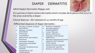
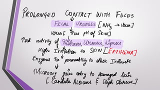
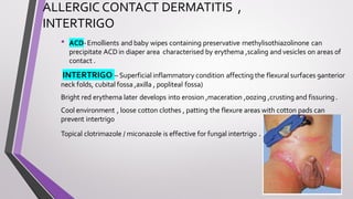
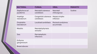
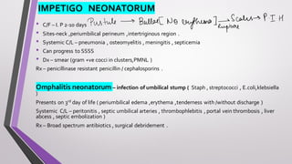
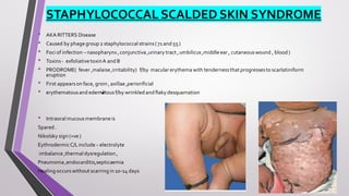
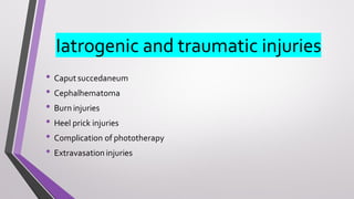
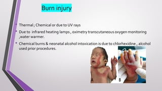
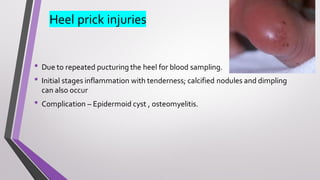
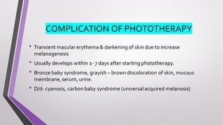
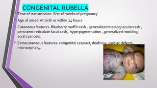
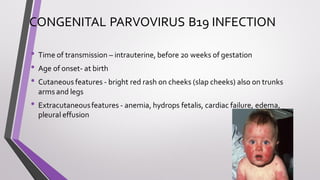
NEONATAL SKIN
DISEASES
DR PRIYANKA DAYALANI
JUNIOR RESIDENT
DR PDMMC AMRAVATI
INDEX
1. DIAPER DERMATITIS
2. ALLERGIC CONTACT DERMATITS
3. BACTERIAL INFECTION
4. FUNGAL INFECTION
5. VIRAL INFECTION
6. PARASITIC INFECTION
7. DISORDERS OF SUBCUTANOUES INFECTION
8. CONGENITAL INFECTIONS
9. IATROGENIC ANDTRAUMATIC INJURIES
DIAPER DERMATITIS
A/K/A Napkin Dermatitis /Nappy rash
It’s a primary irritant contact dermatitis which includes all eruptions that occur in
the area covered by a diaper .
Clinical features – M.C between 6-12 months of age
Differential diagnosis of diaper dermatitis -
PREVENTION AND TREATMENT
ALLERGIC CONTACT DERMATITIS ,
INTERTRIGO
• ACD-Emollients and baby wipes containing preservative methylisothiazolinone can
precipitate ACDin diaper area characterised by erythema ,scaling and vesicles on areas of
contact .
INTERTRIGO – Superficial inflammatory condition affecting the flexural surfaces 9anterior
neck folds, cubital fossa ,axilla , popliteal fossa)
Bright red erythema later develops into erosion ,maceration ,oozing ,crusting and fissuring.
Cool environment , loose cotton clothes , patting the flexure areas with cotton pads can
prevent intertrigo
Topical clotrimazole / miconazole is effective for fungal intertrigo .
BACTERIAL FUNGAL VIRAL PARASITIC
Staphylococcal
pyoderma
Neonatalcutaneous
candidiasis
Neonatal
chikungunya
Scabies
Impetigo
neonatorum
Congential cutaneous
candidiasis
Herpes simplex
infection
Omphalitis
neonatorum
Localisedcandidiasis Perianalcondyloma
accuminata
Mastitis Neonatalpityriasis
verisolor
Ssss Dermatophytic
infections
Ecthyma
gangrenosum
Breast abscess
STAPHYLOCOCCAL PYODERMA,
IMPETIGO NEONATORUM
Staphylococcal pyoderma –Folliculitis d/t Staph aureus ( M.C pustular eruption in neonates )
Can present in 2- 3rd of life as discrete dome shaped pustules on an erythematous base
More common in diaper area and intertriginous areas .
Can lead to formation of an abscess /furuncle
Rx- Mupirocin ointment
Systemic therapy (Ix in case of systemic involvement )
Impetigo neonatorum – aka Bullous impetigo
D/t staph aureus exfoliative toxin A and B
IMPETIGO NEONATORUM
• C/F – I. P 2-10 days
• Sites-neck ,periumbilical perineum ,intertriginous region .
• Systemic C/L – pneumonia , osteomyelitis , meningitis , septicemia
• Can progress to SSSS
• Dx – smear (gram +ve cocci in clusters,PMNL )
Rx – penicillinase resistant penicillin / cephalosporins .
Omphalitis neonatorum – infection of umbilical stump ( Staph , streptococci , E.coli,klebsiella
)
Presents on 3rd day of life ( periumbilical edema ,erythema ,tenderness with /without discharge )
Systemic C/L – peritonitis , septic umbilical arteries , thrombophlebitis , portal vein thrombosis , liver
abcess , septic embolization )
Rx – Broad spectrum antibiotics , surgical debridement .
STAPHYLOCOCCAL SCALDED SKIN SYNDROME
• AKA RITTERS Disease
• Caused by phage group 2 staphylococcal strains(71 and 55 )
• Foci of infection – nasopharynx,conjunctiva ,urinary tract ,umbilicus,middle ear , cutaneouswound , blood )
• Toxins - exfoliative toxinA and B
• PRODROME( fever ,malaise ,irritability) f/by macular erythema with tendernessthat progressesto scarlatiniform
eruption
• First appearson face, groin ,axillae ,periorificial
• erythematousand edematous f/by wrinkled and flaky desquamation
• Intraoral mucousmembrane is
Spared .
Nikolsky sign (+ve )
EythrodermicC/Linclude – electrolyte
imbalance ,thermal dysregulation ,
Pneumonia ,endocarditis,septicaemia
Healing occurswithout scarring in 10-14 days
Treatment -
Ecthyma gangrenosum
• Skin lesiondue invasiveinfectioncaused by pseudomonasaeruginosa
• Seen in infantswith immunodeficiency, bowel surgery ,necrotisingenterocolitis, septicemia
• C/F –
DX – gram stain and culture
HPE( vasculitissurrounded by edema , hemorrhage , necrosiswith minimalinfiltrationbyneutrophils)
Rx – ceftazidimeis the treatment of choice
FUNGAL INFECTIONS
NEONATAL PITYRIASIS VERSICOLOR
• Caused by malassezia species.
• Present within first two weeks of life
• Clinically ; multiple, well defined, hypopigmented to
hyperpigmented macules with branny scaling.
• Sites- face, forehead, nose, neck, trunk.
DERMATOPHYTE INFECTION OF NEWBORN
• Tinea capitis and tinea corporis have been reported
M.C org causing dermatophyte infection in neonates < 6 weeks is trichophyton
rubrumf/by microsporum canis , trichophyton mentagrophytes ,trichophyton
tonsurans
Griseofulvin is the treatment of choice in tinea capitis
But, terbinafine ,fluconazole and itraconazole effective in
dermatophyte infections of newborn
PERIANAL CONDYLOMA ACCUMINATA
• Caused by HPV virus 6, 11, 1-4,16 & 18
• Transmitted through perinatal, postnatal & autoinoculation.
• May associated with laryngeal papilloma.
• Clinically verrucous growth on perianal region is seen.
• D/d- ectopic sebaceous glands , genital papillae (physiological), perianal skin
tags.
NEONATAL CHIKUNGUNYA INFECTION
• Can be transmitted during pregnancyor peripartum period
• I.P – 3-7 days
• Neonatal chikungunya infection aquired in last trimester can present as Fever ,
pain lethargy , sucking difficulties
• Cutaneous lesions can be generalized maculopapular rash, hyperpigmented
rashes, petechial rashes, purpuric ,hemorrhagic vasculitic lesions.
• Hyperpigmentation on nose (chik sign /brownie nose is present )
DIAGNOSIS ANDTREATMENT -
• Dx- Acute Stage( serology /RTPCR)
• IgMANTIBODIES have been detected 3-5 days after the infection and
remain positive for 3 months
• Rx – non specific and only symptomatic
•Scabies –
• Clinically , papules, vesicles, pustules, crusting & scaling.
• Excoriation and burrows uncommon .
• Sites- face ,scalp, extremitis.
• Neonates do not itch, so irritability, excessive crying and poor feeding seen.
DISORDERS OF SUBCUTANEOUS INFECTION
Iatrogenic and traumatic injuries
• Caput succedaneum
• Cephalhematoma
• Burn injuries
• Heel prick injuries
• Complication of phototherapy
• Extravasation injuries
Caput succedaneum
• Extracranialinjury due to mechanical forces during parturition.
• Associated with prolong labour, vacuumdelivery.
• Boggy, localized swelling on scalp with varying degree of bruising and tissue
necrosis.
• Site most commonly occipital midline.
• It crossed the midline and resolve within 48 hours.
Cephalhematoma
• It is periosteal hematoma overlying the calvarium.
• Associated with prolong labour, instrumental delivery, abnormal
presentation, multiple gestation.
• Firm, well demarcated subcutaneous swelling on scalp
• Does not cross midline , as limited to one cranial bone.
• Resolves within few weeks to months
Burn injury
• Thermal; Chemical or due to UV rays
• Due to infrared heating lamps , oximetry transcutaneous oxygen monitoring
,water warmer.
• Chemical burns & neonatal alcohol intoxication is due to chlorhexidine , alcohol
used prior procedures.
Heel prick injuries
• Due to repeated pucturing the heel for blood sampling.
• Initial stages inflammation with tenderness; calcified nodules and dimpling
can also occur
• Complication – Epidermoid cyst , osteomyelitis.
COMPLICATION OF PHOTOTHERAPY
• Transient macular erythema & darkening of skin due to increase
melanogenesis
• Usually develops within 1- 7 days after starting phototherapy.
• Bronze baby syndrome, grayish – brown discoloration of skin, mucous
membrane,serum, urine.
• D/d- cyanosis, carbon baby syndrome (universalacquired melanosis)
CONGENITAL INFECTIONS
• Congenital rubella
• Neonatal herpes
• Neonatal varicella
• Congenital syphilis
• Cytomegalic inclusion disease
• Congenital toxoplasmosis
• Congenital parvovirus B19 infection
CONGENITAL RUBELLA
• Time of transmission- first 16 weeks of pregnancy
• Age of onset- At birth or within 24 hours
• Cutaneous features- Blueberry muffin rash , generalized maculopapular rash ,
persistent reticulate facial rash, hyperpigmentation , generalized mottling,
acral cyanosis.
• Extracutaneousfeatures- congenital cataract,deafness, cardiac defects,
microcephaly,.
NEONATAL HERPES
• Time of maternofetal transmission- Ascending in utero infection or perinatal
transmission or postnatal period
• Age of onset – at birth or within 48 hrs
• Cutaneous features- generalized zosteriform vesicles, (single, grouped,
disseminated) erosions, scars seen.
• Extracutaneousfeatures- fever, respiratory distress, poor feeding,
hypotonia, CNS dysfunction.
NEONATAL VARICELLA
• Time of transmission – intrauterine last weeks, first few days postpartum
• Age of onset- 3-10 days of life.
• Cutaneous features- generalized varicelliform blisters, or scars. Dew drops
on rose petal appearance.
• Extracutaneousfeatures- pneumonia,hepatitis, meningoencephalitis,
coagulopathies
CONGENTIAL SYPHILIS
• Time of maternofetal transmission- intrauterine early third trimester
• Age of onset – within first 2 years
• Cutaneous features- papulosquamousrash , vesiculobullous lesions,
desquamation over palms and soles, targetoid lesions, mucosalpatches ,
condyloma lata.
• Extracutaneousfeatures- nasal snuffles, haemolytic anemia,
thrombocytopenia, hepatosplenomegaly, osteochondritis, deafness.
CONGENITAL TOXOPLASMOSIS
• Time of transmission- transplacental 3rd trimister,.
• Age of onset- at birth or first week of life.
• Cutaneous features- maculopapularrash ,petechiae, purpura,also called
blueberry muffin rash ,subcutaneous nodules, calcifications seen.
• Extracutaneousfeatures- fever, diarrhea, vomiting, cataract,
microopthalmia, pericarditis, seizures
CONGENITAL PARVOVIRUS B19 INFECTION
• Time of transmission – intrauterine, before 20 weeks of gestation
• Age of onset- at birth
• Cutaneous features - bright red rash on cheeks (slap cheeks) also on trunks
arms and legs
• Extracutaneousfeatures - anemia, hydrops fetalis, cardiac failure, edema,
pleural effusion
THANKYOU

NEONATAL SKIN DISEASES 2.pdf

  • 1.
    NEONATAL SKIN DISEASES DR PRIYANKADAYALANI JUNIOR RESIDENT DR PDMMC AMRAVATI
  • 2.
    INDEX 1. DIAPER DERMATITIS 2.ALLERGIC CONTACT DERMATITS 3. BACTERIAL INFECTION 4. FUNGAL INFECTION 5. VIRAL INFECTION 6. PARASITIC INFECTION 7. DISORDERS OF SUBCUTANOUES INFECTION 8. CONGENITAL INFECTIONS 9. IATROGENIC ANDTRAUMATIC INJURIES
  • 3.
    DIAPER DERMATITIS A/K/A NapkinDermatitis /Nappy rash It’s a primary irritant contact dermatitis which includes all eruptions that occur in the area covered by a diaper . Clinical features – M.C between 6-12 months of age Differential diagnosis of diaper dermatitis -
  • 5.
  • 6.
    ALLERGIC CONTACT DERMATITIS, INTERTRIGO • ACD-Emollients and baby wipes containing preservative methylisothiazolinone can precipitate ACDin diaper area characterised by erythema ,scaling and vesicles on areas of contact . INTERTRIGO – Superficial inflammatory condition affecting the flexural surfaces 9anterior neck folds, cubital fossa ,axilla , popliteal fossa) Bright red erythema later develops into erosion ,maceration ,oozing ,crusting and fissuring. Cool environment , loose cotton clothes , patting the flexure areas with cotton pads can prevent intertrigo Topical clotrimazole / miconazole is effective for fungal intertrigo .
  • 7.
    BACTERIAL FUNGAL VIRALPARASITIC Staphylococcal pyoderma Neonatalcutaneous candidiasis Neonatal chikungunya Scabies Impetigo neonatorum Congential cutaneous candidiasis Herpes simplex infection Omphalitis neonatorum Localisedcandidiasis Perianalcondyloma accuminata Mastitis Neonatalpityriasis verisolor Ssss Dermatophytic infections Ecthyma gangrenosum Breast abscess
  • 8.
    STAPHYLOCOCCAL PYODERMA, IMPETIGO NEONATORUM Staphylococcalpyoderma –Folliculitis d/t Staph aureus ( M.C pustular eruption in neonates ) Can present in 2- 3rd of life as discrete dome shaped pustules on an erythematous base More common in diaper area and intertriginous areas . Can lead to formation of an abscess /furuncle Rx- Mupirocin ointment Systemic therapy (Ix in case of systemic involvement ) Impetigo neonatorum – aka Bullous impetigo D/t staph aureus exfoliative toxin A and B
  • 9.
    IMPETIGO NEONATORUM • C/F– I. P 2-10 days • Sites-neck ,periumbilical perineum ,intertriginous region . • Systemic C/L – pneumonia , osteomyelitis , meningitis , septicemia • Can progress to SSSS • Dx – smear (gram +ve cocci in clusters,PMNL ) Rx – penicillinase resistant penicillin / cephalosporins . Omphalitis neonatorum – infection of umbilical stump ( Staph , streptococci , E.coli,klebsiella ) Presents on 3rd day of life ( periumbilical edema ,erythema ,tenderness with /without discharge ) Systemic C/L – peritonitis , septic umbilical arteries , thrombophlebitis , portal vein thrombosis , liver abcess , septic embolization ) Rx – Broad spectrum antibiotics , surgical debridement .
  • 10.
    STAPHYLOCOCCAL SCALDED SKINSYNDROME • AKA RITTERS Disease • Caused by phage group 2 staphylococcal strains(71 and 55 ) • Foci of infection – nasopharynx,conjunctiva ,urinary tract ,umbilicus,middle ear , cutaneouswound , blood ) • Toxins - exfoliative toxinA and B • PRODROME( fever ,malaise ,irritability) f/by macular erythema with tendernessthat progressesto scarlatiniform eruption • First appearson face, groin ,axillae ,periorificial • erythematousand edematous f/by wrinkled and flaky desquamation • Intraoral mucousmembrane is Spared . Nikolsky sign (+ve ) EythrodermicC/Linclude – electrolyte imbalance ,thermal dysregulation , Pneumonia ,endocarditis,septicaemia Healing occurswithout scarring in 10-14 days
  • 11.
  • 12.
    Ecthyma gangrenosum • Skinlesiondue invasiveinfectioncaused by pseudomonasaeruginosa • Seen in infantswith immunodeficiency, bowel surgery ,necrotisingenterocolitis, septicemia • C/F – DX – gram stain and culture HPE( vasculitissurrounded by edema , hemorrhage , necrosiswith minimalinfiltrationbyneutrophils) Rx – ceftazidimeis the treatment of choice
  • 14.
  • 15.
    NEONATAL PITYRIASIS VERSICOLOR •Caused by malassezia species. • Present within first two weeks of life • Clinically ; multiple, well defined, hypopigmented to hyperpigmented macules with branny scaling. • Sites- face, forehead, nose, neck, trunk.
  • 16.
    DERMATOPHYTE INFECTION OFNEWBORN • Tinea capitis and tinea corporis have been reported M.C org causing dermatophyte infection in neonates < 6 weeks is trichophyton rubrumf/by microsporum canis , trichophyton mentagrophytes ,trichophyton tonsurans Griseofulvin is the treatment of choice in tinea capitis But, terbinafine ,fluconazole and itraconazole effective in dermatophyte infections of newborn
  • 18.
    PERIANAL CONDYLOMA ACCUMINATA •Caused by HPV virus 6, 11, 1-4,16 & 18 • Transmitted through perinatal, postnatal & autoinoculation. • May associated with laryngeal papilloma. • Clinically verrucous growth on perianal region is seen. • D/d- ectopic sebaceous glands , genital papillae (physiological), perianal skin tags.
  • 19.
    NEONATAL CHIKUNGUNYA INFECTION •Can be transmitted during pregnancyor peripartum period • I.P – 3-7 days • Neonatal chikungunya infection aquired in last trimester can present as Fever , pain lethargy , sucking difficulties • Cutaneous lesions can be generalized maculopapular rash, hyperpigmented rashes, petechial rashes, purpuric ,hemorrhagic vasculitic lesions. • Hyperpigmentation on nose (chik sign /brownie nose is present )
  • 20.
    DIAGNOSIS ANDTREATMENT - •Dx- Acute Stage( serology /RTPCR) • IgMANTIBODIES have been detected 3-5 days after the infection and remain positive for 3 months • Rx – non specific and only symptomatic
  • 21.
    •Scabies – • Clinically, papules, vesicles, pustules, crusting & scaling. • Excoriation and burrows uncommon . • Sites- face ,scalp, extremitis. • Neonates do not itch, so irritability, excessive crying and poor feeding seen.
  • 22.
  • 23.
    Iatrogenic and traumaticinjuries • Caput succedaneum • Cephalhematoma • Burn injuries • Heel prick injuries • Complication of phototherapy • Extravasation injuries
  • 24.
    Caput succedaneum • Extracranialinjurydue to mechanical forces during parturition. • Associated with prolong labour, vacuumdelivery. • Boggy, localized swelling on scalp with varying degree of bruising and tissue necrosis. • Site most commonly occipital midline. • It crossed the midline and resolve within 48 hours.
  • 25.
    Cephalhematoma • It isperiosteal hematoma overlying the calvarium. • Associated with prolong labour, instrumental delivery, abnormal presentation, multiple gestation. • Firm, well demarcated subcutaneous swelling on scalp • Does not cross midline , as limited to one cranial bone. • Resolves within few weeks to months
  • 26.
    Burn injury • Thermal;Chemical or due to UV rays • Due to infrared heating lamps , oximetry transcutaneous oxygen monitoring ,water warmer. • Chemical burns & neonatal alcohol intoxication is due to chlorhexidine , alcohol used prior procedures.
  • 27.
    Heel prick injuries •Due to repeated pucturing the heel for blood sampling. • Initial stages inflammation with tenderness; calcified nodules and dimpling can also occur • Complication – Epidermoid cyst , osteomyelitis.
  • 28.
    COMPLICATION OF PHOTOTHERAPY •Transient macular erythema & darkening of skin due to increase melanogenesis • Usually develops within 1- 7 days after starting phototherapy. • Bronze baby syndrome, grayish – brown discoloration of skin, mucous membrane,serum, urine. • D/d- cyanosis, carbon baby syndrome (universalacquired melanosis)
  • 29.
    CONGENITAL INFECTIONS • Congenitalrubella • Neonatal herpes • Neonatal varicella • Congenital syphilis • Cytomegalic inclusion disease • Congenital toxoplasmosis • Congenital parvovirus B19 infection
  • 30.
    CONGENITAL RUBELLA • Timeof transmission- first 16 weeks of pregnancy • Age of onset- At birth or within 24 hours • Cutaneous features- Blueberry muffin rash , generalized maculopapular rash , persistent reticulate facial rash, hyperpigmentation , generalized mottling, acral cyanosis. • Extracutaneousfeatures- congenital cataract,deafness, cardiac defects, microcephaly,.
  • 31.
    NEONATAL HERPES • Timeof maternofetal transmission- Ascending in utero infection or perinatal transmission or postnatal period • Age of onset – at birth or within 48 hrs • Cutaneous features- generalized zosteriform vesicles, (single, grouped, disseminated) erosions, scars seen. • Extracutaneousfeatures- fever, respiratory distress, poor feeding, hypotonia, CNS dysfunction.
  • 32.
    NEONATAL VARICELLA • Timeof transmission – intrauterine last weeks, first few days postpartum • Age of onset- 3-10 days of life. • Cutaneous features- generalized varicelliform blisters, or scars. Dew drops on rose petal appearance. • Extracutaneousfeatures- pneumonia,hepatitis, meningoencephalitis, coagulopathies
  • 33.
    CONGENTIAL SYPHILIS • Timeof maternofetal transmission- intrauterine early third trimester • Age of onset – within first 2 years • Cutaneous features- papulosquamousrash , vesiculobullous lesions, desquamation over palms and soles, targetoid lesions, mucosalpatches , condyloma lata. • Extracutaneousfeatures- nasal snuffles, haemolytic anemia, thrombocytopenia, hepatosplenomegaly, osteochondritis, deafness.
  • 34.
    CONGENITAL TOXOPLASMOSIS • Timeof transmission- transplacental 3rd trimister,. • Age of onset- at birth or first week of life. • Cutaneous features- maculopapularrash ,petechiae, purpura,also called blueberry muffin rash ,subcutaneous nodules, calcifications seen. • Extracutaneousfeatures- fever, diarrhea, vomiting, cataract, microopthalmia, pericarditis, seizures
  • 35.
    CONGENITAL PARVOVIRUS B19INFECTION • Time of transmission – intrauterine, before 20 weeks of gestation • Age of onset- at birth • Cutaneous features - bright red rash on cheeks (slap cheeks) also on trunks arms and legs • Extracutaneousfeatures - anemia, hydrops fetalis, cardiac failure, edema, pleural effusion
  • 37.