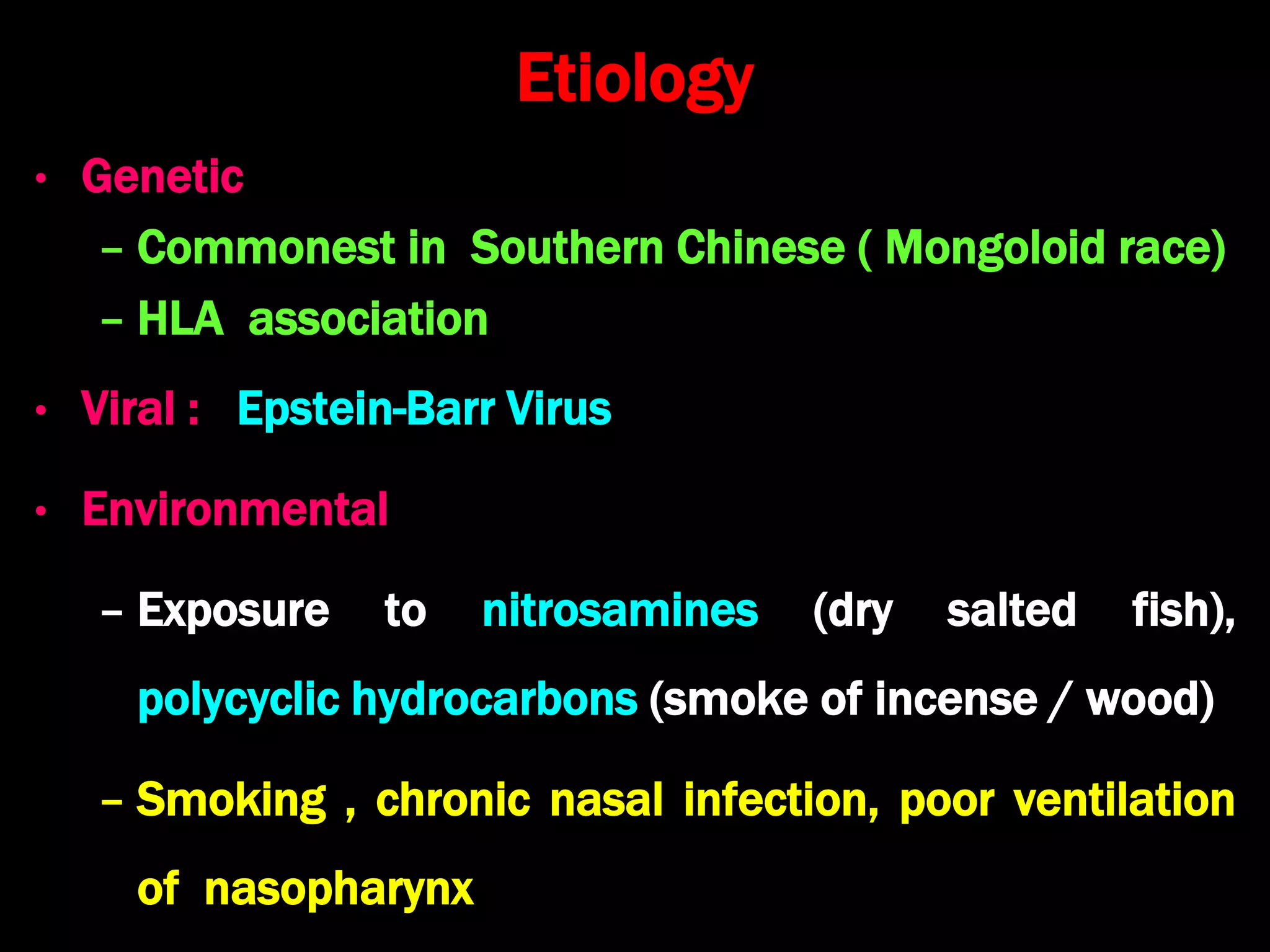
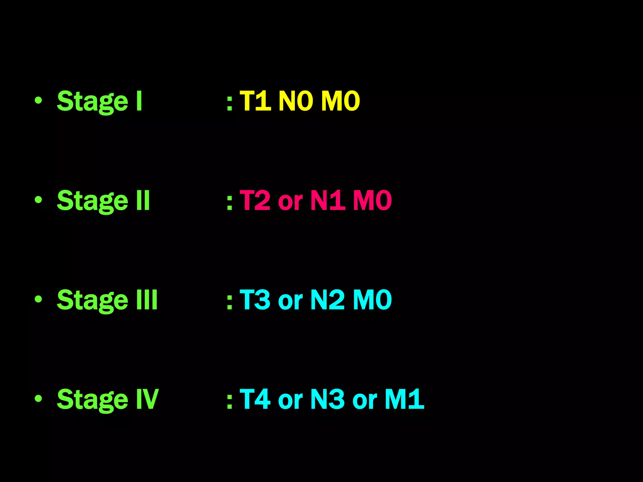
Nasopharyngeal
Carcinoma
Dr. Krishna Koirala
2022-08-08
Introduction
• Non lymphomatous squamous-cell carcinoma
that occurs in the epithelial lining of the
nasopharynx
• Frequently arises from the pharyngeal recess
(fossa of Rosenmüller) posteromedial to the
medial crura of the eustachian tube opening in
the nasopharynx
Fossa of Rosenmuller
Epidemiology
• Accounts for 85% adult nasopharyngeal malignancies
and 30% pediatric nasopharyngeal malignancies
• Common in Chinese and North African people
• Male preponderance of 3:1
• Bimodal age presentation with small peak at 15-25
yrs and a large peak at 55-65 yrs
Etiology
• Genetic
– Commonest in Southern Chinese ( Mongoloid race)
– HLA association
• Viral : Epstein-Barr Virus
• Environmental
– Exposure to nitrosamines (dry salted fish),
polycyclic hydrocarbons (smoke of incense / wood)
– Smoking , chronic nasal infection, poor ventilation
of nasopharynx
W.H.O. Classification (Histological)
• Type 1 :
− Keratinizing squamous cell carcinoma (common
in the older adult population)
• Type 2:
− Non-keratinizing (transitional) carcinoma
• Type 3:
− Undifferentiated carcinoma ( common in
childhood and adolescents , associated with high
EBV Ab titre)
Clinical Features
1. Neck swelling (60%)
• Lateral retropharyngeal LN of Rouviere
• B/L, enlarged jugulodigastric, upper & middle deep
cervical nodes and posterior triangle nodes
2. Nasal (40%)
• Blood stained nasal mucus, epistaxis, nose block,
foul smelling nasal discharge
3. Otologic (30%) : Conductive deafness, tinnitus
4. Ophthalmologic (20%)
• Diplopia & ophthalmoplegia (involvement of CN III,
IV, VI), Proptosis (orbit invasion) & blindness
(involvement of CN II)
5. Neurologic (20 %)
• Jugular foramen syndrome: CN IX, X, XI involved by
lateral retropharyngeal lymph node
• Horner's syndrome: sympathetic chain involvement
6. Severe Headache
• Skull base erosion
7. Trotter's triad
– Conductive deafness: Eustachian Tube block
– Ipsilateral temporo -parietal neuralgia: Trigeminal
nerve involvement
– Ipsilateral palatal paralysis: Vagus nerve involved
8. Distant metastasis
• Bone, lungs & liver
Investigations
1. Nasopharyngoscopy and Diagnostic Nasal Endoscopy
– Mass seen in nasopharynx at fossa of
Rosenmüller
2. Nasopharyngeal tumor biopsy: blind /under vision
3. F.N.A.C. of neck node
4. C.T. scan head and neck : Tumor extent, skull base
erosion, Cervical lymph node metastasis
5. M.R.I. head & neck: for intracranial extension
6. Tests for metastases
− C.T. chest and abdomen, bone scan, P.E.T. scan,
liver function tests
7. Serologic tests
– Immuno-fluorescence for IgA antibodies to Viral
Capsid Antigen, Ig G antibodies to Early Antigen
Diagnostic Nasal Endoscopy
• Gross
–Proliferative
–Ulcerative
–Infiltrative
Computerized Tomogram Scan
Magnetic Resonance Imaging
Endoscopic Biopsy
Whole body bone scan
Positron Emission Tomography
T.N.M. staging
• T1 : confined to nasopharynx
• T2 : soft tissue involvement in oropharynx or nasal
cavity or Parapharyngeal space
• T3 : invasion of bony structures or P.N.S.
• T4 : intracranial, involvement of orbit, cranial nerves,
infratemporal fossa, hypopharynx
N0 : no evidence of regional lymph node involvement
N1 : unilateral
N2 : bilateral (above supraclavicular fossa, < 6 cm)
N3 : > 6 cm or in supraclavicular fossa
M 0 : no evidence of distant metastasis
M 1 : distant metastasis present
• Stage I : T1 N0 M0
• Stage II : T2 or N1 M0
• Stage III : T3 or N2 M0
• Stage IV : T4 or N3 or M1
Treatment modalities
1. Teletherapy or External beam radiotherapy
2. Brachytherapy
3. Chemotherapy
4. Surgery
5. Immunotherapy against E.B.V.
6. Vaccination against EBV: experimental
External beam irradiation
2 lateral fields: nasopharynx, skull base and upper neck
sparing temporal lobe, pituitary and spinal cord
1 anterior field: lower neck; sparing spinal cord & larynx
Brachytherapy
• Treatment of cancer by the insertion of radioactive implants
directly into the tissue
• Used for small tumor, residual or recurrent tumor
– Interstitial: Radioactive source (Radium, Iridium, Iodine,
Gold) inserted into tumor tissue
– Intracavitary: Radioactive source placed inside the
catheter or moulds & inserted into nasopharynx
– High dose rate (HDR): High intensity radiation delivered
with precision under computer guidance
Interstitial Brachytherapy
Intracavitary Brachytherapy
High Dose Rate Brachytherapy
Chemotherapy
• Drugs used
−Cisplatin
−5-Fluorouracil
• Indications
–Radiation failure
–Palliation in distant metastasis
Surgery
1. Nasopharyngectomy, Cryosurgery : for residual or
recurrent tumor
2. Radical neck dissection : for radio-resistant neck
node metastasis
3. Palliative debulking : for T4 tumors
4. Myringotomy & grommet insertion : for persistent
otitis media with effusion
Radical neck dissection & Interstitial
Brachytherapy
Treatment Protocol
T1 : External Radiotherapy (6500 c Gy)
T2 : External Radiotherapy (7000 c Gy)
T3 / T4 : Radiotherapy + Chemotherapy 
Brachytherapy / Salvage surgery if required
N0 : External Radiotherapy (5000 c Gy)
N+ : External Radiotherapy (6000 c Gy) +
Chemotherapy
Prognosis
• W.H.O. Type 2 and 3 carcinomas have good
response to radiotherapy and better survival rates
• Average 5 year survival rates for treated patients
Stage I : 95 – 100 %
Stage II : 60 – 80 %
Stage III : 30 – 60 %
Stage IV : 20 – 30 %
Follow up protocol for ca nasopharynx
• Final assessment (2–3 months after the end of treatment)
– Local and regional exam plus nasopharyngeal endoscopy, FDG-PET/CT
and/or MRI
• First two years
– Local and regional exam plus nasopharyngeal fibroscopy (every 3 to 4
months)
– Chest X-ray, thyroid function test, CT/MRI (yearly)
• Two to five years
– Local and regional exam plus nasopharyngeal fibroscopy
(every 6 months)
– Chest X-ray, thyroid function test, CT/MRI (yearly)

Nasopharyngeal Carcinoma.ppt

  • 1.
  • 2.
    Introduction • Non lymphomatoussquamous-cell carcinoma that occurs in the epithelial lining of the nasopharynx • Frequently arises from the pharyngeal recess (fossa of Rosenmüller) posteromedial to the medial crura of the eustachian tube opening in the nasopharynx
  • 3.
  • 4.
    Epidemiology • Accounts for85% adult nasopharyngeal malignancies and 30% pediatric nasopharyngeal malignancies • Common in Chinese and North African people • Male preponderance of 3:1 • Bimodal age presentation with small peak at 15-25 yrs and a large peak at 55-65 yrs
  • 5.
    Etiology • Genetic – Commonestin Southern Chinese ( Mongoloid race) – HLA association • Viral : Epstein-Barr Virus • Environmental – Exposure to nitrosamines (dry salted fish), polycyclic hydrocarbons (smoke of incense / wood) – Smoking , chronic nasal infection, poor ventilation of nasopharynx
  • 6.
    W.H.O. Classification (Histological) •Type 1 : − Keratinizing squamous cell carcinoma (common in the older adult population) • Type 2: − Non-keratinizing (transitional) carcinoma • Type 3: − Undifferentiated carcinoma ( common in childhood and adolescents , associated with high EBV Ab titre)
  • 7.
    Clinical Features 1. Neckswelling (60%) • Lateral retropharyngeal LN of Rouviere • B/L, enlarged jugulodigastric, upper & middle deep cervical nodes and posterior triangle nodes 2. Nasal (40%) • Blood stained nasal mucus, epistaxis, nose block, foul smelling nasal discharge 3. Otologic (30%) : Conductive deafness, tinnitus
  • 8.
    4. Ophthalmologic (20%) •Diplopia & ophthalmoplegia (involvement of CN III, IV, VI), Proptosis (orbit invasion) & blindness (involvement of CN II) 5. Neurologic (20 %) • Jugular foramen syndrome: CN IX, X, XI involved by lateral retropharyngeal lymph node • Horner's syndrome: sympathetic chain involvement
  • 9.
    6. Severe Headache •Skull base erosion 7. Trotter's triad – Conductive deafness: Eustachian Tube block – Ipsilateral temporo -parietal neuralgia: Trigeminal nerve involvement – Ipsilateral palatal paralysis: Vagus nerve involved 8. Distant metastasis • Bone, lungs & liver
  • 11.
    Investigations 1. Nasopharyngoscopy andDiagnostic Nasal Endoscopy – Mass seen in nasopharynx at fossa of Rosenmüller 2. Nasopharyngeal tumor biopsy: blind /under vision 3. F.N.A.C. of neck node 4. C.T. scan head and neck : Tumor extent, skull base erosion, Cervical lymph node metastasis
  • 12.
    5. M.R.I. head& neck: for intracranial extension 6. Tests for metastases − C.T. chest and abdomen, bone scan, P.E.T. scan, liver function tests 7. Serologic tests – Immuno-fluorescence for IgA antibodies to Viral Capsid Antigen, Ig G antibodies to Early Antigen
  • 13.
    Diagnostic Nasal Endoscopy •Gross –Proliferative –Ulcerative –Infiltrative
  • 14.
  • 15.
  • 16.
  • 17.
  • 18.
  • 19.
    T.N.M. staging • T1: confined to nasopharynx • T2 : soft tissue involvement in oropharynx or nasal cavity or Parapharyngeal space • T3 : invasion of bony structures or P.N.S. • T4 : intracranial, involvement of orbit, cranial nerves, infratemporal fossa, hypopharynx
  • 20.
    N0 : noevidence of regional lymph node involvement N1 : unilateral N2 : bilateral (above supraclavicular fossa, < 6 cm) N3 : > 6 cm or in supraclavicular fossa M 0 : no evidence of distant metastasis M 1 : distant metastasis present
  • 21.
    • Stage I: T1 N0 M0 • Stage II : T2 or N1 M0 • Stage III : T3 or N2 M0 • Stage IV : T4 or N3 or M1
  • 22.
    Treatment modalities 1. Teletherapyor External beam radiotherapy 2. Brachytherapy 3. Chemotherapy 4. Surgery 5. Immunotherapy against E.B.V. 6. Vaccination against EBV: experimental
  • 23.
    External beam irradiation 2lateral fields: nasopharynx, skull base and upper neck sparing temporal lobe, pituitary and spinal cord 1 anterior field: lower neck; sparing spinal cord & larynx
  • 24.
    Brachytherapy • Treatment ofcancer by the insertion of radioactive implants directly into the tissue • Used for small tumor, residual or recurrent tumor – Interstitial: Radioactive source (Radium, Iridium, Iodine, Gold) inserted into tumor tissue – Intracavitary: Radioactive source placed inside the catheter or moulds & inserted into nasopharynx – High dose rate (HDR): High intensity radiation delivered with precision under computer guidance
  • 25.
  • 26.
  • 27.
    High Dose RateBrachytherapy
  • 28.
    Chemotherapy • Drugs used −Cisplatin −5-Fluorouracil •Indications –Radiation failure –Palliation in distant metastasis
  • 29.
    Surgery 1. Nasopharyngectomy, Cryosurgery: for residual or recurrent tumor 2. Radical neck dissection : for radio-resistant neck node metastasis 3. Palliative debulking : for T4 tumors 4. Myringotomy & grommet insertion : for persistent otitis media with effusion
  • 30.
    Radical neck dissection& Interstitial Brachytherapy
  • 31.
    Treatment Protocol T1 :External Radiotherapy (6500 c Gy) T2 : External Radiotherapy (7000 c Gy) T3 / T4 : Radiotherapy + Chemotherapy  Brachytherapy / Salvage surgery if required N0 : External Radiotherapy (5000 c Gy) N+ : External Radiotherapy (6000 c Gy) + Chemotherapy
  • 32.
    Prognosis • W.H.O. Type2 and 3 carcinomas have good response to radiotherapy and better survival rates • Average 5 year survival rates for treated patients Stage I : 95 – 100 % Stage II : 60 – 80 % Stage III : 30 – 60 % Stage IV : 20 – 30 %
  • 33.
    Follow up protocolfor ca nasopharynx • Final assessment (2–3 months after the end of treatment) – Local and regional exam plus nasopharyngeal endoscopy, FDG-PET/CT and/or MRI • First two years – Local and regional exam plus nasopharyngeal fibroscopy (every 3 to 4 months) – Chest X-ray, thyroid function test, CT/MRI (yearly) • Two to five years – Local and regional exam plus nasopharyngeal fibroscopy (every 6 months) – Chest X-ray, thyroid function test, CT/MRI (yearly)

Editor's Notes

  • #4 Fossa of Rossenmuller