
Recommended
More Related Content
What's hot
What's hot (20)
Similar to Nanoliposmes and Nutraceuticals
Similar to Nanoliposmes and Nutraceuticals (20)
Recently uploaded
Recently uploaded (20)
Nanoliposmes and Nutraceuticals
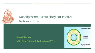
- 1. Nanoliposomal Technology For Food & Nutraceuticals Bharti Sharma MSc. Food Science & Technology (II Yr)
- 2. Food Nanotechnology The goal in food fortification is to ensure that the bioactive compd.(s) are delivered and released at the proper time and location in the body. The bioavailability of many compounds with poor water solubility or low permeability in the small intestine can be improved by appropriate control of food production and consumption. Nanosize allow easier absorption after oral administration as some particles can get absorbed by endocytosis and these nanoparticles can easily move between cells of the gastrointestinal tract . an increase in the surface area due to nanoparticles can explain increased absorption (Source: Rangan et al. 2016)
- 3. Types of Nano Based Delivery System Type Size range Description Limitations Nanoliposomes 10-300nm • Small vesicles surrounded by lipid bilayers • Consists of phospholipids and cholesterol • Physical instability • Chemical degradation Nanocochelates 50-500nm • Small vesicles surrounded by solid lipid bilayer • Consist of phosphatidylserine, cholesterol and calcium ions • Expensive Micelles <100nm • Consist of surfactant molecules with a hydrophobic core and a hydrophilic shell • Large amount of surfactants used Nanoemulsions 10-100nm • Colloidal dispersion of two immiscible liquids (oil and aqueous) with submicron size droplets • Thermodynamically unstable Cocervates 10-600nm • Biopolymer complexes of two oppositely charged protein and/ or polysaccharides via electrostatic interactions • Expensive • Complex structure and procedure Source: Functional Foods and Dietary Supplements: Processing Effects and Health Benefits, Athapol Noomhorm, Imran
- 4. Nanoliposomes Bilayer lipid vesicles- hydrophilic head and hydrophobic fatty acid tail Composed of phospholipids Are useful in areas of drug delivery, as diagnostic agent and in food industries Nanoliposomes- nanometric version of liposomes. Liposome and nanoliposomes – similar in chemical, structural and thermodynamic properties However, nanoliposomes provide more surface area and have the potential to increase solubility, enhance bioavailability, improve controlled release of the encapsulated material to a greater extent.
- 5. Classification of Liposomes On the basis of their size and number of bilayers, liposomes can also be classified as: I. Multilamellar vesicles (MLV)- vesicles have an onion structure, making a multilamellar structure of concentric phospholipid spheres separated by layers of water II. Unilamellar vesicles- the vesicle has a single phospholipid bilayer sphere enclosing the aqueous solution. Unilamellar vesicles can also be classified into two categories: (1) large unilamellar vesicles (LUV >100nm) (2) small unilamellar vesicles (SUV <100nm)
- 6. Method of Preparation Nanoliposomes are colloidal structures formed when phospholipid is mixed into an aqueous solution using high energy input Input of energy (e.g. in the form of sonication, homogenisation, heating, etc.) results in the arrangement of the lipid molecules, in the form of bilayer vesicles, to achieve a thermodynamic equilibrium in the aqueous phase. General methods of preparation All the methods of preparing the liposomes involve four basic stages: 1. Drying down lipids from organic solvent. 2. Dispersing the lipid in aqueous media. 3. Purifying the resultant liposome. 4. Analyzing the final product.
- 7. Sonication Simple method for reducing the size of liposomes and manufacture of nanoliposomes. 2 types of sonicators- 1. Probe Sonicator 2. Bath Sonicator For probe sonication, place the tip of the sonicator in the MLV flask and sonicate the sample for 10–15 min. At this stage, nanoliposomes are formed (SUV). Probe tip sonicators deliver high-energy input to the lipid suspension but suffer from overheating of the lipid suspension causing degradation. Sonication tips also tend to release titanium particles into the lipid suspension, which must be removed by centrifugation prior to use.
- 8. Bath Sonicator • Fill the bath sonicator with room temperature water mixed with a couple of drops of liquid detergent. Using a ring stand and test tube clamp, suspend the MLV flask in the bath sonicator. • The liquid level inside the flask should be equal to that of outside the flask. Sonicate for 20–40 min. • Requires longer process times than probe sonication. • Advantage- can be carried out in a closed container under nitrogen or argon, and does not contaminate the lipid with metal from the probe tip.
- 9. Sonication Dissolve lipids in organic solvents (chloroform) Dry the mixture using rotary evaporator Shake. Formation of micrometric MLV type liposome Add suitable aqueous solution to the dried lipids Remove traces of organic solvent using vacuum pump Formation of nanoliposomes Transfer to sonicator- bath-type or probe (tip) The aqueous medium can contain salts, chelating agents, stabilizers, cryo-protectants (e.g. glycerol) and the drug to be entrapped. Source: Nanoliposomes: Preparation and Analysis- M. R. Mozafari 2009
- 10. Extrusion Method Micrometric liposomes (e.g. MLV) are structurally modified to large unilamellar vesicles (LUV) or nanoliposomes depending on the pore-size of the filters used. Vesicles are physically extruded under pressure through polycarbonate filters of defined pore sizes. Preparation of sample such as MLV is done like before. Set up the extruder and place the polycarbonate filters on it. Load liposomal suspension in one syringe (donor) and place it in one end of the extruder. Place the second syringe (receiver) in another end. Place the extruder stand onto a hot plate.
- 11. • Allow the temperature of the liposome suspension to reach the temperature of the heating block (approximately 5–10 min). • Gently push the plunger of the filled syringe until the liposome suspension is completely transferred to the empty syringe. • Gently push the plunger of the alternate syringe to transfer the suspension back to the original syringe. • Repeat the extrusion process for a minimum of seven passes through the filters. • More passes though the filters, the more homogenous the sample becomes. • Remove the filled syringe and inject the nanoliposome sample into a clean vial. • When MLVs are forced through narrow-pore membrane filters under pressure, membrane rupture and resealing occur and encapsulated content leaks out. Therefore, extrusion is performed in the presence of medium containing the final drug concentration, and external solute is removed only after formation is complete
- 12. Microfluidisation Method of production without using any potentially toxic solvent. Principle: dividing a pressure stream into two parts, passing each part through a fine orifice, and directing the flows at each other inside the chamber of microfluidizer. Uses high pressures (up to l0,000 psi) to guide the flow stream through microchannels. Select the ingredients of the nanoliposomes and their suspension medium (an aqueous phase such as deionized/distilled water or buffer) and mix them. Pass the dispersion through the microfluidizer. The process begins when the product enters the inlet reservoir.
- 13. • An intensifier pump generates extremely high pressures to accelerate the product to the interaction chamber at velocities over 400 m/s. Inside this Y-type chamber, the stream separates into microchannels, which are as narrow as the cross-section of a human hair. • The suspension can be recycled through the equipment in which case the suspension must be cooled because of the temperature increase in the interaction chamber at high operating pressure. Disadvantage: Material loss and the process involves a very high shearing force that can potentially damage the structure of material to be encapsulated.
- 14. Heating Method Method by which liposomes can be prepared using a single instrument without involving any toxic solvent. Hydrate a suitable combination of the phospholipid components in an aqueous medium for a time period of 1–2 h under an inert atmosphere such as nitrogen or argon. Mix the lipid dispersions along with the material to be encapsulated, in a heat-resistant flask such as a pyrex beaker, and add glycerol to a final volume concentration of 3%. Place the flask on a hot-plate stirrer and mix the samples (800-1000rpm) until all lipids are dissolved.
- 15. Depending on the nanoliposomal ingredients, sample volume, type of flask used and its number of baffles, as well as type, speed and duration of mixing, nanometric vesicles can be produced without the need to perform filtration or sonication. The heating method has flexibility for the entrapment of various drugs and other bioactives with respect to their temperature sensitivities, like- • Adding the drug to the reaction medium along with the liposomal ingredients and glycerol; • Adding the drug to the reaction medium when temperature has dropped to a point not lower than the transition temperature (T c) of the lipids
- 16. Visualization of Nanoliposomes Phase Contrast microscopy- detect particles larger than 300 nm and contamination with larger particles. Polarizing Microscope- reveal lamellarity of the liposomes Scanning Probe Microscopy- monolayers of various lipids and lipid attached molecules such as antibody fragments can be studied
- 17. Involvement of Technology In Food Able to incorporate food antimicrobials that could aid in the protection of food products against growth of spoilage and pathogenic microorganisms. One of the first reported liposome applications in food products was in cheese manufacturing. The time and cost of cheese ripening can be reduced by the addition of proteinases to cheese mixes before the isolation of curds. However, enzymes are often inactivated by the conditions of a food system. The addition of free (i.e., unencapsulated) enzymes to the milk causes premature proteolysis, resulting in unfavorable curd consistency and low yields.
- 18. A large proportion of the enzymes are lost in the whey stream, increasing product cost through the requirement for a high initial enzyme concentration and limiting downstream whey processing options. Moreover, the addition of the enzymes directly to curd results in poor enzyme distribution. It is known that encapsulated enzymes have improved stability and activity not only in vivo, but also during food processing in vitro. Liposomes isolate the encapsulated enzymes from the surrounding food environment, providing them with protection under conditions that would otherwise impede activity or even cause denaturation. Liposomes can also be used as a means of controlled release.
- 19. Advantages: sustained-release mechanism can encapsulate lipophilic and hydrophilic material at the same time providing synergistic effect. preserve desirable flavors and aromas or mask unpleasant taste and appearance High bioavailability and absorption compared with other oral forms of supplements. Micronized encapsulation protects against the harsh environment of the GI tract Increased intracellular delivery. Ideal for those for whom swallowing a tablet is not possible. Cost effective by being able to take a lower dose for the same effect. Disadvantages: 1. High current cost. 2. Possibility of poor manufacturing (eg, high particle size, poor ingredients). 3. Possibility of instability. 4. Leakage of encapsulated material.
- 20. • Liposomes, and other lipid particles, are cleared from the blood by the reticulo-endothelial system (RES), also called the mononuclear phagocyte system (MPS). • Clearance is inversely related to size, with the small unilamellar liposomes/vesicles (SUVs) and micronized emulsions circulating the blood the longest. • Small liposomes may be drastically more efficient at intracellular delivery of encapsulated compounds. • In a recent study, with carefully-sized liposomes, cellular uptake increased 9-fold as liposome size was decreased from 236 nm to 97 nm and was 34-fold higher at 64 nm.
- 21. • Modification of membrane surfaces with PEGs further enhances the ability of liposomes and micronized emulsions to avoid detection by the cells of the RES, thus protecting it from liver and spleen clearance and consequently prolonging its circulatory time. • Additionally, Stealth liposomes and micronized emulsions made by covering the carrier surface with hydrophilic chains, such as PEG are more stable in biological environments and can exhibit up to 10-fold higher circulation half-lives than can liposomes without hydrophilic surface coatings.
- 22. • The FDA approved the first liposomal drug, a PEG-lated liposomal formulation of doxorubicin for the treatment of Kaposi’s sarcoma in 1995. • Among nutraceuticals, nanoliposomes suspensions of multichain fatty acids with vitamin C that were prepared and freeze dried for long-term storage show promising approach. • Vitamin D3 was formulated as nanoliposomal product that can be used for fortification of beverages.
- 23. INNOVATIVE DELIVERIES FOR NUTRACEUTICALS NutraLease™, Ltd. (Mishor Adumin, Israel) developed a unique technology to produce micelles, which are self-assembled, structured liquid particles with a diameter of 30 nm or less. These particles are designed to readily penetrate cell membranes and dramatically increase the bioavailability of the phytonutrients carried and protected by the micelles . Proprietary Nutritionals Inc. (Kearny, New Jersey ) launched Benexia™ ALA Powder, neutral-tasting, water-soluble omega-3 microencapsulated powder for numerous food applications LiveTheSource (Ft. Lauderdale, Florida) announced its launch of “daily source,” the first ever nano-encapsulated liquid vitamin, mineral, and herbal supplement. The company claims the herbal compounds in “daily source” will “provide a substantial increase in nutritional value, first, due to their synergy, and second, due to their nano-encapsulation”.
- 24. References Nanotechnology In Food Products: Workshop Summary, Institute Of Medicine (US) Food Forum, Washington (DC): National Academies Press (US); 2009. Liposome: classification, preparation, and applications Abolfazl Akbarzadeh, Rogaie Rezaei-Sadabady, Soodabeh Davaran, Sang Woo Joo, Nosratollah Zargham, Younes Hanifehpour, Mohammad Samiei, Mohammad Kouhi and Kazem Nejati-Koshki Nanoliposomes: Preparation and Analysis- M. R. Mozafari (2009) Nanostructured and nanoencapsulated natural antimicrobials for use in food products: A. Brandelli, T.M. Taylor Trends and Methods for nanobased delivery for nutraceuticals: Anupama Rangan, M.V. Manjula, K.G Satyanarayana Nanodelivery of Nutrients for imporved bioavailability: Anu Bhushani, Udayakumar Harish, Chinnaswamy Anandharamakrishnan 2017