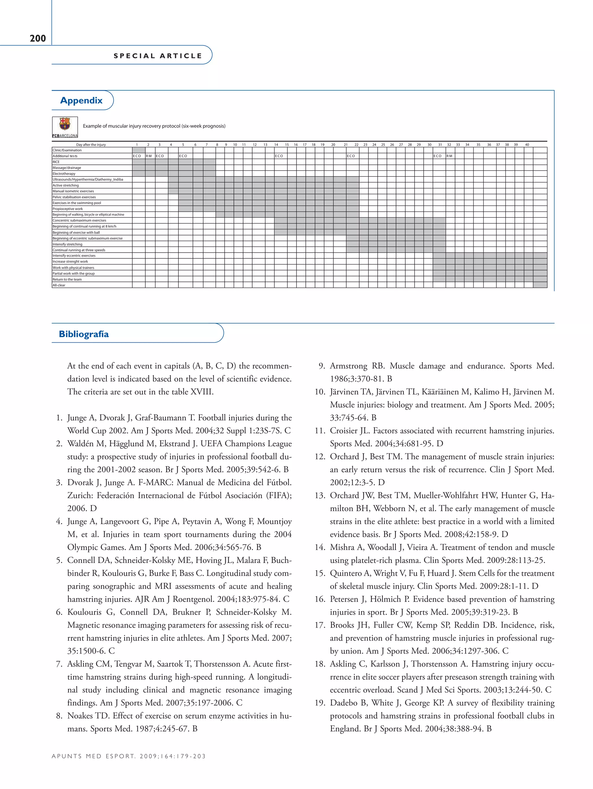
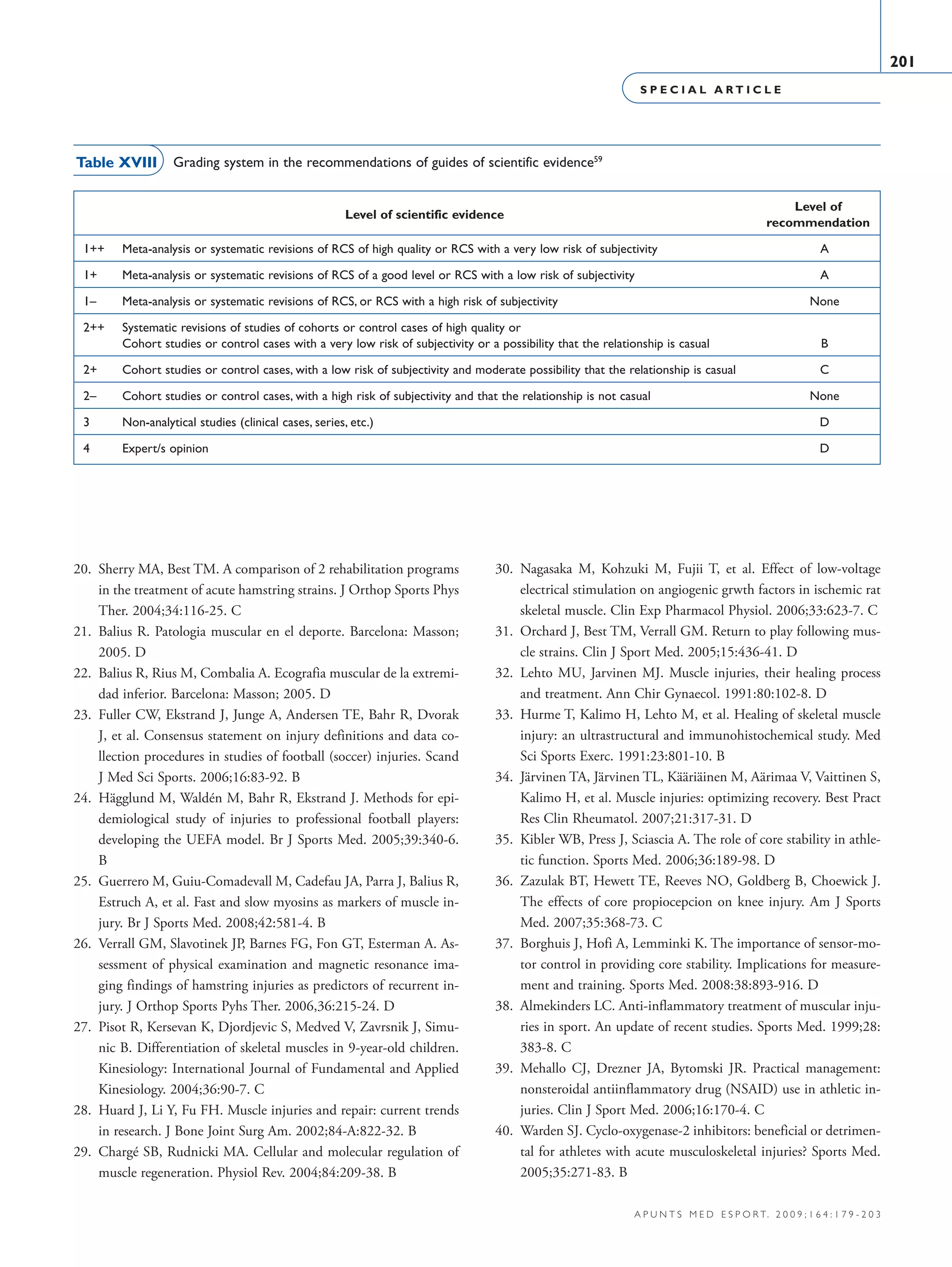
SlideShare a Scribd company logo
1 of 25
Download to read offline
S P E C I A L A R T I C L E
179
a p u n t s m e d e s p o r t. 2 0 0 9 ; 1 6 4 : 1 7 9 - 2 0 3
Clinical Practice Guide for muscular injuries.
Epidemiology, diagnosis, treatment
and prevention
Version: 4.5 (9 February 2009)
Medical Services. Futbol Club Barcelona
Introduction
Muscular injuries are very frequent in the world of sport,
especially in football. The most recent epidemiological studies
show that muscular injuries represent more than 30% of all
injuries (1.8-2.2/1,000 hours of exposure), which means that a
professional football team suffers an average of 12 muscular
injuries per season, equivalent to more than 300 lost sporting
days1-4
. In other professional sports like basketball and handball
the incidence is also high, although not reaching the figures
shown in football.
Despite their high frequency and the interest in finding
solutions, there is little scientific evidence on aspects as
important as prevention and treatment. We will outline some
weak points below:
• The diagnosis of muscular injuries is based on clinical
medicine, fundamentally on symptomatology and especially
on the anamnesis of the injury mechanism and physical
examination. Imaging studies through the musculoskeletal
echography and magnetic resonance are complementary
studies, despite the fact that they could be increasingly more
useful when confirming a diagnosis or especially when giving
a prognosis5-7
. There is no sufficiently specific biochemical
marker available that could help with the diagnosis of the
seriousness and the definitive prognosis of each of the different
muscular injuries8,9
.
• Treatment guidelines for muscular injuries do not follow a
unique model, despite the fact that the different alternatives
have been modified very little10-12
. Finally, new expectations
have been raised thanks to research within the field of
biological repair and regeneration13-15
.
• Certain primary and secondary prevention programmes can
reduce the incidence of suffering muscular injuries, but
scientific evidence is still limited and it has only been possible
to verify them in certain groups of sportsmen16-20
.
The objective of this document is to record the diagnostic,
therapeutic and preventative approaches that should be taken
when faced with the various muscular injuries suffered by the
football players of Barcelona FC. This is not intended as an
exhaustive review of muscular pathology in sport, but rather
a working document that is clear, practical and comprehensive.
The protocols are based on current knowledge from recent
years in the daily work of dealing with these types of
injuries.
Classification
Muscular injuries are traditionally classified, according to
their injury mechanism, as either extrinsic (direct) or intrinsic
(indirect).
• Extrinsic injuries, due to a contusion with an opposing player
or with an object, are classified according to their severity, into
light or benign injuries (grade I), moderate (grade II) or serious
(grade III). These injuries may present lacerations or not.
• Intrinsic injuries, due to stretching, are produced by the
application of a tensional force higher than tissue resistance,
when it is in active contraction (eccentric contraction). The
force and speed with which the tension is applied are variables
that change the viscoelastic properties of the tissue, changing
its susceptibility to breaking. Local fatigue and cellular
temperature may also have an influence. Players notice a
sudden pain, in the form of a pull or a sharp pain and this is
normally related to sprinting, a change in speed or taking a
shot. The classification of intrinsic injuries is more
complex.
06 Articulo especial (179-203).i179 179 9/12/09 12:56:19
Documento descargado de http://www.apunts.org el 20/12/2009. Copia para uso personal, se prohíbe la transmisión de este documento por cualquier medio o formato.
S P E C I A L A R T I C L E
180
a p u n t s m e d e s p o r t. 2 0 0 9 ; 1 6 4 : 1 7 9 - 2 0 3
Following the most up-to-date concepts we propose the
following classification tables for muscular injuries, according
to various criteria (tables I and II)21,22
:
Regarding the prognosis, the days lost are for guidance only
and vary in accordance with the injured muscle, its topography
and the demands made on it afterwards.
Both the musculoskeletal echography and magnetic
resonance enable precise information to be obtained about
the muscular injury in relation to the affected connective
tissue. Although almost all the injuries settle on the
myoconnective junction, according to the type of
myotendinous or interaponeurotic involvement, as well as
the topographic area of each kind of injury, the prognosis
may vary and therefore this must be borne in mind to give
the all-clear to play for return to competitive matches. In this
respect, several studies are beginning to produce clear evidence
about this question, and what appears clear is that the more
involved the connective part and the injury area are, the
worse the prognosis5-7
.
A good clinical history and physical examination, together
with the classifications proposed above must enable the
achievement of a diagnosis. With the objective of being able to
be clearer, we propose the compliance with the following
guideline in order to label each muscular injury.
First, we give the name of the kind of muscular injury
according to anatomopathological criteria. After that, the first
surname, where we give the specific localisation where the
injury is settled and specifically whether there is a relation with
the connective part. Finally, the second surname, where we list
the specific affected muscular group.
Table I Classification of muscular injuries using histopathological criteria. (We understand grade 0 injuries to be injuries in
which very little muscular tissue is affected. In the event that the injury were more extensive, they could present as
more serious injuries and therefore with a worse prognosis)
Nomenclature	 States	 Characteristics	 Prognosis
Contraction and/or DOMS	 Grade 0	 Functional alteration, elevation of proteins and enzymes.Although there is a slight break 	 1-3 days
		  down of the muscular parenchyma, this is considered to be more an adaption
		  mechanism than a real injury
Small fibrillar strain and/or 	 Grade I	 Alterations of few fibres and light injury to connective tissue	 3-15 days
  muscular elongation
Fibrillar strain	 Grade II	 More affected fibres and more connective tissue injuries, with the appearance 	 3 to 8 weeks
		  of a haematoma
Muscular strain	 Grade III	 Major strain or complete displacement.The functionality of the unharmed fibres 	 8 to 12 weeks
		  is completely insufficient
DOMS (Delayed Onset Muscular Soreness) is given to stiffness — not very scientific terms.
Classification of muscular injuries according to imaging criteriaTable II
Nomenclature	 States	 Musculoskeletal echography	 Magnetic resonance
Contraction and DOMS	 Grade 0	 Inconsistent signs. Oedema between fibres and 	 Interstitial and intermuscular oedema. Increase of
		  myofascial and increase in local vascularisation	  the signal in T2 and fat suppression sequences
Small fibrillar break and/or	 Grade I	 Minimum solution of discontinuity, oedema between	 Increase in the interstitial signal and slightly
  muscular elongation		  interfascial fibres and fluid (indirect sign)	  intermuscular
Fibrillar strain	 Grade II	 Clear muscular defect, interfascial fluid and haematoma	 Strong interstitial signal, focal muscular defect,
			  increase in the signal surrounding the tendon
Muscular strain	 Grade III	 Complete muscular disruption and/or of the tendon, 	 Complete muscular and/or tendon strain, with
		  with retraction of the displaced part of the muscle 	  retraction
		  (visible stump)	
06 Articulo especial (179-203).i180 180 9/12/09 12:56:19
Documento descargado de http://www.apunts.org el 20/12/2009. Copia para uso personal, se prohíbe la transmisión de este documento por cualquier medio o formato.
S P E C I A L A R T I C L E
181
a p u n t s m e d e s p o r t. 2 0 0 9 ; 1 6 4 : 1 7 9 - 2 0 3
• Name: Muscular strain grade II
• First surname: of the musculotendinous proximal area
• Second surname: of the biceps femoris
Epidemiological studies
We have studied the incidence of injuries of the first football
team for four years (2003-2007), using the methodology that
various international groups of experts have agreed upon and
which is regulated and governed by the medical committee of
UEFA for the various teams that play in the Champions
League23,24
. This is a prospective study, controlling all injuries
that take place as well as training and competitive hours in
order to discover the risk of suffering the different kinds of
injuries.
The calculation for injury risk will be performed using the
following formula:
Number of injuries/Hours of exposure to training
and/or competition per 1,000 hours
Nowadays this is considered as the universal framework
that enables teams, clubs, sports etc, to be compared. The risk
of suffering any kind of professional football injury is between
6 and 9 injuries per 1,000 hours of exposure, which explains
what this occupational risk would represent in a company of
25 workers that each month had 9 workers off work. The risk
of getting injured during competitive play is between 4 and 6
times more frequent than during training1,2
.
In table III we present the description of the various
parameters regarding injury incidence regarding the muscular
injuries of the first team of Barcelona FC for four seasons.
These are data reviewed by the medical committee of UEFA
and which are published here for the first time.
According to references in the literature, it is estimated that
a professional team of 25 players will have an average of 40-45
injuries per season, of which between 16 and 20 are minor (of
less than one week); between 16 and 20 are moderate (between
1 and 4 weeks), and between 8 and 10 will be serious (more
than one month’s absence). Muscular injuries, 30-40% of all
injuries, the risk of injury is nearly 2 per 1,000 hours of
exposure, each team may have between 10 and 14 muscle
injuries per season.
Description of the main parameters regarding injury incidence of the first team of Barcelona FC in 4 consecutive
seasons. In the table almost all the decimals have been eliminated for practical reasons
Table III
Seasons	 2003/04	 2004/05	 2005/06	 2006/07
Total number of injuries	 63	 44	 31	 37
Injuries/1,000 hours of exposure to training	 7	 3	 3	 3
Injuries/1,000 hours of exposure to competitive play	 34	 25	 15	 15
Muscular injuries 	 22	 6	 14	 14
Minor muscular injuries (1 week)	 8	 2	 2	 3
Moderate muscular injuries (1-4 weeks)	 11	 4	 10	 8
Serious muscular injuries (1 month)	 3	 0	 2	 3
% muscular injuries/total injuries	 35	 14	 45	 33
Total hours of exposure of the team	 5,655	 6,458	 5,719	 7,561
Muscular injuries/1,000 hours of exposure	 4	 1	 2	 2
Muscular injuries/1,000 hours of competitive play	 16	 5	 9	 12
Muscular injuries/1,000 hours of training	 1,7	 0,2	 0,7	 2,2
Total days injured 	 952	 1,404	 657	 685
Days injured due to muscular injury	 317	 84	 264	 324
Training sessions missed due to muscular injury	 203	 46	 173	 186
Matches missed due to muscular injury	 58	 17	 59	 51
06 Articulo especial (179-203).i181 181 9/12/09 12:56:20
Documento descargado de http://www.apunts.org el 20/12/2009. Copia para uso personal, se prohíbe la transmisión de este documento por cualquier medio o formato.
S P E C I A L A R T I C L E
182
a p u n t s m e d e s p o r t. 2 0 0 9 ; 1 6 4 : 1 7 9 - 2 0 3
In tables IV and V we present data on the overall study of
incidence of injury that UEFA has carried out with the majority
of the teams in the Champions League during a period of four
seasons (2003/04/05/06). As can be seen the most frequent
injury is muscular and, more specifically, injuries to the
posterior femoral (hamstring) muscles, of which the biceps
femoris muscle is the most affected.
These data are important because they define very well the
main injuries that occur in professional football and therefore,
where efforts should be directed to plan preventative strategies.
What is measurable is more susceptible to improvement. The
collection of these and other new data will provide us with the
results of preventative measures adopted in reducing the
number of injuries.
Diagnosis
The diagnosis of muscular injuries is mainly clinical, that
is, based on clinical history and physical examination.
In the anamnesis (table VI) it is necessary:
• To collect local and general history: Has the player suffered
similar injuries before?, Does he know of a susceptibility to
suffer injuries?, What pharmacological background does he
have?,...
• To describe the moment of injury: What had the trigger been?,
During work, training or competition?, Was it at the
beginning, the middle or the end of the session?
• To collect the immediate progress: Was the player able to
continue the work session or did he have to abandon it?,
How is the pain progressing?
In the physical examination (table VII) you have to perform
the following:
• Inspection: Is ecchymosis or deformities of the profile of the
muscle mass present?
• Feeling: find points of pain or muscle spasms.
• To request active contraction of the affected muscle, first with the
muscle with stretching, more sensitive in minor injuries, and
afterwards against manual resistance: Is it possible? Is it painful?
What type of contraction (concentric, isometric or
eccentric)?
Type of injury	 Number	 Percentage
  1  Posterior femoral (hamstring) muscles	 396	 14
  2 Adductor muscles	 260	  9
  3  LL Ankle Sprains/strains 	 203	  7
  4  Quadriceps muscles	 160	  6
  5  LL Knee Sprains/strains 	 153	  5
  6 Triceps surae muscles	 124	  4
  7  Lumbar pain	 100	  4
  8 Achilles tendinopathy	  82	  3
  9  Muscular bruising	  82	  3
10  Foot	  74	  3
Table IV Description of the number and percentage
relating to all communicated injuries in the
UEFA study in the 2003-04; 2004-5; 2005-06;
2006-07 seasons
Type of injury	 Total (n = 55)	 Percentage
1  Injury to biceps femoris muscle	 16	 30
2  Injury to adductor medium muscle	 10	 18
3  Injury to triceps surae muscle	  9	 16
4  Injury to quadriceps muscle 	  7	 12
5  Injury to semitendinosus muscle 	  3	  5
6  Others	 10	 19
Table V Description of the number and percentage
relating to muscular injuries of the UEFA study
in the 2003-04; 2004-5; 2005-06; 2006-07 seasons
Anamnesis	 Yes	 No 	 Observations
Prior injury of the same structure			
Other susceptibility to muscle injury			
Training			 Start/half-way/end
Game			 Start/half-way/end
Do you remember the moment			 Shot/sprint/
  of injury?			  jump/ other
Were you able to continue?			
Have you improved?
Table VI Anamnesis
06 Articulo especial (179-203).i182 182 9/12/09 12:56:20
Documento descargado de http://www.apunts.org el 20/12/2009. Copia para uso personal, se prohíbe la transmisión de este documento por cualquier medio o formato.
S P E C I A L A R T I C L E
183
a p u n t s m e d e s p o r t. 2 0 0 9 ; 1 6 4 : 1 7 9 - 2 0 3
• To assess the analytical flexibility of the muscle: whether or not
there is pain with passive stretching.
We have already mentioned that complementary tests such
as musculoskeletal ultrasound and MRI are very helpful in
finding out as soon as possible the degree of injury and, above
all, the prognosis for days off. The MRI is very sensitive and is
precise in identifying the affected structure but on the other
hand the musculoskeletal ultrasound is a dynamic study that
complements the clinical examination, enabling progress
monitoring, the guided evacuation of cavities and is
complemented by the painful echopalpation of a given muscle,
already identified by the ultrasound, a great help with the
topographic diagnosis.
The table VIII presents when it is chronologically most
appropriate to carry out the different complementary tests,
which will be more or less extensive depending on the criteria
of the physician and the availability of resources.
What steps do we follow once a muscle injury is
produced?
• Immediately. Once the injury is produced, through a survey
addressed to the player (What have you noticed?, When?,
How?, Where?, What have you done?) and with a structured
physical examination: inspection, feeling, which movements
cause pain, passive and active, we will be able to make an
initial diagnostic orientation. When the injury is not a major
strain early diagnosis is not easy. It is important and necessary
to know and to wait a few hours to see it develop, as well as
carrying out the appropriate complementary tests.
• At 12 hours. The ultrasound study at this early stage does
not allow for an accurate diagnosis of minor muscular injuries,
but does from grade II injuries. Only highly trained and
specialised personnel in ultrasound analysis are capable of
making a certain diagnostic on the extent of the injury.
Therefore, at 12 hours only a certain diagnosis in injuries of
grade II or higher can be made.
• If the injury is very minor and we are not sure if it is grade 0
or I, the resolution in serum of the protein myosin allows us
to make an early diagnosis of a grade I injury. This
intramuscular contractile protein has a large molecular weight
and does not have to be present in the blood. Its presence
determines fibrillar injury. For kinetic studies it appears to be
that between 12 and 24 hours is the optimum time to
determine this. It is not a test that we can routinely ask for
and its evidence is based on the experience of the working
group that has recently published a primary work that we
believe could be of great interest and in the future, open the
way to new tools in defining muscular injuries25
.
• At 24 hours. This is the time agreed upon by most specialists
in magnetic resonance imaging to establish an appropriate
diagnosis and prognosis. As always, it is necessary that the
personnel who interpret the MRI have extensive experience
in this type of injury. Today, especially in injuries to the
posterior femoral (hamstring) muscles, and specifically, injury
to the proximal muscle tendinous junction of the biceps
femoris muscle, have postulated that the total length of the
injury, the relationship between the isquiatic tuberosity and
the proximal beginning of the injury, and the total affected
area, are the prognosis factors of the time necessary for the
return to competition and the risk of reinjury7,26
.
Examination	 Yes	 No 	 Observations
Time of progress:
Is ecchymosis present?			
Are muscular deformities present?			
Pain points			 Topography:
Muscular spasms			 Which:
Is contraction possible against			 Isometric/
  manual?			  concentric/
			  eccentric
Is the active contraction painful?			 Isometric/
			  concentric/
			  eccentric
Is the capability of passive stretching
  greater?
Is passive stretching painful?
Table VII Examination
	 Clinical 	 Physical 	
Ultrasound	 MRI
	 Biochemical
	 history	 examination			 markers
Immediate	 X	 X			
12 h		 X	 X		 X
24 h		 X	 X	 X	 X
48 h		 X	 X		
Table VIII Timeline for carrying out complementary tests
in the muscular injury
06 Articulo especial (179-203).i183 183 9/12/09 12:56:20
Documento descargado de http://www.apunts.org el 20/12/2009. Copia para uso personal, se prohíbe la transmisión de este documento por cualquier medio o formato.
S P E C I A L A R T I C L E
184
a p u n t s m e d e s p o r t. 2 0 0 9 ; 1 6 4 : 1 7 9 - 2 0 3
• At 48 hours. This is the optimum time to establish a more
appropriate diagnosis and prognosis using ultrasound
analysis.
Finally, a technique has been proposed for assessing the
“muscle tone” called Tensiomyography27
. While so far there is
little scientific evidence from this methodology, perhaps, in the
future, it will be useful to monitor the functional recovery of
the muscle and for it to be a complement to the progress
monitoring of muscular injuries together with musculoskeletal
ultrasound.
Treatment
The basic principle that most authors support is that
muscular injuries should have an early mobilisation and
functionality. Especially after the third day, since it has been
demonstrated that this:
• Rapidly increases vascularisation of compromised muscle
tissue.
• Increases the regeneration of muscular fibres.
• Improves the final reparative phase avoiding fibrous scars.
• Recovers the viscoelastic and contractile characteristics of the
muscles faster, ultimately, the overall functionality of the
muscle28-31
.
Only with regard to the first phase of the treatment of
muscular injuries, between the first and fifth day of the injury,
is there an international consensus thanks to the few clinical
trials that have been able to demonstrate a degree of scientific
evidence10-15
. There is a wide disparity of viewpoints and
proposals by different authors and schools.
In this guide we will give an account of the immediate
standard treatment of muscular injuries. Next, we will explain
what alternatives have been proposed and, finally, we will
present the specific protocols for various muscular injuries that
are underway at Barcelona FC, based on the experience of its
professionals and on the experience of various authors.
First Phase: immediate treatment post injury
Consisting of what is called RICE which in English
means:
Rest.
Ice.
Compression.
Elevation.
This is the most agreed upon treatment for the first three
days32-34
. The immobilisation in the first phase could prevent
future retractions of the strain and make a smaller haematoma.
It has also been shown that the use of cryotherapy has made a
significantly smaller haematoma, reducing inflammation and
accelerating repair. Compression, while reducing the
intramuscular blood flow, appears to have a very potent anti-
inflammatory effect. What is of most interest is to combine the
compression and the cryotherapy at repeated intervals of 15 to
20 minutes approximately every 3 or 4 hours. Regarding this
point it should be explained that:
• This time could be higher depending on the muscular mass
of the muscle involved, for example, a quadriceps muscle of
an adult could require up to 30 to 40 minutes.
• That in muscular injuries by direct contusion (“sandwich”)
the gel will be put in the position “of bearable stretching”.
• Care must be taken with the application of cryotherapy, with
respect to the proposed intervals, and controlling individual
susceptibility, to avoid skin lesions.
Second phase: of the third to the fifth day
• Muscular activation. Early and progressive mobilisation is very
important. If the first phase has passed correctly and without
complications, the following exercises can be started gradually,
taking into account the degree of tolerance to the pain:
– Isometric exercises: Progressively, and taking into account that
they have to be of maximum intensity until the onset of pain.
The pain we are looking for is the bearable discomfort
considered optimum between 5-10% of the visual analogue
scale (VAS). Also it will be important to use progressively
different ranges and to carry them out in different positions
and angles. The protocol we propose in this guide is to perform
isometric exercises on 3 different ranges and with a guideline
of time that can start with 6 seconds of contraction and 2
seconds of relaxation.
• Complementary work. In this phase several matters must be
taken into account:
– Work on stability and movement of the lumbopelvic region,
which is increasingly defined as exercise programmes aimed at
06 Articulo especial (179-203).i184 184 9/12/09 12:56:21
Documento descargado de http://www.apunts.org el 20/12/2009. Copia para uso personal, se prohíbe la transmisión de este documento por cualquier medio o formato.
S P E C I A L A R T I C L E
185
a p u n t s m e d e s p o r t. 2 0 0 9 ; 1 6 4 : 1 7 9 - 2 0 3
improving the “core stability” in order to improve and prevent
muscular injuries of the locomotor apparatus35-37
.
– Physical therapy: this section has always been a controversial
issue. Used worldwide but there is no, or very little scientific
evidence. The physical therapy most commonly used is:
a) Electrotherapy of analgesic effect and muscle decontraction
to promote muscular refunctionalisation.
b) Temperature: ultrasound (this is advisable to be performed
with stretching without passing the point of pain),
hyperthermy (taking into account the depth of the injury
and therefore very important to study the prior ultrasound),
or diathermy. Currently, the physical therapy we are advising
and proposing is diathermy, and we hope in the next version
to have a protocol that has demonstrated its scientific and
clinical evidence.
– Stretching. Stretching of the muscle in this phase has to be
pain free, following the premise of bearable discomfort. We
advise starting with 12 seconds of stretching and 12 seconds of
rest. The advisable and lesser risk stretching is active stretching
by the method of active contraction of antagonistic muscles in
axial rotation, to stretch the injured muscle and improve the
viscoelastic conditions and reduce the risk of fibrous scarring
and reinjury.
– Cardiovascular maintenance, this is another important aspect
to improve muscular recovery and means:
a) Continuing to work on the uninjured structure. Different
types of exercises can be used whether swimming pool, static
bicycle, as permitted by the injured muscle.
b) And with regard to the really injured structure, we must
always respect the principle of reaching the pain or tolerable
discomfort limit, walking to be allowed from the third day
for 30 minutes and to be gradually increased daily by 10
minutes until being able to be added to the run in the
following phase.
Here we wish to make a comment with regard to the latest
fashionable use of “kinesiotape” (neuromuscular bandage) with
the idea of reducing muscle tension by its effect on the fasciae
and having an analgesic effect. This technique requires rigorous
study to verify its effectiveness.
From the fourth-fifth day again to make a clinical and
radiological (musculoskeletal ultrasound) assessment to evaluate
the muscular defect and whether or not there is an intramuscular
haematoma, which is normally the time it takes in forming.
The presence of a haematoma could recommend the carrying
out of an aspiration puncture through ultrasound control, to
maintain two days of compression and again to start the phase
2 protocol.
After about the seventh day, as we have explained, we have
not found an agreed standard treatment and each group or
author is progressively incorporating more intensity and
complexity to the exercises both to increase the continual
running, the stretching, such as strengthening of the muscles
(isometric, concentric and eccentric). We propose in this guide
that once the athlete can walk 1 hour with “bearable
discomfort” he can then move on to continuous running;
initially recommending a submaximal rhythm of 8-10 km/h,
which is precisely the phase higher than fast walking. “The
pain” will return to be the marker that we use to go beyond
this phase.
In the next chapter of this guide we will detail the different
recovery programmes for diagnosed muscular injuries from the
first day until return to competition.
Other treatments
Medication
Precisely at the instant a muscular injury occurs, a sequence
of phases is launched that consists of degeneration,
inflammation, myofibrillar regeneration and finally, the
formation of fibrous tissue15,34
.
The use of analgesics or anti-inflammatories during the
first hours has been a subject of debate since their use would
favour the reduction of the inflammatory phase, when it
seems that thanks to the reactions that occur therein trigger
all of the subsequent mechanisms of the regenerative phase.
There are no control studies to clearly assess the benefit-risk
relation of using non-steroid anti-inflammatory drugs
(NSAID’s) nor glucocorticosteroids, but in any case, it seems
that the use of NSAID’s during the first 48-72 hours is not
harmful and does not interfere with the regeneration phase if
its use is not continued38,39
. Equally, we must remember that
some authors are sure that the indiscriminate use of analgesia
in the first phases of muscular injuries can have a masking
effect of the pain, hindering diagnosis and encouraging a
relapse40,41
.
Moreover, the use of antifibrotic medication such as the
range of interferon, suramin and decorin, which are still in the
clinical trials phase, may in the future be a good complementary
treatment in the treatment of muscular injury42,43
. Finally, a
06 Articulo especial (179-203).i185 185 9/12/09 12:56:21
Documento descargado de http://www.apunts.org el 20/12/2009. Copia para uso personal, se prohíbe la transmisión de este documento por cualquier medio o formato.
S P E C I A L A R T I C L E
186
a p u n t s m e d e s p o r t. 2 0 0 9 ; 1 6 4 : 1 7 9 - 2 0 3
diuretic, losartan, has also shown to have antifibrotic
properties44
.
The hyperoxygenation of the injured tissue by means of
instruments that facilitate the spreading of oxygen such as
hyperbaric are under research. It seems that they are effective
but difficult to implement45
.
Infiltration and use of PRP
Some schools have used the infiltrations on the focus of the
injury with different products, such as corticosteroids and the
combination of homeopathic products (Traumeel®
) or others of
dubious origin (Actovegin®
)46-49
. In recent years, moreover,
what has become popular in Spain is the use of Platelet-Rich
Plasma (PRP), also known as “growth factors”. It seems useful
that if in each phase of muscular repair: first, destruction and
inflammation; second repair and regeneration, and third, final
restructuring, we were capable of adding most appropriate
growth factor we would be able to get better results. This is a
highly topical clinical and scientific debate50
. There is a lack of
rigorous clinical trials showing that this type of treatment
improves recovery time of muscular injuries and reduces the
risk of relapse. In the absence of such studies, the medical
service of Barcelona FC has agreed that the indication of
infiltration of PRP will be made when complying with criterion
1 together with criterion 2 or 3.
1. Athletes over 18 years old suffering an injury in a high
demand muscle by the sport that is practiced.
2. Recurring intramuscular or myofascial cavity injuries such as
the rupture of the distal myotendinous of the anterior rectal
muscle or that of the Gastrocnemius muscle (so-called
“tennis leg”).
3. Progress of complex and chronic injuries.
In this sense, we can now use PRP thanks to the clinical
trial that we conducted on muscular injuries that affect the
musculature of the leg (triceps, biceps, sural) known as “tennis
leg” (FIS_ research project awarded by the “Instituto de Salud
Carlos III” of the Ministry of Science and Innovation”).
Surgical treatment
Muscular injuries should be treated conservatively, but
there are some consensual general surgical indications10,34
.
• Muscular injury of grade III, total strain.
• Muscular injury with partial (50 %) or total tendinous
retraction.
• Muscular injury of grade II, with more than 50% of the
abdominal muscle affected.
• When there is a very large intramuscular haematoma that
does not respond to repeated aspiration puncture.
Likewise, different specificities should be taken into
account, and the surgical option could be prescribed in the case
of recurrent muscular injuries that form significant fibrous
scars and that generate adhesions with neural involvement.
Moreover, in contrast, in some type of muscles such as the long
adductor muscle, in a grade II-III injury, surgical treatment
may not be needed as it has already been seen progressing
favourably with conservative treatment.
It is not the reason for this guide, but we must remember
that after this type of surgery a very careful, programmed and
long rehabilitation is required. It is estimated that the return to
sport could take, depending on the muscle, from a few months
up to a year.
Specific recovery protocols for muscular injuries
In this section we present, comprehensively, the various
protocols that we recommend for the more common muscles
in our environment.
Protocol 1. Grade I injury to the MTJ of the proximal
portion of the biceps femoris muscle (table IX)
This is the injury that affects the shared tendon of the
hamstrings m., also known in our field as the “upper rack”.
This is a complex injury that occurs between the biceps femoris
muscle and in a close link with the semitendinosus muscle and
this has a worse prognosis, as we commented previously, the
longer the injury is, the larger the area occupied and the closer
to the ischial tuberosity6,7,26
. It is in this injury where the rule
“so many centimetres long, so many weeks laid off” has empirical
evidence and as we have commented on certain scientific
evidence. So here we propose in protocol 1 a grade 1 type
injury that could be an injury of 1 to 2 cm. long of the biceps
femoris muscle, and in the next protocol we will see the
protocol to follow with a more extensive injury.
In both cases clinical progress monitoring and by ultrasound
will have to be made to pass to the next phase and in order not
to give a premature all-clear, as it is an injury that relapses easily.
06 Articulo especial (179-203).i186 186 9/12/09 12:56:21
Documento descargado de http://www.apunts.org el 20/12/2009. Copia para uso personal, se prohíbe la transmisión de este documento por cualquier medio o formato.
S P E C I A L A R T I C L E
187
a p u n t s m e d e s p o r t. 2 0 0 9 ; 1 6 4 : 1 7 9 - 2 0 3
Protocol 2. Grade II injury to the MTJ
of the proximal portion of the biceps femoris muscle
(table X)
The protocol in a second grade injury, with a recovery
prognosis of some 6-8 weeks approximately. The protocol
we propose is very similar to that proposed by various
authors10-12
.
Comments
We do isometric exercise from the third day and the number
of series, the time of contraction and the work of at least three
ranges of reference will be done according to the pain. This
isometric exercise and the active stretching will help to reduce
the oedema, enable the injury to heal correctly and improve
the functionality and viscoelastic capacity of the muscle. In
injuries of the hamstring musculature we give more importance
to the work of eccentric reinforcement due to its etiology. One
author10
proposes that at 4 weeks to conduct an analytical
study of the strength and/or muscular power to analyse possible
strength deficits. Finally, personalised work has to be set out for
each type of player in the last phase depending on their
characteristics.
Protocol 3. Grade II injury to the MTJ of the distal
portion of the femoris biceps muscle (table XI)
The protocol of recovery is very similar to protocol 2. This
injury is known in our field as “distal rack”, with a recovery
prognosis of approximately 4-6 weeks.
Comments
In general the prognosis of this type of injury is better than
that of the proximal 1/3. Often the long and short portions of
the biceps femoris muscle are affected, causing the “rack” effect
as we have commented previously. At times it could leave a
residual myofascial haematoma that does not necessarily affect
return to competition, providing that its functionality is
complete.
For the purpose of the double innervation of the two heads
of the biceps femoris, it is interesting to note that it is necessary
to work with internal and external rotation of the leg to
selectively activate the two heads.
Protocol 4. Grade II injury to the adductor medium
muscle (table XII)
It is important to distinguish this type of injury from the
injury to the periostic tendon junction, of a chronic nature.
This is an acute injury that creates an immediate functional
impotence, which usually creates a rapid haematoma.
Sonographically, a laminar defect can be seen below the
fascial expansion of the adductor medium muscle. It
progresses well, but if the injury is extensive, of more than 3
From 0 to 3 days
  • RICE.
  • Electrotherapy.
  • Massage/Drainage.
From 3 to 7 days
  • Ultrasounds daily or Hyperthermy alternate days, or diathermy
daily.
  • Assessment and begin working with lumbo-pelvic stabilisation
exercises.
  • Beginning of manual isometric exercises (4 series of
10 repetitions in 3 different ranges and with times of progressive
contraction, e.g.: 6 seconds of contraction and 2 seconds
of relaxation).
  • Active stretching from work with the antagonistic muscles in
axial rotation following a guide of 12 seconds maintaining the
stretching and 12 seconds rest.
  • Propioceptive work.
  • Begin physiotherapy work in the swimming pool.
  • Physiotherapy work in the field (walking forward and backward,
active stretching exercises, lateral movements, etc.).
From 7 to 14 days
  • Start of the manual strength work by means of the eccentric
submaximum method (of low intensity) of 4 to 6 series and
of 8 to 10 repetitions.
  • Isometric reinforcement, concentric and eccentric carried out by
the athlete (Russian belt...).
  • Continue with field work (introduce the ball).
  • Continue with guidance in the gym (lumbar-pelvic
strengthening...).
  • Continue with field work and, as appropriate, transfer
to rehabilitation.
  • Start partial work with the group and specifically with fitness
trainers.
Day 15 approx.
  • All-clear
Table IX
06 Articulo especial (179-203).i187 187 9/12/09 12:56:22
Documento descargado de http://www.apunts.org el 20/12/2009. Copia para uso personal, se prohíbe la transmisión de este documento por cualquier medio o formato.
S P E C I A L A R T I C L E
188
a p u n t s m e d e s p o r t. 2 0 0 9 ; 1 6 4 : 1 7 9 - 2 0 3
cm, the football player must wait for a recovery of between
4 and 6 weeks.
Comments
With this type of injury it is very important to make a
proper assessment of the lumbar-pelvic stability in relation to
the frequent pubis problems, and more specifically with
dynamic osteopathy of pubis (DOP). Therefore it is necessary
to assess by ultrasound or MRI the pubis symphysis, as if
affected, this may delay the return to normal training. It is not
uncommon to find players who, after suffering an injury of
this type begin a pubalgia that is probably a consequence of
stopping training, which destabilises the symphysis and the
entire lumbar-pelvic ring, therefore it will be very important to
be alert to this fact. So it will be very important to influence
the active stretching exercises and avoid excessive strengthening
of the adductor medium muscle that will cause an imbalance
at the level of pubis symphysis with the consequent risk of the
appearance of a DOP.
Moreover, we must remember that this muscle allows for
many exercises without discomfort, but on the other hand,
  • Dynamic propioceptive exercises.
  • Massage therapy post exercise when necessary.
  • Transfer to physical therapists to meet sporting movement needs.
From 30 to 45 days
  • Follow-up with combined isometric/concentric and eccentric
reinforcement, especially eccentric work of the hamstring
muscles.
  • Correction of possible deficiencies of flexibility and strength.
  • Field work to improve the conditional capacities, coordinated
with physical trainers.
  • Partial progressive work with the group (without explosive
exercises....) and specifically with fitness trainers.
  • Massage therapy all-clear.
  • Full work with the group.
45 days
  • All-clear
  • Pass on to work on injury prevention.
Table X (continuation)
From 0 to 3 days
  • RICE.
  • Electrotherapy.
  • Massage/Drainage.
From 4 to 7 days
  • Ultrasounds daily or hyperthermy alternate days, or diathermy
daily.
  • Assessment and lumbo-pelvic stabilisation exercises.
  • Beginning of manual isometric exercises of 4 series and 10
repetitions with 3 different ranges and with times of progressive
contraction starting by 6 seconds of contraction and 2 seconds
of relaxation.
  •Active stretching using the antagonist muscle with a time
of contraction relaxation 12 seconds: 12 seconds.
  • Start swimming pool work.
  • Guidelines in the gym using non-injurious structures
and prevention planning.
From 7 to 14 days
  • Daily ultrasound with stretching or diathermy daily.
  • Follow-up manual isometric reinforcement.
  • Begin work with bicycle, or elliptical machine or to walk
approximately 30 minutes.
  • Follow-up with active stretching.
  • Lumbar-pelvic stability guidelines
  • Propioceptive work.
  •Gym work (for injury and healthy structures).
From 14 to 21 days
  • Follow-up with manual isometric reinforcement, stretching
and propioceptive work.
  • Begin manual strength work by concentric submaximum
method of 4 to 6 series and of 8 to 10 repetitions.
  • Start progressive continual running.
  • Follow-up lumbar-pelvic stability guidelines.
  • Start field work with ball
  • Massage therapy
From 21 to 30 days
  • Follow-up with manual concentric reinforcement.
  • Begin manual strength work by eccentric submaximum method.
From 4 to 6 series and of 6 to 8 repetitions.
  • Intensify stretching exercises.
  • Progressive running in different rhythms without sprinting.
Table X
06 Articulo especial (179-203).i188 188 9/12/09 12:56:22
Documento descargado de http://www.apunts.org el 20/12/2009. Copia para uso personal, se prohíbe la transmisión de este documento por cualquier medio o formato.
S P E C I A L A R T I C L E
189
a p u n t s m e d e s p o r t. 2 0 0 9 ; 1 6 4 : 1 7 9 - 2 0 3
such as actively taking part in specific moves of football such as
the shot, changes of direction, etc. it will be necessary to
lengthen the phase of functional rehabilitation to sporting
movement before allowing return to competition.
Protocol 5. Grade I injury to the soleus muscle
(table XIII)
In this type of injury a small puncture in the calf is typical,
but the player can usually continue doing submaximum
exercise, which sometimes delays the final diagnosis. It is
important to make differential diagnosis with all injuries called
“tennis leg”. The diagnosis must be confirmed by MRI more
than by ultrasound.
Comments
In this type of injury and from the point of view of the
physiotherapist, we understand that it is very important to
work progressively, extensively and for a long period with
isometric exercises. We need to ensure good tissue repair and a
complete lack of discomfort to the making of any movement
or exercise by the player. Reinjury is very frequent.
We need to ensure a guideline of concentric and eccentric
exercises daily and although there is a significant clinical
improvement the first few days, we should not accelerate the
From 0 to 3 days
  • RICE.
  • Electrotherapy.
  • Massage/Drainage.
From 4 to 7 days
  • Ultrasound daily or Hyperthermy on alternate days or diathermy
daily.
  • Assessment and lumbar-pelvic stabilisation exercises.
  • Beginning of manual isometric exercises of 4 series and 10
repetitions with 3 different ranges and with times of progressive
contraction by starting 6 seconds of contraction and 2 seconds
of rest.
  • Active stretching exercises (12 seconds contraction
and 12 seconds relaxation).
  • Start of physiotherapy work in the swimming pool.
  • Post work drainage.
  • Guidance in the gym with non-injurious structures
and prevention planning.
From 7 to 14 days
  • Ultrasound with stretching or diathermy daily.
  • Continue manual isometric reinforcement of 4 series
and 10 repetitions in 3 ranges.
  • Begin work in the field (walking, active stretching...).
  • Bicycle, elliptical machine or 30 minutes walking approximately.
  • Propioceptive work..
  • Continue lumbar-pelvic stability guidance work.
  • Continue with swimming pool work.
  • Continue with reinforcement (isometric). Continue with guidance
in the gym for injured and healthy structures.
From 14 to 21 days
  • Continue with manual isometric reinforcement.
  • Begin manual strength work by concentric submaximum method
of 4 to 6 series and of 8 to 10 repetitions.
  • Begin progressive continual running.
  • Begin field work with ball.
  • Massage therapy.
From 21 to 30 days
  • Continue with combined isometric/concentric reinforcement.
  • Begin manual strength work by eccentric submaximum method
of 4 to 6 series and of 6 to 8 repetitions.
  • Intensify stretching exercises.
  • Progressive running with different rhythms without sprinting.
  • Propioceptive exercises.
  • Post exercise massage therapy when necessary.
  • Transfer to physical therapists to meet sporting movement needs.
From 30 to 45 days
  • Continue with general and specific reinforcement, especially
eccentric work of the hamstring muscles.
  • Correction of possible flexibility and strength deficits.
  • Massage therapy all-clear when necessary.
  • Field work for improvement of the conditional capacities,
coordinated with the fitness trainers.
  • Partial progressive work with the group (without explosive
exercises...) and specifically with fitness trainers.
  • Full work with the group.
Table XI (continuation)Table XI
06 Articulo especial (179-203).i189 189 9/12/09 12:56:23
Documento descargado de http://www.apunts.org el 20/12/2009. Copia para uso personal, se prohíbe la transmisión de este documento por cualquier medio o formato.
S P E C I A L A R T I C L E
190
a p u n t s m e d e s p o r t. 2 0 0 9 ; 1 6 4 : 1 7 9 - 2 0 3
phases nor the return to competition, as the muscle is not yet
ready to make fully guaranteed explosive activities for up to
three weeks. And remember that if it is a Grade II injury return
to competition could extended by up to 6 weeks.
Osteopathic work on the correct position and mobility of
the foot, ankle and knee joints, and assessment by a podiatrist,
can help (taking into account changes in the athlete’s footwear,
changes of surface, etc.).
Protocol 6. Grade II injury to the inner calf muscle
(table XIV)
This injury consists of the partial sprain of the medial head
of the inner calf muscle along with tendinous muscle, and is
popularly known as “tennis leg”. Care must be taken with this
type of injury and it will be very important to perform periodic
ultrasound monitoring to control the repair with a laminar
scar or if there is a myofascial haematoma (or even
myoaponeurotic) between the inner calf and the soleus muscle,
because if this haematoma appears, progress will be slower and
will extend the deadline for returning to training by at least 8
to 10 weeks.
  • Intensify stretching work.
  • Dynamic propioceptive exercises.
  • Post exercise massage therapy when necessary.
  • Transfer to physical therapists to meet sporting movement needs.
  • Can start work with physical fitness trainers in any conditional
and/or coordinated capacity.
From 30 to 45 days
  • Intensify eccentric exercise.
  • Continue working for improvement of the conditional capacities,
coordinated with the physical fitness trainers.
  • Progressive partial work with the group (without aggressive
changes of direction, specific repetitive shots except with trainers.
  • Massage therapy post exercise when necessary.
  • Complete work with the group.
45 days
  • All-clear.
  • Passing to work on injury prevention.
Table XII (continuation)
From 0 to 3 days
  • RICE.
  • Electrotherapy.
  • Massage/Drainage.
From 4 to 7 days
  • Ultrasound daily or Hyperthermy alternate days or diathermy
daily.
  • Assessment and lumbar-pelvic stability exercises.
  • Begin manual isometric exercises of 4 series and 10 repetitions
with 3 different ranges and with times of progressive contraction
starting by 6 seconds of contraction and 2 seconds of rest.
  • Active stretching exercises (12 seconds of contraction
and 12 seconds of relaxation).
  • Begin physiotherapy exercise in the swimming pool.
  • Post exercise drainage.
  • Guidance in the gym for uninjured structures and prevention
guidelines.
From 7 to 14 days
  • Ultrasound with stretching or diathermy daily.
  • Continue strengthening manual isometric exercises of 4 series
and 10 repetitions in 3 ranges.
  • Begin field work (walk approximately 30 minutes).
  • Continue with active stretching exercises.
  • Propioceptive work.
  • Continue with guidance in the gym for injured and healthy
structure.
  • To start pubis guidelines (pelvic stabilisers, lumbar and abdominal
flexibility exercises, as well as the gluteus).
  • Continue with swimming pool exercises.
From 14 to 21 days
  • Continue with reinforcement (gym and manual).
  • Begin manual strength work by concentric submaximum method
of 4 to 6 series and of 8 to 10 repetitions.
  • Continue with active stretching exercises.
  • Begin progressive continual running.
  • Continue propioceptive work and lumbar-pelvic stabilisation.
  • Begin field work with ball.
From 21 to 30 days
  • Continue with concentric strengthening work.
  • Begin manual strength work by eccentric submaximum method
of 4 to 6 series and of 8 to 10 repetitions.
Table XII
06 Articulo especial (179-203).i190 190 9/12/09 12:56:23
Documento descargado de http://www.apunts.org el 20/12/2009. Copia para uso personal, se prohíbe la transmisión de este documento por cualquier medio o formato.
S P E C I A L A R T I C L E
191
a p u n t s m e d e s p o r t. 2 0 0 9 ; 1 6 4 : 1 7 9 - 2 0 3
From 0 to 3 days
  • RICE.
  • Electrotherapy.
  • Massage/Drainage.
From 3 to 7 days
  • Ultrasound daily or diathermy daily.
  • Assessment and lumbar-pelvic stability exercises.
  • Beginning of manual isometric exercises of 4 series and 10
repetitions with 3 different ranges and with times of progressive
contraction, of 6 seconds of contraction and 2 seconds of rest.
  • Active stretching exercise (12 seconds of contraction and
12 seconds of relaxation) having a lot of respect to the threshold
of “bearable pain”.
  • Start physiotherapy exercise in the swimming pool.
  • Post exercise drainage.
  • Guidance in the gym for uninjured structures and prevention
guidelines.
From 7 to 14 days
  • Ultrasound with stretching or diathermy daily.
  • Continue manual isometric strengthening work and active
stretching exercises.
  • Begin manual strength exercises by eccentric submaximum
method of 4 to 6 series and of 8 to 10 repetitions.
  • Assessment when starting continual running, in a very progressive
manner.
  • Guidance on propioceptive and lumbar-pelvic stabilisation exercises.
  • Can start work in the field with ball.
From 14 to 21 days
  • Begin manual strength exercises by eccentric submaximum
method of 6 to 8 series and of 12 to 15 repetitions.
  • Continue with isometric strengthening combined with concentric
and start working with eccentric exercises.
  • Increase intensity of continual running and adding changes
of direction and rhythm.
  • Continue propioceptive and lumbar-pelvic stabilisation exercises.
  • Massage therapy drainage/post exercise all-clear.
  • Progressive work with physical fitness trainers in conditional and
coordinated capacity.
From day 21
  • Long period of increased intensity of all physical exercises, general
and specific. It is difficult to give an all-clear date as this type of
injury is very special with a large variability between individuals in
terms of the return to competition.
Table XIII
Comments
In this type of injury we recommend, as well as RICE
during the first days, a bandage to prevent the possible
haematoma that occurs between inner calf muscle and the
soleus muscle51
. The presence of an interfacial haematoma
counter-indicates hyperthermy. In cases where progress is not
good and there is a myofascial haematoma we recommend a
treatment based on aspiration puncture of the haematoma and
in difficult cases a puncture may be recommended together
with the introduction of platelet-rich plasma, as discussed
previously.
Another aspect to consider in this type of injury is, when
performing the ultrasound, to rule out deep vein thrombosis
that is sometimes associated with it52
.
Protocol 7. Grade II injury to the intermuscular
septum of the anterior rectus muscle (table XV)
The injuries from the anterior rectus muscle are classified
as injuries from the intramuscular septum, from the direct and
superficial musculotendinous and from the deep MTD
junction. The injury of the septum is a typical football injury
suffered during a shot with the ball, and has a high risk of
relapse and of becoming chronic. The prognosis of an extensive
injury of the septum should not be less than 6 weeks.
Comments
The most important recommendations are:
• Monitoring progress of the injury using a periodic ultrasound
study. The proximal injuries and in relation to the sartorius
muscle are of a worse prognosis53
.
• Be careful with the beginning of eccentric exercises. Until we
have an image of formation of fibrous or connective tissue we
do not begin the phase of strengthening with eccentric
exercises. These could cause a “whipping” on the muscular
structure that surrounds the septum and causes mechanical
friction that does not allow muscle tissue repair.
• This action can generate a “soft” scar that facilitates relapses.
• Also be very careful when it is time to add field work, jumping
type exercises and repetitive shots.
• It is very useful, from the start, to monitor the mechanics of
shots and exercises in braking on the pitch.
• As always, there is additional work to strengthen and stabilise
the lumbar pelvic and rotator muscles of the hip.
06 Articulo especial (179-203).i191 191 9/12/09 12:56:24
Documento descargado de http://www.apunts.org el 20/12/2009. Copia para uso personal, se prohíbe la transmisión de este documento por cualquier medio o formato.
S P E C I A L A R T I C L E
192
a p u n t s m e d e s p o r t. 2 0 0 9 ; 1 6 4 : 1 7 9 - 2 0 3
Sequential management model of the treatment
of muscular injuries (fig. 1)
In this section we present our model of sequential treatment
ofmuscularinjuries.Thismodelisveryimportanttounderstand
how different professionals are progressively included in the
recovery of each muscular injury.
It is important that the muscle injury of a player is checked
and treated by few professionals, but with professional
equipment, where a multidisciplinary team with specific skills,
where it is even more important to be very rigorous in the
inclusion of and transfer of the player to each of the professionals
involved.
Normally, the team doctor must be the principal manager
and coordinate the other professionals such as physiotherapists,
physical fitness trainers and rehabilitation trainers. The
rehabilitation professionals are very common in professional
football teams that have specialised in optimising the transfer
of players “from the stretcher to the pitch”, so that then the
physical fitness trainers are able to work the conditional and
coordinated capacities with maximum safety.
Initially, the multidisciplinary team agrees to carry out a
recovery programme, taking into account the basic protocols
described previously. Usually a series of assessment checks and
a possible date for a medical all-clear and the all-clear to play.
(A planning model is attached at the end of this document).
  • Continue propioceptive exercise guidelines, active stretching
and lumbar-pelvic stabilisation exercises.
From 30 to 45 days
  • Intensify eccentric exercise.
  • Intensified work with physical fitness trainers adding exercises
to improve conditional and coordinated capacity.
  • Massage therapy all-clear when necessary.
  • Progressive partial work with the group (without aggressive
changes of direction, specific repetitive shots except with
trainers).
  • Full work with the group.
45 days
  • Medical all-clear.
  • Passing to work on prevention of injuries.
Table XIV (continuation)
From 0 from 3 days
  • RICE.
  • Electrotherapy.
  • Massage/drainage.
From 4 to 7 days
  • Ultrasound daily or hyperthermy alternate days or diathermy daily.
  • Assessment and lumbar-pelvic stability exercises.
  • Start of manual isometric exercises of 4 series and 10
repetitions with 3 different ranges and with times of progressive
contraction, beginning with 6 seconds of contraction
and 2 seconds of rest.
  • Active stretching exercises (12 seconds of contraction
and 12 seconds of relaxation).
  • Begin physiotherapy exercise in the swimming pool.
  • Guidance in the gym for uninjured structures and prevention
guidelines.
From 7 to 14 days
  • Ultrasound with stretching exercises or diathermy daily.
  • Continue with swimming pool exercises.
  • Continue with the progressive strengthening of isometric
exercises.
  • Continue propioceptive exercise guidelines, active stretching
and lumbar-pelvic stabilisation exercises.
  • Walking 30 minutes, bicycle and elliptical machine.
  • Continue with lumbar-pelvic stabilisation exercises.
From 14 to 21 days
  • Ultrasound with stretching or diathermy daily.
  • Begin manual strength exercises by concentric submaximum
method of 6 to 8 series and of 12 to 15 repetitions.
  • Begin continuous running from 7-8 km/h.
  • Start field exercises with the ball.
  • Continue propioceptive exercise guidance, active stretching
and lumbar-pelvic stabilisation exercises.
From 21 to 30 days
  • Continue with the strengthening of combined isometric/
concentric exercises.
  • Begin manual strength exercises by eccentric submaximum
method of 4 to 6 series and of 8 to 10 repetitions.
  • Increase of intensity of continuous running at different rhythms.
  • Transfer to physical therapists to meet sporting movement needs.
  • Start work with physical fitness trainers adding exercises
to improve conditional and coordinated capacity.
Table XIV
06 Articulo especial (179-203).i192 192 9/12/09 12:56:24
Documento descargado de http://www.apunts.org el 20/12/2009. Copia para uso personal, se prohíbe la transmisión de este documento por cualquier medio o formato.
S P E C I A L A R T I C L E
193
a p u n t s m e d e s p o r t. 2 0 0 9 ; 1 6 4 : 1 7 9 - 2 0 3
The protocol must always be personalised and must check
that the objectives of each phase have been achieved. In this
way the perception of the player is good, since there is good
coordination.
We understand that the concepts of medical all-clear
and all-clear to play are clear, but in reality they are confused.
Normally, when we give the medical all-clear we also give
the all-clear to play, in which the player is now working
with the group with absolute normality. He therefore trains
for a few days, and if all goes well, he is fit again to play in
matches.
Criteria in order to give the all-clear to play
When the injured player has completed the process of
rehabilitation and regaining fitness, he will start training with
the team. We will have to take a decision on when he can
return to play with absolute assurance that he is not going to
return to injury. The risk of a reinjury in the same place is very
high in muscular injuries, 14 -16% over the two months after
being given the all-clear31
.
The decision is usually taken based on experience, both of
the player and of the trainer, physician and physiotherapist,
and the carrying out of a test of strength, or on the field, and
some imaging evidence, such as an ultrasound or an MRI.
  • Post exercise massage therapy when necessary.
  • Transfer to physical therapists to meet sporting movement needs.
  • Able to start work with physical fitness trainers in any conditional
and/or coordinated capacity.
From 30 to 45 days
  • Intensify eccentric exercise.
  • Continue working for improvement of the conditional
and coordinated capacities with the physical fitness trainers.
  • Progressive partial work with the group (without aggressive
changes of direction, specific repetitive shots except with
trainers).
  • Massage therapy all-clear when necessary.
  • Full work with the group.
45 days
  • Medical all-clear.
  • Passing to work on prevention of injuries.
Table XV (continuation)
From 0 to 3 days
  • RICE.
  • Electrotherapy.
  • Massage/Drainage.
From 4 to 7 days
  • Ultrasound daily or Hyperthermy alternate days or diathermy
daily.
  • Assessment and lumbar-pelvic stability exercises.
  • Start of manual isometric exercises of 4 series and 10 repetitions
with 3 different ranges and with times of progressive contraction,
beginning with 6 seconds of contraction and 2 seconds of rest.
  • Active stretching exercises, very progressive (12 seconds
of contraction and 12 seconds of relaxation).
  • Begin physiotherapy exercise in the swimming pool.
  • Post exercise drainage.
  • Guidance in the gym for uninjured structures and prevention
guidelines.
From 7 to 14 days
  • Ultrasound with stretching or diathermy daily.
  • Continue with manual isometric strengthening exercises
of 4 series and 10 repetitions in 3 ranges.
  • Start field work (approximately 30 minutes walking).
  • Continue with active stretching exercise.
  • Propioceptive exercises.
  • Continue with guidance in the gym for injured and healthy
structures.
  • Continue with swimming pool exercises.
From 14 to 21 days
  • Continue with strengthening (gym and manual).
  • Begin manual strength exercises by concentric submaximum
method of 4 to 6 series and of 8 to 10 repetitions.
  • Continue with active stretching exercise.
  • Start progressive continual running.
  • Continue propioceptive and lumbar-pelvic stabilisation exercises.
  • Start field work with the ball.
From 21 to 30 days
  • Continue with concentric strengthening exercises.
  • Begin manual strength exercises by eccentric submaximum
method of 4 to 6 series and of 8 to 10 repetitions.
  • Intensify stretching exercise.
  • Dynamic propioceptive exercises.
Table XV
06 Articulo especial (179-203).i193 193 9/12/09 12:56:24
Documento descargado de http://www.apunts.org el 20/12/2009. Copia para uso personal, se prohíbe la transmisión de este documento por cualquier medio o formato.
S P E C I A L A R T I C L E
194
a p u n t s m e d e s p o r t. 2 0 0 9 ; 1 6 4 : 1 7 9 - 2 0 3
We have not found studies that show a clear scientific
evidence regarding the following of certain strategies in the
literature reviewed.
We recommend to continue, obviously, with the experience
of everyone, but we have some criteria that could be very useful
in taking the best decision. This is shown in the tables XVI and
XVII, modified from Orchard31
.
With respect to this table XVII some things need to be
clarified:
The criteria of an image as a marker to give the all-clear to
play, has to be qualified and is not normally determinant.
Many times, and above all in myofascial injuries, we may give
the all-clear to play although images remain of interaponeurotic
haematomas.
However, what is of great importance is strength and
flexibility. When these two conditional capacities are the same
as before the appearance of the injury, we can be very unworried.
Obviously, there are other factors that do not appear in the
tables XVI and XVII and usually have to be taken into account.
They are very hard to specify but carry the experience of the
professionals we have around the player. We mention this so
that each person can reflect and assess each situation in which
they find themselves:
• Contractual labour situation.
• Psycho-emotional state: anxious, hyper motivation, fears.
• Veteran or novice status with the team.
• Sport, and in this sense various factors are involved, from the
characteristics of the player, his own game, the sporting
movement, the pitch, etc., as an example a second grade
injury to the biceps femoris muscle could have a return to
competition that could range from 3 weeks in a basketball
player up to 6 weeks in a footballer.
Then, finally it must be decided when it is best to return to play:
Home game or away? Start in the first half or the second? Etc.
As final objective criteria to allow incorporation into the
sport, we propose the following points:
• Clinical criteria: clinical and physical examination
• Imaging criteria: ultrasound
• Functional criteria:
– Test of strength (isokinetic study, lab muscle etc.).
– General physical test.
– Specific physical test.
Also, we will never be 100% certain that a player will not
return to reinjury and the risks must be considered according
to specific circumstances.
As always in our environment (professional sport) we are
unable to be conservative but we can be sensible, and the more
Phase 1 Phase 2 Phase 3 Phase 4 Phase 5
Injury
Physiotherapists
Osteopaths
Physical
trainers
Medical
treatment and
physiotherapy
Improve physical
condition, flexibility
and strenath
Recovery of the
injured structure
and readaptation
of the sporting
movement
Readaptators
Extra specific
individual
work
Physical
trainers
Full retorn
to the team
Doctor
1. Diagnosis
2. Design of the plan
Trainers
Medical
all-clear
All-clear
to play
Recovery Program.Figure 1
06 Articulo especial (179-203).i194 194 9/12/09 12:56:25
Documento descargado de http://www.apunts.org el 20/12/2009. Copia para uso personal, se prohíbe la transmisión de este documento por cualquier medio o formato.
S P E C I A L A R T I C L E
195
a p u n t s m e d e s p o r t. 2 0 0 9 ; 1 6 4 : 1 7 9 - 2 0 3
knowledge and experience we have, the better we are able to
take the final decision.
Prevention strategies (fig. 2)
Recent years have seen a qualitative step forward in the
field of prevention of sports injuries by incorporating scientific
methods, with the objective of evaluating if the different
strategies being made to reduce the incidence of injury are
effective or not. In this regard, we present the Van Mechelen
scheme that synthesises this model54
, the first and most
straightforward, although current researchers in this
environment have taken into account many more factors that
have to be borne in mind for the future55,56
.
It is clear that the first thing to do is a proper epidemiological
injury study to know what the real scale of the problem is.
Then, assessing the most obvious risk factors and taking into
account the mechanisms of each injury, an adequate prevention
protocol must be designed for each injury type. This protocol
should have well-defined objectives and each one of the
exercises that must be done, duration, number of repetitions
per week, etc. Once designed, it should be applicable to a
particular population and checked to see if it is sufficiently
sensitive and effective to introduce positive changes in the
incidence of injury.
Until now, and if we focus on muscular injuries, there are
very few scientific studies that demonstrate that a determined
preventative protocol has been effective and that, therefore, has
caused a statistically significant decrease in muscular injuries.
To give an example, even today we have no scientific
evidence to recommend passive stretching exercises as the
standard preventative method of muscular injuries.
In the case of prevention of injuries to the hamstring
muscles, for example, there is very little research based on
evidence. Only a few protocols combine certain types of
stretching exercises to improve flexibility and some protocols
with types of eccentric exercises have demonstrated a clear
decline in the incidence of injuries in hamstring muscles.
Therefore, based on the few studies published to date, and
taking into account our experience, we propose a protocol for
the prevention of the most common muscle injuries in football,
although we have been able to adapt it to other sports such as
basketball with high expectations for the time being.
1. Scale
of the problem
2.To establish risk
factors and
mechanism of injury
4.To establish the
effectiveness
of the program
3.To introduce
preventive
measures
Causal model of prevention
by Van Mechelen54
.
Figure 2
Factors indicative of more conservative attitude before
reinstatement
There remains a lack of strength in comparison with the uninjured leg
There remains a lack of flexibility with respect to the injured leg
Inability to complete training without pain or limited to carrying
  out certain movements
Persistence in the Ultrasound or MRI study of abnormal signal
Features of sprinters, forwards
Veteran player
At the beginning and the middle of the season
Injuries in high risk areas such as: biceps femoris muscle,
 intermuscular septum of the anterior rectus muscle, inner calf
muscle and soleus muscle and adductor medium muscle
Prior injury (3 months)
Table XVI Conservative criteria for optimum return
to competition
Positive factors for rapid reinstatement
No lack of strength in comparison with the uninjured leg
No lack of flexibility in comparison with the uninjured leg
No problem in doing more than one training session with the team
Ultrasound and/or MRI are normal
Player of low risk, few muscular injuries
Young player with experience of managing injuries
Good prognosis of the injured area, such as the semimembranosus
  muscle, medial and lateral, gluteal muscle, external calf muscle
Table XVII Positive and negative criteria for optimum
return to competition
MRI: magnetic resonance imaging.
06 Articulo especial (179-203).i195 195 9/12/09 12:56:26
Documento descargado de http://www.apunts.org el 20/12/2009. Copia para uso personal, se prohíbe la transmisión de este documento por cualquier medio o formato.
S P E C I A L A R T I C L E
196
a p u n t s m e d e s p o r t. 2 0 0 9 ; 1 6 4 : 1 7 9 - 2 0 3
We will not go into a discussion about measures of a general
type such as hygienic-dietetics, type of warm-up, etc, that we
consider basic and essential and that we understand are carried
out correctly.
The protocol we propose (fig. 3) is based on the programme
we know as “F-MARC 11”, created by the medical centre of
assessment and investigation of FIFA (F-MARC) in cooperation
with a group of international experts3
. They are currently
conducting various prospective longitudinal studies in different
sports to verify its effectiveness57
. For example, Arnason 200858
haspublishedtheresultsofastudyusingpartofthisprogramme,
where he found a lower incidence of injuries of the hamstring
muscles in professional footballers. It is a simple preventative
programme, which is attractive, very effective and requires no
special equipment except one ball and can be done in 15
minutes. The main objectives of this programme of exercises
are, basically, the lumbopelvic stabilisation, neuromuscular
control, polymetrics and agility.
This programme will be completed at each training session,
after warm-up and stretching exercises of the main muscular
groups. It is very important that the exercises are performed as
they are designed. They are described as follows:
1st exercise: Supporting oneself on the forearm
(fig. 4)
Initial position: We are in ventral position and with the
upper part of the body with arms at right angles. The feet must
be vertical to the ground as shown in the photo.
Action: Lift abdomen, hips and the knees so that the body
forms a straight line, from shoulders to heels. Elbows should
be positioned vertically, below the shoulders. Contract
abdominal and gluteal muscles. Shoulder blades pressed
inward. Lift right leg a few centimetres off the ground and
maintain the position for about 15 seconds. Return to the
initial position, relax and repeat the exercise with other leg.
Repeat the exercise 3 times.
2nd exercise: Supporting oneself on the forearm in a
lateral position (fig. 5)
Initial position: Place yourself sideways, position torso
and one arm in such a way that elbow is in a vertical position
at the same height as shoulder and with the forearm in contact
with the ground. Bend lower knee around 90°.
Action: Raise upper leg and hips until they are at the same
height as the shoulders, forming a straight line and parallel to
the ground. Maintain the position for 15 seconds. Return to
the initial position, relax and repeat the exercise 3 times.
3rd exercise: Hamstring muscles (fig. 6)
Initial position: Place knees on the ground, keeping body
straight. The space between knees must be the same as the
width of hips. Cross arms in front of the chest. A team-mate
will have to take hold of the ankles behind and hold against the
ground with two hands.
Action: Lean slightly forward with torso straight and hips
vertical. Legs, hips and torso form a single block. Maintain body
in this straight position as long as you can while falling forward
until stopped by your hands. Repeat the exercise 5 times.
4th exercise: Cross-country skiing (fig. 7)
Initial position: Stand on one leg, the right, and relax the
other without it touching the ground. Slightly bend knee and
Poster F-MARC program of FIFA.Figure 3
06 Articulo especial (179-203).i196 196 9/12/09 12:56:27
Documento descargado de http://www.apunts.org el 20/12/2009. Copia para uso personal, se prohíbe la transmisión de este documento por cualquier medio o formato.
S P E C I A L A R T I C L E
197
a p u n t s m e d e s p o r t. 2 0 0 9 ; 1 6 4 : 1 7 9 - 2 0 3
hips so that the torso is tilted forward. If seen from the front,
the hip, knee and foot of the supporting leg have to be in a
straight line.
Action: Make movements with the supported leg, while
alternatively swinging the arms. Flex knee to the maximum.
Weight to be distributed across the sole of the foot. While the
leg is extended you must not keep the knee stiff. The pelvis and
the torso must be in balance and inclined slightly forward.
Repeat 15 times.
Supporting oneself on the forearm.Figure 4
Supporting oneself on the forearm in a lateral
position.
Figure 5
Hamstring muscles.Figure 6
Cross-country skiing.Figure 7
Standing on one leg with throwing.Figure 8
06 Articulo especial (179-203).i197 197 9/12/09 12:56:30
Documento descargado de http://www.apunts.org el 20/12/2009. Copia para uso personal, se prohíbe la transmisión de este documento por cualquier medio o formato.
S P E C I A L A R T I C L E
198
a p u n t s m e d e s p o r t. 2 0 0 9 ; 1 6 4 : 1 7 9 - 2 0 3
5th exercise: Standing on one leg with throwing (fig. 8)
Initial position: Position yourselves face to face with a
team-mate at a distance of approximately 3 metres, both
standing on the right leg. Knees and hips slightly bent. Maintain
body weight over the centre of the foot. If seen from the front,
the hip, knee and foot of the supported leg must form a straight
line.
Action: Throw the ball backwards and forwards. If
supported by the right leg, throw the ball with the left arm and
vice versa. Catch the ball with two hands and return it with
only one. The faster the ball is passed the more effective the
exercise will be. Repeat 10 times with each leg.
6th exercise: Standing on one leg and flexing
the torso (fig. 9)
Initial position: The same as in exercise 5, face to face with
a team-mate at a distance of 3 metres, both on the right leg.
Action: The same as in exercise 5, throw the ball backwards
and forwards, but before returning it touch the ground with
the ball without using force. Repeat 10 times with each leg.
7th exercise: Standing on one leg making “figure
of eight” movements (fig. 10)
Initial position: The same as in exercise 5, face to face with
a team-mate at a distance of 3 metres, both on the right leg.
Action: The same as in exercise 5, throw the ball backwards
and forwards, but before returning it make “figure of eights”
between the legs, first round the supporting leg, inclining the
torso forward, and then around the driving leg, keeping it as
stiff as possible. Repeat 10 times with each leg.
8th exercise: Jump with both legs (fig. 11)
Initial position: Stop, taking into account that the
separation between the knees and lower leg has to be the same
as the width of the hips, at approximately 20 centimetres to the
side of a line. Slightly bending knees and hips so that the torso
leans forward a little. If seen from the front, the hip, knee and
the foot of the supporting leg should form a straight line. Arms
should be slightly bent and close to the body.
Action: Jump with both legs together, sideways, above the
line and returning to the original site as straight as possible.
Land gently on the tips of the toes with knees slightly bent.
Repeat 10 times.
Standing on one leg and flexing the torso.Figure 9
Standing on one leg making “figure of eight”
movements.
Figure 10
Jump with both legs.Figure 11
06 Articulo especial (179-203).i198 198 9/12/09 12:56:31
Documento descargado de http://www.apunts.org el 20/12/2009. Copia para uso personal, se prohíbe la transmisión de este documento por cualquier medio o formato.
S P E C I A L A R T I C L E
199
a p u n t s m e d e s p o r t. 2 0 0 9 ; 1 6 4 : 1 7 9 - 2 0 3
9th exercise: Zigzag jump (fig. 12)
Initial position: Stand at the beginning of the zigzag path
(6 markers at a distance of 10 × 20 metres), with legs separated
at the same distance as the width of the hips. Bend knees and
hips so that the torso leans forward during the exercise. One
shoulder should be in the same direction as the movement.
Action: Zigzag sideways until the next marker, turn so that
shoulder is in the direction of the following marker and
continue the distance in zigzag as quickly as possible. Always
jumping and landing on toes. Repeat the whole distance
twice..
10th exercise: Long and high jumps (fig. 13)
Initial position: Stand on the driving leg with the torso
stiff. The arm on the same side as the driving leg should be in
front of the body. Seen from the front, the hip, knee and the
foot of the supporting leg should form a straight line.
Action: Jump as high as possible with the driving leg and
also lifting the knee of the rear leg as high as possible, bending
the opposite arm at the same time and passing it in front of the
body. With the knee slightly bent, land gently on the front part
of the foot. Cover this distance of approximately 30 metres
twice.
Long and high jumps.Figure 13
Zigzag jump.Figure 12
06 Articulo especial (179-203).i199 199 9/12/09 12:56:32
Documento descargado de http://www.apunts.org el 20/12/2009. Copia para uso personal, se prohíbe la transmisión de este documento por cualquier medio o formato.
S P E C I A L A R T I C L E
200
a p u n t s m e d e s p o r t. 2 0 0 9 ; 1 6 4 : 1 7 9 - 2 0 3
Appendix
Example of muscular injury recovery protocol (six-week prognosis)
Day after the injury 1 2 3 4 5 6 7 8 9 10 11 12 13 14 15 16 17 18 19 20 21 22 23 24 25 26 27 28 29 30 31 32 33 34 35 36 37 38 39 40
Clinic/Examination
MROCEOCEOCEOCEOCEMROCEstsetlanoitiddA
RICE
Massage/drainage
Electrotherapy
Ultrasounds/Hyperthermia/Diathermy_Indiba
Active stretching
Manual isometric exercises
Pelvic stabilisation exercises
Exercises in the swimming pool
Propioceptive work
Beginning of walking, bicycle or elliptical machine
Concentric submaximum exercises
Beginning of continual running at 8 km/h
Beginning of exercise with ball
Beginning of eccentric submaximum exercise
Intensify stretching
Continual running at three speeds
Intensify eccentric exercises
Increase strenght work
Work with physical trainers
Partial work with the group
Return to the team
All-clear
Bibliografía
	 At the end of each event in capitals (A, B, C, D) the recommen-
dation level is indicated based on the level of scientific evidence.
The criteria are set out in the table XVIII.
1.	 Junge A, Dvorak J, Graf-Baumann T. Football injuries during the
World Cup 2002. Am J Sports Med. 2004;32 Suppl 1:23S-7S. C
2.	 Waldén M, Hägglund M, Ekstrand J. UEFA Champions League
study: a prospective study of injuries in professional football du-
ring the 2001-2002 season. Br J Sports Med. 2005;39:542-6. B
3.	 Dvorak J, Junge A. F-MARC: Manual de Medicina del Fútbol.
Zurich: Federación Internacional de Fútbol Asociación (FIFA);
2006. D
4.	 Junge A, Langevoort G, Pipe A, Peytavin A, Wong F, Mountjoy
M, et al. Injuries in team sport tournaments during the 2004
Olympic Games. Am J Sports Med. 2006;34:565-76. B
5.	 Connell DA, Schneider-Kolsky ME, Hoving JL, Malara F, Buch-
binder R, Koulouris G, Burke F, Bass C. Longitudinal study com-
paring sonographic and MRI assessments of acute and healing
hamstring injuries. AJR Am J Roentgenol. 2004;183:975-84. C
6.	 Koulouris G, Connell DA, Brukner P, Schneider-Kolsky M.
Magnetic resonance imaging parameters for assessing risk of recu-
rrent hamstring injuries in elite athletes. Am J Sports Med. 2007;
35:1500-6. C
7.	 Askling CM, Tengvar M, Saartok T, Thorstensson A. Acute first-
time hamstring strains during high-speed running. A longitudi-
nal study including clinical and magnetic resonance imaging
findings. Am J Sports Med. 2007;35:197-2006. C
8.	 Noakes TD. Effect of exercise on serum enzyme activities in hu-
mans. Sports Med. 1987;4:245-67. B
9.	 Armstrong RB. Muscle damage and endurance. Sports Med.
1986;3:370-81. B
10.	 Järvinen TA, Järvinen TL, Kääriäinen M, Kalimo H, Järvinen M.
Muscle injuries: biology and treatment. Am J Sports Med. 2005;
33:745-64. B
11.	 Croisier JL. Factors associated with recurrent hamstring injuries.
Sports Med. 2004;34:681-95. D
12.	 Orchard J, Best TM. The management of muscle strain injuries:
an early return versus the risk of recurrence. Clin J Sport Med.
2002;12:3-5. D
13.	 Orchard JW, Best TM, Mueller-Wohlfahrt HW, Hunter G, Ha-
milton BH, Webborn N, et al. The early management of muscle
strains in the elite athlete: best practice in a world with a limited
evidence basis. Br J Sports Med. 2008;42:158-9. D
14.	 Mishra A, Woodall J, Vieira A. Treatment of tendon and muscle
using platelet-rich plasma. Clin Sports Med. 2009:28:113-25.
15.	 Quintero A, Wright V, Fu F, Huard J. Stem Cells for the treatment
of skeletal muscle injury. Clin Sports Med. 2009:28:1-11. D
16.	 Petersen J, Hölmich P. Evidence based prevention of hamstring
injuries in sport. Br J Sports Med. 2005;39:319-23. B
17.	 Brooks JH, Fuller CW, Kemp SP, Reddin DB. Incidence, risk,
and prevention of hamstring muscle injuries in professional rug-
by union. Am J Sports Med. 2006;34:1297-306. C
18.	 Askling C, Karlsson J, Thorstensson A. Hamstring injury occu-
rrence in elite soccer players after preseason strength training with
eccentric overload. Scand J Med Sci Sports. 2003;13:244-50. C
19.	 Dadebo B, White J, George KP. A survey of flexibility training
protocols and hamstring strains in professional football clubs in
England. Br J Sports Med. 2004;38:388-94. B
06 Articulo especial (179-203).i200 200 9/12/09 12:56:33
Documento descargado de http://www.apunts.org el 20/12/2009. Copia para uso personal, se prohíbe la transmisión de este documento por cualquier medio o formato.
S P E C I A L A R T I C L E
201
a p u n t s m e d e s p o r t. 2 0 0 9 ; 1 6 4 : 1 7 9 - 2 0 3
	
Level of scientific evidence
	 Level of
		 recommendation
1++	 Meta-analysis or systematic revisions of RCS of high quality or RCS with a very low risk of subjectivity 	 A
1+	 Meta-analysis or systematic revisions of RCS of a good level or RCS with a low risk of subjectivity	 A
1–	 Meta-analysis or systematic revisions of RCS, or RCS with a high risk of subjectivity	 None
2++	 Systematic revisions of studies of cohorts or control cases of high quality or
	 Cohort studies or control cases with a very low risk of subjectivity or a possibility that the relationship is casual 	 B
2+	 Cohort studies or control cases, with a low risk of subjectivity and moderate possibility that the relationship is casual	 C
2–	 Cohort studies or control cases, with a high risk of subjectivity and that the relationship is not casual	 None
3	 Non-analytical studies (clinical cases, series, etc.)	 D
4	 Expert/s opinion	 D
Table XVIII Grading system in the recommendations of guides of scientific evidence59
20.	 Sherry MA, Best TM. A comparison of 2 rehabilitation programs
in the treatment of acute hamstring strains. J Orthop Sports Phys
Ther. 2004;34:116-25. C
21.	 Balius R. Patologia muscular en el deporte. Barcelona: Masson;
2005. D
22.	 Balius R, Rius M, Combalia A. Ecografia muscular de la extremi-
dad inferior. Barcelona: Masson; 2005. D
23.	 Fuller CW, Ekstrand J, Junge A, Andersen TE, Bahr R, Dvorak
J, et al. Consensus statement on injury definitions and data co-
llection procedures in studies of football (soccer) injuries. Scand
J Med Sci Sports. 2006;16:83-92. B
24.	 Hägglund M, Waldén M, Bahr R, Ekstrand J. Methods for epi-
demiological study of injuries to professional football players:
developing the UEFA model. Br J Sports Med. 2005;39:340-6.
B
25.	 Guerrero M, Guiu-Comadevall M, Cadefau JA, Parra J, Balius R,
Estruch A, et al. Fast and slow myosins as markers of muscle in-
jury. Br J Sports Med. 2008;42:581-4. B
26.	 Verrall GM, Slavotinek JP, Barnes FG, Fon GT, Esterman A. As-
sessment of physical examination and magnetic resonance ima-
ging findings of hamstring injuries as predictors of recurrent in-
jury. J Orthop Sports Pyhs Ther. 2006,36:215-24. D
27.	 Pisot R, Kersevan K, Djordjevic S, Medved V, Zavrsnik J, Simu-
nic B. Differentiation of skeletal muscles in 9-year-old children.
Kinesiology: International Journal of Fundamental and Applied
Kinesiology. 2004;36:90-7. C
28.	 Huard J, Li Y, Fu FH. Muscle injuries and repair: current trends
in research. J Bone Joint Surg Am. 2002;84-A:822-32. B
29.	 Chargé SB, Rudnicki MA. Cellular and molecular regulation of
muscle regeneration. Physiol Rev. 2004;84:209-38. B
30.	 Nagasaka M, Kohzuki M, Fujii T, et al. Effect of low-voltage
electrical stimulation on angiogenic grwth factors in ischemic rat
skeletal muscle. Clin Exp Pharmacol Physiol. 2006;33:623-7. C
31.	 Orchard J, Best TM, Verrall GM. Return to play following mus-
cle strains. Clin J Sport Med. 2005;15:436-41. D
32.	 Lehto MU, Jarvinen MJ. Muscle injuries, their healing process
and treatment. Ann Chir Gynaecol. 1991:80:102-8. D
33.	 Hurme T, Kalimo H, Lehto M, et al. Healing of skeletal muscle
injury: an ultrastructural and immunohistochemical study. Med
Sci Sports Exerc. 1991:23:801-10. B
34.	 Järvinen TA, Järvinen TL, Kääriäinen M, Aärimaa V, Vaittinen S,
Kalimo H, et al. Muscle injuries: optimizing recovery. Best Pract
Res Clin Rheumatol. 2007;21:317-31. D
35.	 Kibler WB, Press J, Sciascia A. The role of core stability in athle-
tic function. Sports Med. 2006;36:189-98. D
36.	 Zazulak BT, Hewett TE, Reeves NO, Goldberg B, Choewick J.
The effects of core propiocepcion on knee injury. Am J Sports
Med. 2007;35:368-73. C
37.	 Borghuis J, Hofi A, Lemminki K. The importance of sensor-mo-
tor control in providing core stability. Implications for measure-
ment and training. Sports Med. 2008:38:893-916. D
38.	 Almekinders LC. Anti-inflammatory treatment of muscular inju-
ries in sport. An update of recent studies. Sports Med. 1999;28:
383-8. C
39.	 Mehallo CJ, Drezner JA, Bytomski JR. Practical management:
nonsteroidal antiinflammatory drug (NSAID) use in athletic in-
juries. Clin J Sport Med. 2006;16:170-4. C
40.	 Warden SJ. Cyclo-oxygenase-2 inhibitors: beneficial or detrimen-
tal for athletes with acute musculoskeletal injuries? Sports Med.
2005;35:271-83. B
06 Articulo especial (179-203).i201 201 9/12/09 12:56:33
Documento descargado de http://www.apunts.org el 20/12/2009. Copia para uso personal, se prohíbe la transmisión de este documento por cualquier medio o formato.
S P E C I A L A R T I C L E
202
a p u n t s m e d e s p o r t. 2 0 0 9 ; 1 6 4 : 1 7 9 - 2 0 3
41.	 Baldwin Lanier A. Use of non steroidal anti-inflammatory drugs
following exercise-induced muscle injury. Sports Med. 2003;33:
177-85. C
42.	 Foster W, Li Y, Usas A, et al. Gamma interferon as antifibrosis
agent in skeletal muscle. J Orthop Res. 2003;21:798-804. C
43.	 Sato K, Li Y, Foster W, Fukushima K, Badlani N, Adachi N, et
al. Improvement of muscle healing through enhancement of
muscle regeneration and prevention of fibrosis. Muscle Nerve.
2003;28:365-72. C
44.	 Bedair HS, Karthikeyan T, Quintero AJ, et al. Angiotensin II
receptor blockade administered after injury improves muscle re-
generation and decreases fibrosis in normal skeletal muscle. Am J
Sports Med. 2008;36:1548-55. B
45.	 Ishii Y, Deie M, Adachi N, Yasunaga Y, Sharman P, Miyanaga Y,
et al. Hyperbaric oxygen as an adjuvant for athletes. Sports Med.
2005;35:739-46. C
46.	 Wright-Carpenter T, Klein P, Schäferhoff P, Appell HJ, Mir LM,
Wehling P. Treatment of muscle injuries by local administration
of autologous conditioned serum: a pilot study on sportsmen
with muscle strains. Int J Sports Med. 2004;25:588-93. D
47.	 Wright-Carpenter T, Klein P, Schäferhoff P, Appell HJ, Mir LM,
Wehling P. Treatment of muscle injuries by local administration
of autologous conditioned serum: animal experiments using a
muscle contusion model. Int J Sports Med. 2004;25:582-7. C
48.	 Dvorak J, Feddermann N, Grimm K. Glucocorticosteroids in
football: use and misuse. Br J Sports Med. 2006;40 Suppl 1:i48-
54. D
49.	 Shneider C, Schneider B, Hanisch J, van Haselen R. The role of
a homoeopathic preparation compared with conventional thera-
py in the treatment of injuries: An observational cohort study.
Complement Ther Med. 2008;16:22-7. D
50.	 Mishra A, Woddall J, Vieira A. Treatment of tendon and muscle
using platelet-rich plasma. Clin Sports Med. 2009:28:113-25. D
51.	 Kwak H, Lee K, Han Y. Ruptures of the medial head of the gas-
trocnemius (tennis leg). Journal of Clinical Imaging. 2006;30:48-
53. D
52.	 Delgado GJ, Chung C, Lektrakul N, et al. Tennis leg: Clinical
US Study of 141 patients and anatomic investigation of four ca-
davers with MR imaging and US. Radiology. 2002;224:112-9.
D
53.	 Balius R, Maestro A, Pedret C, Estruch A, Mota J, Rodríguez L,
et al. Central aponeurosis tears of the rectus femoris practical so-
nographic prognosis. Brtish J Sports Med, 2008; 052332. Epub.
D
54.	 Van Mechelen W, Hlobil H, Kemper HC. Incidence, severity,
aetiology and prevention of sports injuries. A review of concepts.
Sports Med. 1992;14:82-99. D
55.	 Finch C. A new framework for research leading to sports injury
prevention. J Sci Med Sport. 2006;9:3-9. D
56.	 VanTiggelen D, Wickes S, Stevens V, Roosen P, Witvrouw E.
Effective prevention of sports injuries: a model integrating effica-
cy, efficiency, compliance and risk-taking behavior. Br J Sport
Med. 2008;42:648-52. D
57.	 Soligard T, Myklebust G, Steffen K, Holme I, Silvers H, Bizzini
M, et al. Comprehensive warm-up programme to prevent injuries
in young female footballers: cluster randomized controlled trial.
BMJ. 2008;337;a2469. C
58.	 Arnason A, Andersen TE, Holme I, Engebretsen L, Bahr R. Pre-
vention of hamstring strains in elite soccer: an intervention study.
Scand J Med Sci Sports, 2008:18:40-8. C
59.	 Harbour R, Miller J. A new system for grading recommendations
in evidence based guidelines. BMJ. 2001;323:334-6. A
06 Articulo especial (179-203).i202 202 9/12/09 12:56:33
Documento descargado de http://www.apunts.org el 20/12/2009. Copia para uso personal, se prohíbe la transmisión de este documento por cualquier medio o formato.
S P E C I A L A R T I C L E
203
a p u n t s m e d e s p o r t. 2 0 0 9 ; 1 6 4 : 1 7 9 - 2 0 3
Authors and collaborators
Authors. Medical Services. Barcelona FC
Gil Rodas, doctor of medicine and sports doctor
Ricard Pruna, sports doctor
Lluís Til, traumatologist and sports doctor
Carles Martín, physiotherapist
Collaborators from the medical service
at Barcelona FC:
Josep Antoni Gutiérrez, sports doctor
Jordi Puigdellívol, sports doctor and traumatologist
Franchek Drobnic, doctor of medicine and sports doctor
Miquel Albanell, doctor of sports and business
Ramon Pi, sports doctor
Xavier Valle, sports doctor
Dani Medina, sports doctor
Jordi Marcos, sports doctor
Xavier Yanguas, sports doctor
Gemma Hernandez, sports doctor
Miquel Andolz, traumatology and sports doctor
Paco Seirul·lo, graduate in Physical Education
Toni Caparrós, graduate in Physical Education
Lorenzo Buenaventura, graduate in Physical Education
Toni Bové, physiotherapist and nurse
Ferran Arnedo, physiotherapist and nurse
Salvador Hellín, physiotherapist and nurse
Albert Andreu, physiotherapist
Jaume Jardí, physiotherapist
Emili Ricart, physiotherapist
Gabriel Layunta, physiotherapist
Jordi Parés, physiotherapist
Jaume Munill, physiotherapist
David Alvarez, physiotherapist
Juanjo Brau, physiotherapist
Albert Altarriba, physiotherapist
Jaume Langa, physiotherapist and nurse
Roger Gironés, physiotherapist
Members of the Medical Commission of Barcelona FC
Joan Nardi, doctor of medicine and traumatologist
Enric Càceres, doctor of medicine and traumatologist
External Collaborators
Ramon Balius, doctor of medicine and sports doctor
Ramon Cugat, doctor of medicine and traumatologist
Àngel Sánchez, doctor of medicine and rehabilitation doctor
Lluís Orozco, doctor of medicine and traumatologist
Robert Soler, doctor of medicine and rheumatologist doctor
Esteban Gorostiaga, doctor of medicine and sports doctor
Carles Pedret, sports doctor
Marta Rius, sports doctor
Alfons Mascaró, physiotherapist
06 Articulo especial (179-203).i203 203 9/12/09 12:56:34
Documento descargado de http://www.apunts.org el 20/12/2009. Copia para uso personal, se prohíbe la transmisión de este documento por cualquier medio o formato.

More Related Content

What's hot

Nikos Malliaropoulos - Rehabilitation of hamstring injuries
Nikos Malliaropoulos - Rehabilitation of hamstring injuries Nikos Malliaropoulos - Rehabilitation of hamstring injuries
Nikos Malliaropoulos - Rehabilitation of hamstring injuries MuscleTech Network
 
Bryan Heiderscheit - MRI findings
Bryan Heiderscheit - MRI findingsBryan Heiderscheit - MRI findings
Bryan Heiderscheit - MRI findingsMuscleTech Network
 
George Kouloris: MR Imaging of the Quadricepc Muscle Complex
George Kouloris: MR Imaging of the Quadricepc Muscle ComplexGeorge Kouloris: MR Imaging of the Quadricepc Muscle Complex
George Kouloris: MR Imaging of the Quadricepc Muscle ComplexMuscleTech Network
 
Bryan English - classification of muscle injuries in sport
Bryan English - classification of muscle injuries in sportBryan English - classification of muscle injuries in sport
Bryan English - classification of muscle injuries in sportMuscleTech Network
 
Ramon Balius - Quadriceps Muscle Injuries Diagnosis
Ramon Balius - Quadriceps Muscle Injuries DiagnosisRamon Balius - Quadriceps Muscle Injuries Diagnosis
Ramon Balius - Quadriceps Muscle Injuries DiagnosisMuscleTech Network
 
Fabrizio Tencone - experiences in hamstrings
Fabrizio Tencone - experiences in hamstrings Fabrizio Tencone - experiences in hamstrings
Fabrizio Tencone - experiences in hamstrings MuscleTech Network
 
2 tmg injury rehab
2 tmg injury rehab2 tmg injury rehab
2 tmg injury rehabTomaz Brinec
 
Bryan Heiderscheit _hamstring-mechanics
Bryan Heiderscheit _hamstring-mechanicsBryan Heiderscheit _hamstring-mechanics
Bryan Heiderscheit _hamstring-mechanicsMuscleTech Network
 
Taddio Isokinetic London 2012: Football Medicine Strategies for Knee Injuries
Taddio Isokinetic London 2012: Football Medicine Strategies for Knee InjuriesTaddio Isokinetic London 2012: Football Medicine Strategies for Knee Injuries
Taddio Isokinetic London 2012: Football Medicine Strategies for Knee InjuriesNicola Taddio
 
Taddio Nicola Knee Remplacement Rehabilitation The Battle 2018 Cattolica (ITA)
Taddio Nicola Knee Remplacement Rehabilitation The Battle 2018 Cattolica (ITA)Taddio Nicola Knee Remplacement Rehabilitation The Battle 2018 Cattolica (ITA)
Taddio Nicola Knee Remplacement Rehabilitation The Battle 2018 Cattolica (ITA)Nicola Taddio
 
Anthony Shield - is nmi a risk factor for hamstring strain injury
Anthony Shield - is nmi a risk factor for hamstring strain injury Anthony Shield - is nmi a risk factor for hamstring strain injury
Anthony Shield - is nmi a risk factor for hamstring strain injury MuscleTech Network
 
Interventions for prevention and rehabilitation of hamstring injuries
Interventions for prevention and rehabilitation of hamstring injuriesInterventions for prevention and rehabilitation of hamstring injuries
Interventions for prevention and rehabilitation of hamstring injuriesReza Rahimi-Moghaddam
 
Miguel Khoury. M.D. University of Buenos Aires.
Miguel Khoury. M.D. University of Buenos Aires.	Miguel Khoury. M.D. University of Buenos Aires.
Miguel Khoury. M.D. University of Buenos Aires. MuscleTech Network
 
Lasse Lempainen - Hamstring injuries: When is surgery indicated?
Lasse Lempainen - Hamstring injuries: When is surgery indicated? Lasse Lempainen - Hamstring injuries: When is surgery indicated?
Lasse Lempainen - Hamstring injuries: When is surgery indicated? MuscleTech Network
 
Erik Witvrouw - Hamstring Injuries
Erik Witvrouw - Hamstring InjuriesErik Witvrouw - Hamstring Injuries
Erik Witvrouw - Hamstring InjuriesMuscleTech Network
 

What's hot (20)

International Journal of Sports Science & Medicine
International Journal of Sports Science & MedicineInternational Journal of Sports Science & Medicine
International Journal of Sports Science & Medicine
 
Mark Sherry - hamstring
Mark Sherry - hamstring Mark Sherry - hamstring
Mark Sherry - hamstring
 
Nikos Malliaropoulos - Rehabilitation of hamstring injuries
Nikos Malliaropoulos - Rehabilitation of hamstring injuries Nikos Malliaropoulos - Rehabilitation of hamstring injuries
Nikos Malliaropoulos - Rehabilitation of hamstring injuries
 
Bryan Heiderscheit - MRI findings
Bryan Heiderscheit - MRI findingsBryan Heiderscheit - MRI findings
Bryan Heiderscheit - MRI findings
 
George Kouloris: MR Imaging of the Quadricepc Muscle Complex
George Kouloris: MR Imaging of the Quadricepc Muscle ComplexGeorge Kouloris: MR Imaging of the Quadricepc Muscle Complex
George Kouloris: MR Imaging of the Quadricepc Muscle Complex
 
Bryan English - classification of muscle injuries in sport
Bryan English - classification of muscle injuries in sportBryan English - classification of muscle injuries in sport
Bryan English - classification of muscle injuries in sport
 
Ramon Balius - Quadriceps Muscle Injuries Diagnosis
Ramon Balius - Quadriceps Muscle Injuries DiagnosisRamon Balius - Quadriceps Muscle Injuries Diagnosis
Ramon Balius - Quadriceps Muscle Injuries Diagnosis
 
Fabrizio Tencone - experiences in hamstrings
Fabrizio Tencone - experiences in hamstrings Fabrizio Tencone - experiences in hamstrings
Fabrizio Tencone - experiences in hamstrings
 
2 tmg injury rehab
2 tmg injury rehab2 tmg injury rehab
2 tmg injury rehab
 
Ricard pruna - Return To Play
Ricard pruna - Return To PlayRicard pruna - Return To Play
Ricard pruna - Return To Play
 
Hs rehab 2
Hs rehab 2Hs rehab 2
Hs rehab 2
 
Bryan Heiderscheit _hamstring-mechanics
Bryan Heiderscheit _hamstring-mechanicsBryan Heiderscheit _hamstring-mechanics
Bryan Heiderscheit _hamstring-mechanics
 
Muscle tear presentation
Muscle tear presentationMuscle tear presentation
Muscle tear presentation
 
Taddio Isokinetic London 2012: Football Medicine Strategies for Knee Injuries
Taddio Isokinetic London 2012: Football Medicine Strategies for Knee InjuriesTaddio Isokinetic London 2012: Football Medicine Strategies for Knee Injuries
Taddio Isokinetic London 2012: Football Medicine Strategies for Knee Injuries
 
Taddio Nicola Knee Remplacement Rehabilitation The Battle 2018 Cattolica (ITA)
Taddio Nicola Knee Remplacement Rehabilitation The Battle 2018 Cattolica (ITA)Taddio Nicola Knee Remplacement Rehabilitation The Battle 2018 Cattolica (ITA)
Taddio Nicola Knee Remplacement Rehabilitation The Battle 2018 Cattolica (ITA)
 
Anthony Shield - is nmi a risk factor for hamstring strain injury
Anthony Shield - is nmi a risk factor for hamstring strain injury Anthony Shield - is nmi a risk factor for hamstring strain injury
Anthony Shield - is nmi a risk factor for hamstring strain injury
 
Interventions for prevention and rehabilitation of hamstring injuries
Interventions for prevention and rehabilitation of hamstring injuriesInterventions for prevention and rehabilitation of hamstring injuries
Interventions for prevention and rehabilitation of hamstring injuries
 
Miguel Khoury. M.D. University of Buenos Aires.
Miguel Khoury. M.D. University of Buenos Aires.	Miguel Khoury. M.D. University of Buenos Aires.
Miguel Khoury. M.D. University of Buenos Aires.
 
Lasse Lempainen - Hamstring injuries: When is surgery indicated?
Lasse Lempainen - Hamstring injuries: When is surgery indicated? Lasse Lempainen - Hamstring injuries: When is surgery indicated?
Lasse Lempainen - Hamstring injuries: When is surgery indicated?
 
Erik Witvrouw - Hamstring Injuries
Erik Witvrouw - Hamstring InjuriesErik Witvrouw - Hamstring Injuries
Erik Witvrouw - Hamstring Injuries
 

Viewers also liked

Guia de Pràctica Clínica de les lesions musculars. Epidemiologia, diagnòstic,...
Guia de Pràctica Clínica de les lesions musculars. Epidemiologia, diagnòstic,...Guia de Pràctica Clínica de les lesions musculars. Epidemiologia, diagnòstic,...
Guia de Pràctica Clínica de les lesions musculars. Epidemiologia, diagnòstic,...MuscleTech Network
 
Jan Ekstrand - Epidemiology of hamstring injuries
Jan Ekstrand - Epidemiology of hamstring injuriesJan Ekstrand - Epidemiology of hamstring injuries
Jan Ekstrand - Epidemiology of hamstring injuriesMuscleTech Network
 
8th Muscletech Network Workshop
8th Muscletech Network Workshop 8th Muscletech Network Workshop
8th Muscletech Network Workshop MuscleTech Network
 
Faculty Profiles 8th MTN Workshop
Faculty Profiles 8th MTN WorkshopFaculty Profiles 8th MTN Workshop
Faculty Profiles 8th MTN WorkshopMuscleTech Network
 
7th MTN Workshop and 4th ECOSEP Congress
7th MTN Workshop and 4th ECOSEP Congress7th MTN Workshop and 4th ECOSEP Congress
7th MTN Workshop and 4th ECOSEP CongressMuscleTech Network
 
Rectus Femoris Injuries: what and when? Sakari Orava
Rectus Femoris Injuries: what and when? Sakari OravaRectus Femoris Injuries: what and when? Sakari Orava
Rectus Femoris Injuries: what and when? Sakari OravaMuscleTech Network
 
Eduard Alentorn Geli - Risk factors for rectus femoris muscle injuries
Eduard Alentorn Geli - Risk factors for rectus femoris muscle injuriesEduard Alentorn Geli - Risk factors for rectus femoris muscle injuries
Eduard Alentorn Geli - Risk factors for rectus femoris muscle injuriesMuscleTech Network
 
Rectus Femoris Injuries: what and when? Ramon Cugat
Rectus Femoris Injuries: what and when? Ramon CugatRectus Femoris Injuries: what and when? Ramon Cugat
Rectus Femoris Injuries: what and when? Ramon CugatMuscleTech Network
 
Andreas Serner - Rectus Femoris Injuries Experiencies (Aspetar)
Andreas Serner - Rectus Femoris Injuries Experiencies (Aspetar)Andreas Serner - Rectus Femoris Injuries Experiencies (Aspetar)
Andreas Serner - Rectus Femoris Injuries Experiencies (Aspetar)MuscleTech Network
 
Rectus Femoris Injuries: what and when? William Garret
Rectus Femoris Injuries: what and when? William GarretRectus Femoris Injuries: what and when? William Garret
Rectus Femoris Injuries: what and when? William GarretMuscleTech Network
 
Rectus Femoris Injuries. Experience at Sevilla FC
Rectus Femoris Injuries. Experience at Sevilla FCRectus Femoris Injuries. Experience at Sevilla FC
Rectus Femoris Injuries. Experience at Sevilla FCMuscleTech Network
 
Rehabilitation of rectus femoris injuries. Experience at Barcelona FC
Rehabilitation of rectus femoris injuries. Experience at Barcelona FCRehabilitation of rectus femoris injuries. Experience at Barcelona FC
Rehabilitation of rectus femoris injuries. Experience at Barcelona FCMuscleTech Network
 
Neuromuscular plasticity in quadriceps functions in response to training
Neuromuscular plasticity in quadriceps functions in response to trainingNeuromuscular plasticity in quadriceps functions in response to training
Neuromuscular plasticity in quadriceps functions in response to trainingMuscleTech Network
 
Return to play in rectus femoris muscle injuries. Our experience with profess...
Return to play in rectus femoris muscle injuries. Our experience with profess...Return to play in rectus femoris muscle injuries. Our experience with profess...
Return to play in rectus femoris muscle injuries. Our experience with profess...MuscleTech Network
 
Presentation of research projects. Bioibérica-FCB-Leitat
Presentation of research projects. Bioibérica-FCB-LeitatPresentation of research projects. Bioibérica-FCB-Leitat
Presentation of research projects. Bioibérica-FCB-LeitatMuscleTech Network
 

Viewers also liked (17)

Guia de Pràctica Clínica de les lesions musculars. Epidemiologia, diagnòstic,...
Guia de Pràctica Clínica de les lesions musculars. Epidemiologia, diagnòstic,...Guia de Pràctica Clínica de les lesions musculars. Epidemiologia, diagnòstic,...
Guia de Pràctica Clínica de les lesions musculars. Epidemiologia, diagnòstic,...
 
Jan Ekstrand - Epidemiology of hamstring injuries
Jan Ekstrand - Epidemiology of hamstring injuriesJan Ekstrand - Epidemiology of hamstring injuries
Jan Ekstrand - Epidemiology of hamstring injuries
 
Abstract guideline #MTNECOSEP
Abstract guideline #MTNECOSEPAbstract guideline #MTNECOSEP
Abstract guideline #MTNECOSEP
 
8th Muscletech Network Workshop
8th Muscletech Network Workshop 8th Muscletech Network Workshop
8th Muscletech Network Workshop
 
Faculty Profiles 8th MTN Workshop
Faculty Profiles 8th MTN WorkshopFaculty Profiles 8th MTN Workshop
Faculty Profiles 8th MTN Workshop
 
7th MTN Workshop and 4th ECOSEP Congress
7th MTN Workshop and 4th ECOSEP Congress7th MTN Workshop and 4th ECOSEP Congress
7th MTN Workshop and 4th ECOSEP Congress
 
Rectus Femoris Injuries: what and when? Sakari Orava
Rectus Femoris Injuries: what and when? Sakari OravaRectus Femoris Injuries: what and when? Sakari Orava
Rectus Femoris Injuries: what and when? Sakari Orava
 
Eduard Alentorn Geli - Risk factors for rectus femoris muscle injuries
Eduard Alentorn Geli - Risk factors for rectus femoris muscle injuriesEduard Alentorn Geli - Risk factors for rectus femoris muscle injuries
Eduard Alentorn Geli - Risk factors for rectus femoris muscle injuries
 
Sponsorship & Exhibition
Sponsorship & ExhibitionSponsorship & Exhibition
Sponsorship & Exhibition
 
Rectus Femoris Injuries: what and when? Ramon Cugat
Rectus Femoris Injuries: what and when? Ramon CugatRectus Femoris Injuries: what and when? Ramon Cugat
Rectus Femoris Injuries: what and when? Ramon Cugat
 
Andreas Serner - Rectus Femoris Injuries Experiencies (Aspetar)
Andreas Serner - Rectus Femoris Injuries Experiencies (Aspetar)Andreas Serner - Rectus Femoris Injuries Experiencies (Aspetar)
Andreas Serner - Rectus Femoris Injuries Experiencies (Aspetar)
 
Rectus Femoris Injuries: what and when? William Garret
Rectus Femoris Injuries: what and when? William GarretRectus Femoris Injuries: what and when? William Garret
Rectus Femoris Injuries: what and when? William Garret
 
Rectus Femoris Injuries. Experience at Sevilla FC
Rectus Femoris Injuries. Experience at Sevilla FCRectus Femoris Injuries. Experience at Sevilla FC
Rectus Femoris Injuries. Experience at Sevilla FC
 
Rehabilitation of rectus femoris injuries. Experience at Barcelona FC
Rehabilitation of rectus femoris injuries. Experience at Barcelona FCRehabilitation of rectus femoris injuries. Experience at Barcelona FC
Rehabilitation of rectus femoris injuries. Experience at Barcelona FC
 
Neuromuscular plasticity in quadriceps functions in response to training
Neuromuscular plasticity in quadriceps functions in response to trainingNeuromuscular plasticity in quadriceps functions in response to training
Neuromuscular plasticity in quadriceps functions in response to training
 
Return to play in rectus femoris muscle injuries. Our experience with profess...
Return to play in rectus femoris muscle injuries. Our experience with profess...Return to play in rectus femoris muscle injuries. Our experience with profess...
Return to play in rectus femoris muscle injuries. Our experience with profess...
 
Presentation of research projects. Bioibérica-FCB-Leitat
Presentation of research projects. Bioibérica-FCB-LeitatPresentation of research projects. Bioibérica-FCB-Leitat
Presentation of research projects. Bioibérica-FCB-Leitat
 

Similar to Clinical Practice Guide for muscular injuries. Epidemiology, diagnosis, treatment and prevention

Fivb medical injury_prevention
Fivb medical injury_preventionFivb medical injury_prevention
Fivb medical injury_preventionDickdick Maulana
 
Hamstring injuries have increased by 4% annually
Hamstring injuries have increased by 4% annuallyHamstring injuries have increased by 4% annually
Hamstring injuries have increased by 4% annuallyFernando Farias
 
TeamArthritisFinalReport
TeamArthritisFinalReportTeamArthritisFinalReport
TeamArthritisFinalReportHelion Dhrimaj
 
Cerebral concussion
Cerebral concussionCerebral concussion
Cerebral concussionjarmaidaalih
 
International Epidemiology of Anterior Cruciate Ligament Injuries- Crimson Pu...
International Epidemiology of Anterior Cruciate Ligament Injuries- Crimson Pu...International Epidemiology of Anterior Cruciate Ligament Injuries- Crimson Pu...
International Epidemiology of Anterior Cruciate Ligament Injuries- Crimson Pu...CrimsonPublishersOPROJ
 
The Risk Factors that Affecting the Anterior Cruciate Ligament (ACL) Injury i...
The Risk Factors that Affecting the Anterior Cruciate Ligament (ACL) Injury i...The Risk Factors that Affecting the Anterior Cruciate Ligament (ACL) Injury i...
The Risk Factors that Affecting the Anterior Cruciate Ligament (ACL) Injury i...Crimsonpublisherscojnh
 
Clinico-Demographic Profile of Traumatic Spinal Injury in a Tertiary Hospital
Clinico-Demographic Profile of Traumatic Spinal Injury in a Tertiary HospitalClinico-Demographic Profile of Traumatic Spinal Injury in a Tertiary Hospital
Clinico-Demographic Profile of Traumatic Spinal Injury in a Tertiary HospitalLemuelJohnTonogan
 
Classification of chronic osteomyelitis
Classification of chronic osteomyelitisClassification of chronic osteomyelitis
Classification of chronic osteomyelitisDr-Anuj Nigam
 
Use of PRP-HA for the Treatment of Articular and Peri-Articular Diseases By ...
Use of PRP-HA for the Treatment of Articular  and Peri-Articular Diseases By ...Use of PRP-HA for the Treatment of Articular  and Peri-Articular Diseases By ...
Use of PRP-HA for the Treatment of Articular and Peri-Articular Diseases By ...Regenlab
 
Hamstring strain prevention in elite soccer players
Hamstring strain prevention in elite soccer playersHamstring strain prevention in elite soccer players
Hamstring strain prevention in elite soccer playersFernando Farias
 
Prevention of sports injuries
Prevention of sports injuriesPrevention of sports injuries
Prevention of sports injuriesDr Usha (Physio)
 
[TRAUMATOLOGY] SOFT TISSUE MANAGEMENT AND RECONSTRUCTION IN ORTHOPAEDICS EMER...
[TRAUMATOLOGY] SOFT TISSUE MANAGEMENT AND RECONSTRUCTION IN ORTHOPAEDICS EMER...[TRAUMATOLOGY] SOFT TISSUE MANAGEMENT AND RECONSTRUCTION IN ORTHOPAEDICS EMER...
[TRAUMATOLOGY] SOFT TISSUE MANAGEMENT AND RECONSTRUCTION IN ORTHOPAEDICS EMER...Bethwell Radiro
 
Consensus statement on concussion in sport
Consensus statement on concussion in sportConsensus statement on concussion in sport
Consensus statement on concussion in sportccicalifornia
 
Consensus statement on concussion in sport
Consensus statement on concussion in sportConsensus statement on concussion in sport
Consensus statement on concussion in sportccicalifornia
 
Sports Medicine
Sports MedicineSports Medicine
Sports MedicineMiami Dade
 

Similar to Clinical Practice Guide for muscular injuries. Epidemiology, diagnosis, treatment and prevention (20)

Fivb medical injury_prevention
Fivb medical injury_preventionFivb medical injury_prevention
Fivb medical injury_prevention
 
Hamstring injuries have increased by 4% annually
Hamstring injuries have increased by 4% annuallyHamstring injuries have increased by 4% annually
Hamstring injuries have increased by 4% annually
 
TeamArthritisFinalReport
TeamArthritisFinalReportTeamArthritisFinalReport
TeamArthritisFinalReport
 
Cerebral concussion
Cerebral concussionCerebral concussion
Cerebral concussion
 
Atlas of Imaging in Sports Medicine
Atlas of Imaging in Sports Medicine Atlas of Imaging in Sports Medicine
Atlas of Imaging in Sports Medicine
 
International Epidemiology of Anterior Cruciate Ligament Injuries- Crimson Pu...
International Epidemiology of Anterior Cruciate Ligament Injuries- Crimson Pu...International Epidemiology of Anterior Cruciate Ligament Injuries- Crimson Pu...
International Epidemiology of Anterior Cruciate Ligament Injuries- Crimson Pu...
 
Hamstrings injuries
Hamstrings injuriesHamstrings injuries
Hamstrings injuries
 
The Risk Factors that Affecting the Anterior Cruciate Ligament (ACL) Injury i...
The Risk Factors that Affecting the Anterior Cruciate Ligament (ACL) Injury i...The Risk Factors that Affecting the Anterior Cruciate Ligament (ACL) Injury i...
The Risk Factors that Affecting the Anterior Cruciate Ligament (ACL) Injury i...
 
Clinico-Demographic Profile of Traumatic Spinal Injury in a Tertiary Hospital
Clinico-Demographic Profile of Traumatic Spinal Injury in a Tertiary HospitalClinico-Demographic Profile of Traumatic Spinal Injury in a Tertiary Hospital
Clinico-Demographic Profile of Traumatic Spinal Injury in a Tertiary Hospital
 
Multiple Trauma
Multiple TraumaMultiple Trauma
Multiple Trauma
 
Classification of chronic osteomyelitis
Classification of chronic osteomyelitisClassification of chronic osteomyelitis
Classification of chronic osteomyelitis
 
Use of PRP-HA for the Treatment of Articular and Peri-Articular Diseases By ...
Use of PRP-HA for the Treatment of Articular  and Peri-Articular Diseases By ...Use of PRP-HA for the Treatment of Articular  and Peri-Articular Diseases By ...
Use of PRP-HA for the Treatment of Articular and Peri-Articular Diseases By ...
 
Hamstring strain prevention in elite soccer players
Hamstring strain prevention in elite soccer playersHamstring strain prevention in elite soccer players
Hamstring strain prevention in elite soccer players
 
Prevention of sports injuries
Prevention of sports injuriesPrevention of sports injuries
Prevention of sports injuries
 
Floating Knee
Floating KneeFloating Knee
Floating Knee
 
[TRAUMATOLOGY] SOFT TISSUE MANAGEMENT AND RECONSTRUCTION IN ORTHOPAEDICS EMER...
[TRAUMATOLOGY] SOFT TISSUE MANAGEMENT AND RECONSTRUCTION IN ORTHOPAEDICS EMER...[TRAUMATOLOGY] SOFT TISSUE MANAGEMENT AND RECONSTRUCTION IN ORTHOPAEDICS EMER...
[TRAUMATOLOGY] SOFT TISSUE MANAGEMENT AND RECONSTRUCTION IN ORTHOPAEDICS EMER...
 
Consensus statement on concussion in sport
Consensus statement on concussion in sportConsensus statement on concussion in sport
Consensus statement on concussion in sport
 
Consensus statement on concussion in sport
Consensus statement on concussion in sportConsensus statement on concussion in sport
Consensus statement on concussion in sport
 
Sports Medicine
Sports MedicineSports Medicine
Sports Medicine
 
Multiple Trauma 3
Multiple Trauma 3Multiple Trauma 3
Multiple Trauma 3
 

More from MuscleTech Network

Quadriceps Muscle Injuries: James Moore
Quadriceps Muscle Injuries: James MooreQuadriceps Muscle Injuries: James Moore
Quadriceps Muscle Injuries: James MooreMuscleTech Network
 
Quadriceps Muscle Injuries - William Garret (Eduard Alentor-Geli)
Quadriceps Muscle Injuries - William Garret (Eduard Alentor-Geli)Quadriceps Muscle Injuries - William Garret (Eduard Alentor-Geli)
Quadriceps Muscle Injuries - William Garret (Eduard Alentor-Geli)MuscleTech Network
 
Stefano Della Villa - Presentation of the Isokinetic Congress, May 2017
Stefano Della Villa - Presentation of the Isokinetic Congress, May 2017Stefano Della Villa - Presentation of the Isokinetic Congress, May 2017
Stefano Della Villa - Presentation of the Isokinetic Congress, May 2017MuscleTech Network
 
Ara Kassarjian: MRI of the Rectus Femoris - PRP
Ara Kassarjian: MRI of the Rectus Femoris - PRPAra Kassarjian: MRI of the Rectus Femoris - PRP
Ara Kassarjian: MRI of the Rectus Femoris - PRPMuscleTech Network
 
Isabel Miguel: Quadriceps muscle anatomy Cadaver study - PRP
Isabel Miguel: Quadriceps muscle anatomy Cadaver study - PRPIsabel Miguel: Quadriceps muscle anatomy Cadaver study - PRP
Isabel Miguel: Quadriceps muscle anatomy Cadaver study - PRPMuscleTech Network
 
John Orchard: Mechanism of Rectus Femoris Injuries - PRP
John Orchard: Mechanism of Rectus Femoris Injuries - PRPJohn Orchard: Mechanism of Rectus Femoris Injuries - PRP
John Orchard: Mechanism of Rectus Femoris Injuries - PRPMuscleTech Network
 
Guía de Práctica Clínica de las lesiones musculares. Epidemiología, diagnósti...
Guía de Práctica Clínica de las lesiones musculares. Epidemiología, diagnósti...Guía de Práctica Clínica de las lesiones musculares. Epidemiología, diagnósti...
Guía de Práctica Clínica de las lesiones musculares. Epidemiología, diagnósti...MuscleTech Network
 

More from MuscleTech Network (8)

Quadriceps Muscle Injuries: James Moore
Quadriceps Muscle Injuries: James MooreQuadriceps Muscle Injuries: James Moore
Quadriceps Muscle Injuries: James Moore
 
Quadriceps Muscle Injuries - William Garret (Eduard Alentor-Geli)
Quadriceps Muscle Injuries - William Garret (Eduard Alentor-Geli)Quadriceps Muscle Injuries - William Garret (Eduard Alentor-Geli)
Quadriceps Muscle Injuries - William Garret (Eduard Alentor-Geli)
 
Stefano Della Villa - Presentation of the Isokinetic Congress, May 2017
Stefano Della Villa - Presentation of the Isokinetic Congress, May 2017Stefano Della Villa - Presentation of the Isokinetic Congress, May 2017
Stefano Della Villa - Presentation of the Isokinetic Congress, May 2017
 
Ara Kassarjian: MRI of the Rectus Femoris - PRP
Ara Kassarjian: MRI of the Rectus Femoris - PRPAra Kassarjian: MRI of the Rectus Femoris - PRP
Ara Kassarjian: MRI of the Rectus Femoris - PRP
 
Isabel Miguel: Quadriceps muscle anatomy Cadaver study - PRP
Isabel Miguel: Quadriceps muscle anatomy Cadaver study - PRPIsabel Miguel: Quadriceps muscle anatomy Cadaver study - PRP
Isabel Miguel: Quadriceps muscle anatomy Cadaver study - PRP
 
John Orchard: Mechanism of Rectus Femoris Injuries - PRP
John Orchard: Mechanism of Rectus Femoris Injuries - PRPJohn Orchard: Mechanism of Rectus Femoris Injuries - PRP
John Orchard: Mechanism of Rectus Femoris Injuries - PRP
 
Guía de Práctica Clínica de las lesiones musculares. Epidemiología, diagnósti...
Guía de Práctica Clínica de las lesiones musculares. Epidemiología, diagnósti...Guía de Práctica Clínica de las lesiones musculares. Epidemiología, diagnósti...
Guía de Práctica Clínica de las lesiones musculares. Epidemiología, diagnósti...
 
Mario Bizzini - Fifa 11+
Mario Bizzini - Fifa 11+Mario Bizzini - Fifa 11+
Mario Bizzini - Fifa 11+
 

Clinical Practice Guide for muscular injuries. Epidemiology, diagnosis, treatment and prevention

  • 1. S P E C I A L A R T I C L E 179 a p u n t s m e d e s p o r t. 2 0 0 9 ; 1 6 4 : 1 7 9 - 2 0 3 Clinical Practice Guide for muscular injuries. Epidemiology, diagnosis, treatment and prevention Version: 4.5 (9 February 2009) Medical Services. Futbol Club Barcelona Introduction Muscular injuries are very frequent in the world of sport, especially in football. The most recent epidemiological studies show that muscular injuries represent more than 30% of all injuries (1.8-2.2/1,000 hours of exposure), which means that a professional football team suffers an average of 12 muscular injuries per season, equivalent to more than 300 lost sporting days1-4 . In other professional sports like basketball and handball the incidence is also high, although not reaching the figures shown in football. Despite their high frequency and the interest in finding solutions, there is little scientific evidence on aspects as important as prevention and treatment. We will outline some weak points below: • The diagnosis of muscular injuries is based on clinical medicine, fundamentally on symptomatology and especially on the anamnesis of the injury mechanism and physical examination. Imaging studies through the musculoskeletal echography and magnetic resonance are complementary studies, despite the fact that they could be increasingly more useful when confirming a diagnosis or especially when giving a prognosis5-7 . There is no sufficiently specific biochemical marker available that could help with the diagnosis of the seriousness and the definitive prognosis of each of the different muscular injuries8,9 . • Treatment guidelines for muscular injuries do not follow a unique model, despite the fact that the different alternatives have been modified very little10-12 . Finally, new expectations have been raised thanks to research within the field of biological repair and regeneration13-15 . • Certain primary and secondary prevention programmes can reduce the incidence of suffering muscular injuries, but scientific evidence is still limited and it has only been possible to verify them in certain groups of sportsmen16-20 . The objective of this document is to record the diagnostic, therapeutic and preventative approaches that should be taken when faced with the various muscular injuries suffered by the football players of Barcelona FC. This is not intended as an exhaustive review of muscular pathology in sport, but rather a working document that is clear, practical and comprehensive. The protocols are based on current knowledge from recent years in the daily work of dealing with these types of injuries. Classification Muscular injuries are traditionally classified, according to their injury mechanism, as either extrinsic (direct) or intrinsic (indirect). • Extrinsic injuries, due to a contusion with an opposing player or with an object, are classified according to their severity, into light or benign injuries (grade I), moderate (grade II) or serious (grade III). These injuries may present lacerations or not. • Intrinsic injuries, due to stretching, are produced by the application of a tensional force higher than tissue resistance, when it is in active contraction (eccentric contraction). The force and speed with which the tension is applied are variables that change the viscoelastic properties of the tissue, changing its susceptibility to breaking. Local fatigue and cellular temperature may also have an influence. Players notice a sudden pain, in the form of a pull or a sharp pain and this is normally related to sprinting, a change in speed or taking a shot. The classification of intrinsic injuries is more complex. 06 Articulo especial (179-203).i179 179 9/12/09 12:56:19 Documento descargado de http://www.apunts.org el 20/12/2009. Copia para uso personal, se prohíbe la transmisión de este documento por cualquier medio o formato.
  • 2. S P E C I A L A R T I C L E 180 a p u n t s m e d e s p o r t. 2 0 0 9 ; 1 6 4 : 1 7 9 - 2 0 3 Following the most up-to-date concepts we propose the following classification tables for muscular injuries, according to various criteria (tables I and II)21,22 : Regarding the prognosis, the days lost are for guidance only and vary in accordance with the injured muscle, its topography and the demands made on it afterwards. Both the musculoskeletal echography and magnetic resonance enable precise information to be obtained about the muscular injury in relation to the affected connective tissue. Although almost all the injuries settle on the myoconnective junction, according to the type of myotendinous or interaponeurotic involvement, as well as the topographic area of each kind of injury, the prognosis may vary and therefore this must be borne in mind to give the all-clear to play for return to competitive matches. In this respect, several studies are beginning to produce clear evidence about this question, and what appears clear is that the more involved the connective part and the injury area are, the worse the prognosis5-7 . A good clinical history and physical examination, together with the classifications proposed above must enable the achievement of a diagnosis. With the objective of being able to be clearer, we propose the compliance with the following guideline in order to label each muscular injury. First, we give the name of the kind of muscular injury according to anatomopathological criteria. After that, the first surname, where we give the specific localisation where the injury is settled and specifically whether there is a relation with the connective part. Finally, the second surname, where we list the specific affected muscular group. Table I Classification of muscular injuries using histopathological criteria. (We understand grade 0 injuries to be injuries in which very little muscular tissue is affected. In the event that the injury were more extensive, they could present as more serious injuries and therefore with a worse prognosis) Nomenclature States Characteristics Prognosis Contraction and/or DOMS Grade 0 Functional alteration, elevation of proteins and enzymes.Although there is a slight break 1-3 days   down of the muscular parenchyma, this is considered to be more an adaption   mechanism than a real injury Small fibrillar strain and/or Grade I Alterations of few fibres and light injury to connective tissue 3-15 days   muscular elongation Fibrillar strain Grade II More affected fibres and more connective tissue injuries, with the appearance 3 to 8 weeks   of a haematoma Muscular strain Grade III Major strain or complete displacement.The functionality of the unharmed fibres 8 to 12 weeks   is completely insufficient DOMS (Delayed Onset Muscular Soreness) is given to stiffness — not very scientific terms. DOMS (Delayed Onset Muscular Soreness) is given to stiffness — not very scientific terms. Classification of muscular injuries according to imaging criteriaTable II Nomenclature States Musculoskeletal echography Magnetic resonance Contraction and DOMS Grade 0 Inconsistent signs. Oedema between fibres and Interstitial and intermuscular oedema. Increase of   myofascial and increase in local vascularisation   the signal in T2 and fat suppression sequences Small fibrillar break and/or Grade I Minimum solution of discontinuity, oedema between Increase in the interstitial signal and slightly   muscular elongation   interfascial fibres and fluid (indirect sign)   intermuscular Fibrillar strain Grade II Clear muscular defect, interfascial fluid and haematoma Strong interstitial signal, focal muscular defect,   increase in the signal surrounding the tendon Muscular strain Grade III Complete muscular disruption and/or of the tendon, Complete muscular and/or tendon strain, with   with retraction of the displaced part of the muscle   retraction   (visible stump) 06 Articulo especial (179-203).i180 180 9/12/09 12:56:19 Documento descargado de http://www.apunts.org el 20/12/2009. Copia para uso personal, se prohíbe la transmisión de este documento por cualquier medio o formato.
  • 3. S P E C I A L A R T I C L E 181 a p u n t s m e d e s p o r t. 2 0 0 9 ; 1 6 4 : 1 7 9 - 2 0 3 • Name: Muscular strain grade II • First surname: of the musculotendinous proximal area • Second surname: of the biceps femoris Epidemiological studies We have studied the incidence of injuries of the first football team for four years (2003-2007), using the methodology that various international groups of experts have agreed upon and which is regulated and governed by the medical committee of UEFA for the various teams that play in the Champions League23,24 . This is a prospective study, controlling all injuries that take place as well as training and competitive hours in order to discover the risk of suffering the different kinds of injuries. The calculation for injury risk will be performed using the following formula: Number of injuries/Hours of exposure to training and/or competition per 1,000 hours Nowadays this is considered as the universal framework that enables teams, clubs, sports etc, to be compared. The risk of suffering any kind of professional football injury is between 6 and 9 injuries per 1,000 hours of exposure, which explains what this occupational risk would represent in a company of 25 workers that each month had 9 workers off work. The risk of getting injured during competitive play is between 4 and 6 times more frequent than during training1,2 . In table III we present the description of the various parameters regarding injury incidence regarding the muscular injuries of the first team of Barcelona FC for four seasons. These are data reviewed by the medical committee of UEFA and which are published here for the first time. According to references in the literature, it is estimated that a professional team of 25 players will have an average of 40-45 injuries per season, of which between 16 and 20 are minor (of less than one week); between 16 and 20 are moderate (between 1 and 4 weeks), and between 8 and 10 will be serious (more than one month’s absence). Muscular injuries, 30-40% of all injuries, the risk of injury is nearly 2 per 1,000 hours of exposure, each team may have between 10 and 14 muscle injuries per season. Description of the main parameters regarding injury incidence of the first team of Barcelona FC in 4 consecutive seasons. In the table almost all the decimals have been eliminated for practical reasons Table III Seasons 2003/04 2004/05 2005/06 2006/07 Total number of injuries 63 44 31 37 Injuries/1,000 hours of exposure to training 7 3 3 3 Injuries/1,000 hours of exposure to competitive play 34 25 15 15 Muscular injuries 22 6 14 14 Minor muscular injuries (1 week) 8 2 2 3 Moderate muscular injuries (1-4 weeks) 11 4 10 8 Serious muscular injuries (1 month) 3 0 2 3 % muscular injuries/total injuries 35 14 45 33 Total hours of exposure of the team 5,655 6,458 5,719 7,561 Muscular injuries/1,000 hours of exposure 4 1 2 2 Muscular injuries/1,000 hours of competitive play 16 5 9 12 Muscular injuries/1,000 hours of training 1,7 0,2 0,7 2,2 Total days injured 952 1,404 657 685 Days injured due to muscular injury 317 84 264 324 Training sessions missed due to muscular injury 203 46 173 186 Matches missed due to muscular injury 58 17 59 51 06 Articulo especial (179-203).i181 181 9/12/09 12:56:20 Documento descargado de http://www.apunts.org el 20/12/2009. Copia para uso personal, se prohíbe la transmisión de este documento por cualquier medio o formato.
  • 4. S P E C I A L A R T I C L E 182 a p u n t s m e d e s p o r t. 2 0 0 9 ; 1 6 4 : 1 7 9 - 2 0 3 In tables IV and V we present data on the overall study of incidence of injury that UEFA has carried out with the majority of the teams in the Champions League during a period of four seasons (2003/04/05/06). As can be seen the most frequent injury is muscular and, more specifically, injuries to the posterior femoral (hamstring) muscles, of which the biceps femoris muscle is the most affected. These data are important because they define very well the main injuries that occur in professional football and therefore, where efforts should be directed to plan preventative strategies. What is measurable is more susceptible to improvement. The collection of these and other new data will provide us with the results of preventative measures adopted in reducing the number of injuries. Diagnosis The diagnosis of muscular injuries is mainly clinical, that is, based on clinical history and physical examination. In the anamnesis (table VI) it is necessary: • To collect local and general history: Has the player suffered similar injuries before?, Does he know of a susceptibility to suffer injuries?, What pharmacological background does he have?,... • To describe the moment of injury: What had the trigger been?, During work, training or competition?, Was it at the beginning, the middle or the end of the session? • To collect the immediate progress: Was the player able to continue the work session or did he have to abandon it?, How is the pain progressing? In the physical examination (table VII) you have to perform the following: • Inspection: Is ecchymosis or deformities of the profile of the muscle mass present? • Feeling: find points of pain or muscle spasms. • To request active contraction of the affected muscle, first with the muscle with stretching, more sensitive in minor injuries, and afterwards against manual resistance: Is it possible? Is it painful? What type of contraction (concentric, isometric or eccentric)? Type of injury Number Percentage   1  Posterior femoral (hamstring) muscles 396 14   2 Adductor muscles 260   9   3  LL Ankle Sprains/strains 203   7   4  Quadriceps muscles 160   6   5  LL Knee Sprains/strains 153   5   6 Triceps surae muscles 124   4   7  Lumbar pain 100   4   8 Achilles tendinopathy   82   3   9  Muscular bruising   82   3 10  Foot   74   3 Table IV Description of the number and percentage relating to all communicated injuries in the UEFA study in the 2003-04; 2004-5; 2005-06; 2006-07 seasons Type of injury Total (n = 55) Percentage 1  Injury to biceps femoris muscle 16 30 2  Injury to adductor medium muscle 10 18 3  Injury to triceps surae muscle   9 16 4  Injury to quadriceps muscle   7 12 5  Injury to semitendinosus muscle   3   5 6  Others 10 19 Table V Description of the number and percentage relating to muscular injuries of the UEFA study in the 2003-04; 2004-5; 2005-06; 2006-07 seasons Anamnesis Yes No Observations Prior injury of the same structure Other susceptibility to muscle injury Training Start/half-way/end Game Start/half-way/end Do you remember the moment Shot/sprint/   of injury?   jump/ other Were you able to continue? Have you improved? Table VI Anamnesis 06 Articulo especial (179-203).i182 182 9/12/09 12:56:20 Documento descargado de http://www.apunts.org el 20/12/2009. Copia para uso personal, se prohíbe la transmisión de este documento por cualquier medio o formato.
  • 5. S P E C I A L A R T I C L E 183 a p u n t s m e d e s p o r t. 2 0 0 9 ; 1 6 4 : 1 7 9 - 2 0 3 • To assess the analytical flexibility of the muscle: whether or not there is pain with passive stretching. We have already mentioned that complementary tests such as musculoskeletal ultrasound and MRI are very helpful in finding out as soon as possible the degree of injury and, above all, the prognosis for days off. The MRI is very sensitive and is precise in identifying the affected structure but on the other hand the musculoskeletal ultrasound is a dynamic study that complements the clinical examination, enabling progress monitoring, the guided evacuation of cavities and is complemented by the painful echopalpation of a given muscle, already identified by the ultrasound, a great help with the topographic diagnosis. The table VIII presents when it is chronologically most appropriate to carry out the different complementary tests, which will be more or less extensive depending on the criteria of the physician and the availability of resources. What steps do we follow once a muscle injury is produced? • Immediately. Once the injury is produced, through a survey addressed to the player (What have you noticed?, When?, How?, Where?, What have you done?) and with a structured physical examination: inspection, feeling, which movements cause pain, passive and active, we will be able to make an initial diagnostic orientation. When the injury is not a major strain early diagnosis is not easy. It is important and necessary to know and to wait a few hours to see it develop, as well as carrying out the appropriate complementary tests. • At 12 hours. The ultrasound study at this early stage does not allow for an accurate diagnosis of minor muscular injuries, but does from grade II injuries. Only highly trained and specialised personnel in ultrasound analysis are capable of making a certain diagnostic on the extent of the injury. Therefore, at 12 hours only a certain diagnosis in injuries of grade II or higher can be made. • If the injury is very minor and we are not sure if it is grade 0 or I, the resolution in serum of the protein myosin allows us to make an early diagnosis of a grade I injury. This intramuscular contractile protein has a large molecular weight and does not have to be present in the blood. Its presence determines fibrillar injury. For kinetic studies it appears to be that between 12 and 24 hours is the optimum time to determine this. It is not a test that we can routinely ask for and its evidence is based on the experience of the working group that has recently published a primary work that we believe could be of great interest and in the future, open the way to new tools in defining muscular injuries25 . • At 24 hours. This is the time agreed upon by most specialists in magnetic resonance imaging to establish an appropriate diagnosis and prognosis. As always, it is necessary that the personnel who interpret the MRI have extensive experience in this type of injury. Today, especially in injuries to the posterior femoral (hamstring) muscles, and specifically, injury to the proximal muscle tendinous junction of the biceps femoris muscle, have postulated that the total length of the injury, the relationship between the isquiatic tuberosity and the proximal beginning of the injury, and the total affected area, are the prognosis factors of the time necessary for the return to competition and the risk of reinjury7,26 . Examination Yes No Observations Time of progress: Is ecchymosis present? Are muscular deformities present? Pain points Topography: Muscular spasms Which: Is contraction possible against Isometric/   manual?   concentric/   eccentric Is the active contraction painful? Isometric/   concentric/   eccentric Is the capability of passive stretching   greater? Is passive stretching painful? Table VII Examination Clinical Physical Ultrasound MRI Biochemical history examination markers Immediate X X 12 h X X X 24 h X X X X 48 h X X Table VIII Timeline for carrying out complementary tests in the muscular injury 06 Articulo especial (179-203).i183 183 9/12/09 12:56:20 Documento descargado de http://www.apunts.org el 20/12/2009. Copia para uso personal, se prohíbe la transmisión de este documento por cualquier medio o formato.
  • 6. S P E C I A L A R T I C L E 184 a p u n t s m e d e s p o r t. 2 0 0 9 ; 1 6 4 : 1 7 9 - 2 0 3 • At 48 hours. This is the optimum time to establish a more appropriate diagnosis and prognosis using ultrasound analysis. Finally, a technique has been proposed for assessing the “muscle tone” called Tensiomyography27 . While so far there is little scientific evidence from this methodology, perhaps, in the future, it will be useful to monitor the functional recovery of the muscle and for it to be a complement to the progress monitoring of muscular injuries together with musculoskeletal ultrasound. Treatment The basic principle that most authors support is that muscular injuries should have an early mobilisation and functionality. Especially after the third day, since it has been demonstrated that this: • Rapidly increases vascularisation of compromised muscle tissue. • Increases the regeneration of muscular fibres. • Improves the final reparative phase avoiding fibrous scars. • Recovers the viscoelastic and contractile characteristics of the muscles faster, ultimately, the overall functionality of the muscle28-31 . Only with regard to the first phase of the treatment of muscular injuries, between the first and fifth day of the injury, is there an international consensus thanks to the few clinical trials that have been able to demonstrate a degree of scientific evidence10-15 . There is a wide disparity of viewpoints and proposals by different authors and schools. In this guide we will give an account of the immediate standard treatment of muscular injuries. Next, we will explain what alternatives have been proposed and, finally, we will present the specific protocols for various muscular injuries that are underway at Barcelona FC, based on the experience of its professionals and on the experience of various authors. First Phase: immediate treatment post injury Consisting of what is called RICE which in English means: Rest. Ice. Compression. Elevation. This is the most agreed upon treatment for the first three days32-34 . The immobilisation in the first phase could prevent future retractions of the strain and make a smaller haematoma. It has also been shown that the use of cryotherapy has made a significantly smaller haematoma, reducing inflammation and accelerating repair. Compression, while reducing the intramuscular blood flow, appears to have a very potent anti- inflammatory effect. What is of most interest is to combine the compression and the cryotherapy at repeated intervals of 15 to 20 minutes approximately every 3 or 4 hours. Regarding this point it should be explained that: • This time could be higher depending on the muscular mass of the muscle involved, for example, a quadriceps muscle of an adult could require up to 30 to 40 minutes. • That in muscular injuries by direct contusion (“sandwich”) the gel will be put in the position “of bearable stretching”. • Care must be taken with the application of cryotherapy, with respect to the proposed intervals, and controlling individual susceptibility, to avoid skin lesions. Second phase: of the third to the fifth day • Muscular activation. Early and progressive mobilisation is very important. If the first phase has passed correctly and without complications, the following exercises can be started gradually, taking into account the degree of tolerance to the pain: – Isometric exercises: Progressively, and taking into account that they have to be of maximum intensity until the onset of pain. The pain we are looking for is the bearable discomfort considered optimum between 5-10% of the visual analogue scale (VAS). Also it will be important to use progressively different ranges and to carry them out in different positions and angles. The protocol we propose in this guide is to perform isometric exercises on 3 different ranges and with a guideline of time that can start with 6 seconds of contraction and 2 seconds of relaxation. • Complementary work. In this phase several matters must be taken into account: – Work on stability and movement of the lumbopelvic region, which is increasingly defined as exercise programmes aimed at 06 Articulo especial (179-203).i184 184 9/12/09 12:56:21 Documento descargado de http://www.apunts.org el 20/12/2009. Copia para uso personal, se prohíbe la transmisión de este documento por cualquier medio o formato.
  • 7. S P E C I A L A R T I C L E 185 a p u n t s m e d e s p o r t. 2 0 0 9 ; 1 6 4 : 1 7 9 - 2 0 3 improving the “core stability” in order to improve and prevent muscular injuries of the locomotor apparatus35-37 . – Physical therapy: this section has always been a controversial issue. Used worldwide but there is no, or very little scientific evidence. The physical therapy most commonly used is: a) Electrotherapy of analgesic effect and muscle decontraction to promote muscular refunctionalisation. b) Temperature: ultrasound (this is advisable to be performed with stretching without passing the point of pain), hyperthermy (taking into account the depth of the injury and therefore very important to study the prior ultrasound), or diathermy. Currently, the physical therapy we are advising and proposing is diathermy, and we hope in the next version to have a protocol that has demonstrated its scientific and clinical evidence. – Stretching. Stretching of the muscle in this phase has to be pain free, following the premise of bearable discomfort. We advise starting with 12 seconds of stretching and 12 seconds of rest. The advisable and lesser risk stretching is active stretching by the method of active contraction of antagonistic muscles in axial rotation, to stretch the injured muscle and improve the viscoelastic conditions and reduce the risk of fibrous scarring and reinjury. – Cardiovascular maintenance, this is another important aspect to improve muscular recovery and means: a) Continuing to work on the uninjured structure. Different types of exercises can be used whether swimming pool, static bicycle, as permitted by the injured muscle. b) And with regard to the really injured structure, we must always respect the principle of reaching the pain or tolerable discomfort limit, walking to be allowed from the third day for 30 minutes and to be gradually increased daily by 10 minutes until being able to be added to the run in the following phase. Here we wish to make a comment with regard to the latest fashionable use of “kinesiotape” (neuromuscular bandage) with the idea of reducing muscle tension by its effect on the fasciae and having an analgesic effect. This technique requires rigorous study to verify its effectiveness. From the fourth-fifth day again to make a clinical and radiological (musculoskeletal ultrasound) assessment to evaluate the muscular defect and whether or not there is an intramuscular haematoma, which is normally the time it takes in forming. The presence of a haematoma could recommend the carrying out of an aspiration puncture through ultrasound control, to maintain two days of compression and again to start the phase 2 protocol. After about the seventh day, as we have explained, we have not found an agreed standard treatment and each group or author is progressively incorporating more intensity and complexity to the exercises both to increase the continual running, the stretching, such as strengthening of the muscles (isometric, concentric and eccentric). We propose in this guide that once the athlete can walk 1 hour with “bearable discomfort” he can then move on to continuous running; initially recommending a submaximal rhythm of 8-10 km/h, which is precisely the phase higher than fast walking. “The pain” will return to be the marker that we use to go beyond this phase. In the next chapter of this guide we will detail the different recovery programmes for diagnosed muscular injuries from the first day until return to competition. Other treatments Medication Precisely at the instant a muscular injury occurs, a sequence of phases is launched that consists of degeneration, inflammation, myofibrillar regeneration and finally, the formation of fibrous tissue15,34 . The use of analgesics or anti-inflammatories during the first hours has been a subject of debate since their use would favour the reduction of the inflammatory phase, when it seems that thanks to the reactions that occur therein trigger all of the subsequent mechanisms of the regenerative phase. There are no control studies to clearly assess the benefit-risk relation of using non-steroid anti-inflammatory drugs (NSAID’s) nor glucocorticosteroids, but in any case, it seems that the use of NSAID’s during the first 48-72 hours is not harmful and does not interfere with the regeneration phase if its use is not continued38,39 . Equally, we must remember that some authors are sure that the indiscriminate use of analgesia in the first phases of muscular injuries can have a masking effect of the pain, hindering diagnosis and encouraging a relapse40,41 . Moreover, the use of antifibrotic medication such as the range of interferon, suramin and decorin, which are still in the clinical trials phase, may in the future be a good complementary treatment in the treatment of muscular injury42,43 . Finally, a 06 Articulo especial (179-203).i185 185 9/12/09 12:56:21 Documento descargado de http://www.apunts.org el 20/12/2009. Copia para uso personal, se prohíbe la transmisión de este documento por cualquier medio o formato.
  • 8. S P E C I A L A R T I C L E 186 a p u n t s m e d e s p o r t. 2 0 0 9 ; 1 6 4 : 1 7 9 - 2 0 3 diuretic, losartan, has also shown to have antifibrotic properties44 . The hyperoxygenation of the injured tissue by means of instruments that facilitate the spreading of oxygen such as hyperbaric are under research. It seems that they are effective but difficult to implement45 . Infiltration and use of PRP Some schools have used the infiltrations on the focus of the injury with different products, such as corticosteroids and the combination of homeopathic products (Traumeel® ) or others of dubious origin (Actovegin® )46-49 . In recent years, moreover, what has become popular in Spain is the use of Platelet-Rich Plasma (PRP), also known as “growth factors”. It seems useful that if in each phase of muscular repair: first, destruction and inflammation; second repair and regeneration, and third, final restructuring, we were capable of adding most appropriate growth factor we would be able to get better results. This is a highly topical clinical and scientific debate50 . There is a lack of rigorous clinical trials showing that this type of treatment improves recovery time of muscular injuries and reduces the risk of relapse. In the absence of such studies, the medical service of Barcelona FC has agreed that the indication of infiltration of PRP will be made when complying with criterion 1 together with criterion 2 or 3. 1. Athletes over 18 years old suffering an injury in a high demand muscle by the sport that is practiced. 2. Recurring intramuscular or myofascial cavity injuries such as the rupture of the distal myotendinous of the anterior rectal muscle or that of the Gastrocnemius muscle (so-called “tennis leg”). 3. Progress of complex and chronic injuries. In this sense, we can now use PRP thanks to the clinical trial that we conducted on muscular injuries that affect the musculature of the leg (triceps, biceps, sural) known as “tennis leg” (FIS_ research project awarded by the “Instituto de Salud Carlos III” of the Ministry of Science and Innovation”). Surgical treatment Muscular injuries should be treated conservatively, but there are some consensual general surgical indications10,34 . • Muscular injury of grade III, total strain. • Muscular injury with partial (50 %) or total tendinous retraction. • Muscular injury of grade II, with more than 50% of the abdominal muscle affected. • When there is a very large intramuscular haematoma that does not respond to repeated aspiration puncture. Likewise, different specificities should be taken into account, and the surgical option could be prescribed in the case of recurrent muscular injuries that form significant fibrous scars and that generate adhesions with neural involvement. Moreover, in contrast, in some type of muscles such as the long adductor muscle, in a grade II-III injury, surgical treatment may not be needed as it has already been seen progressing favourably with conservative treatment. It is not the reason for this guide, but we must remember that after this type of surgery a very careful, programmed and long rehabilitation is required. It is estimated that the return to sport could take, depending on the muscle, from a few months up to a year. Specific recovery protocols for muscular injuries In this section we present, comprehensively, the various protocols that we recommend for the more common muscles in our environment. Protocol 1. Grade I injury to the MTJ of the proximal portion of the biceps femoris muscle (table IX) This is the injury that affects the shared tendon of the hamstrings m., also known in our field as the “upper rack”. This is a complex injury that occurs between the biceps femoris muscle and in a close link with the semitendinosus muscle and this has a worse prognosis, as we commented previously, the longer the injury is, the larger the area occupied and the closer to the ischial tuberosity6,7,26 . It is in this injury where the rule “so many centimetres long, so many weeks laid off” has empirical evidence and as we have commented on certain scientific evidence. So here we propose in protocol 1 a grade 1 type injury that could be an injury of 1 to 2 cm. long of the biceps femoris muscle, and in the next protocol we will see the protocol to follow with a more extensive injury. In both cases clinical progress monitoring and by ultrasound will have to be made to pass to the next phase and in order not to give a premature all-clear, as it is an injury that relapses easily. 06 Articulo especial (179-203).i186 186 9/12/09 12:56:21 Documento descargado de http://www.apunts.org el 20/12/2009. Copia para uso personal, se prohíbe la transmisión de este documento por cualquier medio o formato.
  • 9. S P E C I A L A R T I C L E 187 a p u n t s m e d e s p o r t. 2 0 0 9 ; 1 6 4 : 1 7 9 - 2 0 3 Protocol 2. Grade II injury to the MTJ of the proximal portion of the biceps femoris muscle (table X) The protocol in a second grade injury, with a recovery prognosis of some 6-8 weeks approximately. The protocol we propose is very similar to that proposed by various authors10-12 . Comments We do isometric exercise from the third day and the number of series, the time of contraction and the work of at least three ranges of reference will be done according to the pain. This isometric exercise and the active stretching will help to reduce the oedema, enable the injury to heal correctly and improve the functionality and viscoelastic capacity of the muscle. In injuries of the hamstring musculature we give more importance to the work of eccentric reinforcement due to its etiology. One author10 proposes that at 4 weeks to conduct an analytical study of the strength and/or muscular power to analyse possible strength deficits. Finally, personalised work has to be set out for each type of player in the last phase depending on their characteristics. Protocol 3. Grade II injury to the MTJ of the distal portion of the femoris biceps muscle (table XI) The protocol of recovery is very similar to protocol 2. This injury is known in our field as “distal rack”, with a recovery prognosis of approximately 4-6 weeks. Comments In general the prognosis of this type of injury is better than that of the proximal 1/3. Often the long and short portions of the biceps femoris muscle are affected, causing the “rack” effect as we have commented previously. At times it could leave a residual myofascial haematoma that does not necessarily affect return to competition, providing that its functionality is complete. For the purpose of the double innervation of the two heads of the biceps femoris, it is interesting to note that it is necessary to work with internal and external rotation of the leg to selectively activate the two heads. Protocol 4. Grade II injury to the adductor medium muscle (table XII) It is important to distinguish this type of injury from the injury to the periostic tendon junction, of a chronic nature. This is an acute injury that creates an immediate functional impotence, which usually creates a rapid haematoma. Sonographically, a laminar defect can be seen below the fascial expansion of the adductor medium muscle. It progresses well, but if the injury is extensive, of more than 3 From 0 to 3 days   • RICE.   • Electrotherapy.   • Massage/Drainage. From 3 to 7 days   • Ultrasounds daily or Hyperthermy alternate days, or diathermy daily.   • Assessment and begin working with lumbo-pelvic stabilisation exercises.   • Beginning of manual isometric exercises (4 series of 10 repetitions in 3 different ranges and with times of progressive contraction, e.g.: 6 seconds of contraction and 2 seconds of relaxation).   • Active stretching from work with the antagonistic muscles in axial rotation following a guide of 12 seconds maintaining the stretching and 12 seconds rest.   • Propioceptive work.   • Begin physiotherapy work in the swimming pool.   • Physiotherapy work in the field (walking forward and backward, active stretching exercises, lateral movements, etc.). From 7 to 14 days   • Start of the manual strength work by means of the eccentric submaximum method (of low intensity) of 4 to 6 series and of 8 to 10 repetitions.   • Isometric reinforcement, concentric and eccentric carried out by the athlete (Russian belt...).   • Continue with field work (introduce the ball).   • Continue with guidance in the gym (lumbar-pelvic strengthening...).   • Continue with field work and, as appropriate, transfer to rehabilitation.   • Start partial work with the group and specifically with fitness trainers. Day 15 approx.   • All-clear Table IX 06 Articulo especial (179-203).i187 187 9/12/09 12:56:22 Documento descargado de http://www.apunts.org el 20/12/2009. Copia para uso personal, se prohíbe la transmisión de este documento por cualquier medio o formato.
  • 10. S P E C I A L A R T I C L E 188 a p u n t s m e d e s p o r t. 2 0 0 9 ; 1 6 4 : 1 7 9 - 2 0 3 cm, the football player must wait for a recovery of between 4 and 6 weeks. Comments With this type of injury it is very important to make a proper assessment of the lumbar-pelvic stability in relation to the frequent pubis problems, and more specifically with dynamic osteopathy of pubis (DOP). Therefore it is necessary to assess by ultrasound or MRI the pubis symphysis, as if affected, this may delay the return to normal training. It is not uncommon to find players who, after suffering an injury of this type begin a pubalgia that is probably a consequence of stopping training, which destabilises the symphysis and the entire lumbar-pelvic ring, therefore it will be very important to be alert to this fact. So it will be very important to influence the active stretching exercises and avoid excessive strengthening of the adductor medium muscle that will cause an imbalance at the level of pubis symphysis with the consequent risk of the appearance of a DOP. Moreover, we must remember that this muscle allows for many exercises without discomfort, but on the other hand,   • Dynamic propioceptive exercises.   • Massage therapy post exercise when necessary.   • Transfer to physical therapists to meet sporting movement needs. From 30 to 45 days   • Follow-up with combined isometric/concentric and eccentric reinforcement, especially eccentric work of the hamstring muscles.   • Correction of possible deficiencies of flexibility and strength.   • Field work to improve the conditional capacities, coordinated with physical trainers.   • Partial progressive work with the group (without explosive exercises....) and specifically with fitness trainers.   • Massage therapy all-clear.   • Full work with the group. 45 days   • All-clear   • Pass on to work on injury prevention. Table X (continuation) From 0 to 3 days   • RICE.   • Electrotherapy.   • Massage/Drainage. From 4 to 7 days   • Ultrasounds daily or hyperthermy alternate days, or diathermy daily.   • Assessment and lumbo-pelvic stabilisation exercises.   • Beginning of manual isometric exercises of 4 series and 10 repetitions with 3 different ranges and with times of progressive contraction starting by 6 seconds of contraction and 2 seconds of relaxation.   •Active stretching using the antagonist muscle with a time of contraction relaxation 12 seconds: 12 seconds.   • Start swimming pool work.   • Guidelines in the gym using non-injurious structures and prevention planning. From 7 to 14 days   • Daily ultrasound with stretching or diathermy daily.   • Follow-up manual isometric reinforcement.   • Begin work with bicycle, or elliptical machine or to walk approximately 30 minutes.   • Follow-up with active stretching.   • Lumbar-pelvic stability guidelines   • Propioceptive work.   •Gym work (for injury and healthy structures). From 14 to 21 days   • Follow-up with manual isometric reinforcement, stretching and propioceptive work.   • Begin manual strength work by concentric submaximum method of 4 to 6 series and of 8 to 10 repetitions.   • Start progressive continual running.   • Follow-up lumbar-pelvic stability guidelines.   • Start field work with ball   • Massage therapy From 21 to 30 days   • Follow-up with manual concentric reinforcement.   • Begin manual strength work by eccentric submaximum method. From 4 to 6 series and of 6 to 8 repetitions.   • Intensify stretching exercises.   • Progressive running in different rhythms without sprinting. Table X 06 Articulo especial (179-203).i188 188 9/12/09 12:56:22 Documento descargado de http://www.apunts.org el 20/12/2009. Copia para uso personal, se prohíbe la transmisión de este documento por cualquier medio o formato.
  • 11. S P E C I A L A R T I C L E 189 a p u n t s m e d e s p o r t. 2 0 0 9 ; 1 6 4 : 1 7 9 - 2 0 3 such as actively taking part in specific moves of football such as the shot, changes of direction, etc. it will be necessary to lengthen the phase of functional rehabilitation to sporting movement before allowing return to competition. Protocol 5. Grade I injury to the soleus muscle (table XIII) In this type of injury a small puncture in the calf is typical, but the player can usually continue doing submaximum exercise, which sometimes delays the final diagnosis. It is important to make differential diagnosis with all injuries called “tennis leg”. The diagnosis must be confirmed by MRI more than by ultrasound. Comments In this type of injury and from the point of view of the physiotherapist, we understand that it is very important to work progressively, extensively and for a long period with isometric exercises. We need to ensure good tissue repair and a complete lack of discomfort to the making of any movement or exercise by the player. Reinjury is very frequent. We need to ensure a guideline of concentric and eccentric exercises daily and although there is a significant clinical improvement the first few days, we should not accelerate the From 0 to 3 days   • RICE.   • Electrotherapy.   • Massage/Drainage. From 4 to 7 days   • Ultrasound daily or Hyperthermy on alternate days or diathermy daily.   • Assessment and lumbar-pelvic stabilisation exercises.   • Beginning of manual isometric exercises of 4 series and 10 repetitions with 3 different ranges and with times of progressive contraction by starting 6 seconds of contraction and 2 seconds of rest.   • Active stretching exercises (12 seconds contraction and 12 seconds relaxation).   • Start of physiotherapy work in the swimming pool.   • Post work drainage.   • Guidance in the gym with non-injurious structures and prevention planning. From 7 to 14 days   • Ultrasound with stretching or diathermy daily.   • Continue manual isometric reinforcement of 4 series and 10 repetitions in 3 ranges.   • Begin work in the field (walking, active stretching...).   • Bicycle, elliptical machine or 30 minutes walking approximately.   • Propioceptive work..   • Continue lumbar-pelvic stability guidance work.   • Continue with swimming pool work.   • Continue with reinforcement (isometric). Continue with guidance in the gym for injured and healthy structures. From 14 to 21 days   • Continue with manual isometric reinforcement.   • Begin manual strength work by concentric submaximum method of 4 to 6 series and of 8 to 10 repetitions.   • Begin progressive continual running.   • Begin field work with ball.   • Massage therapy. From 21 to 30 days   • Continue with combined isometric/concentric reinforcement.   • Begin manual strength work by eccentric submaximum method of 4 to 6 series and of 6 to 8 repetitions.   • Intensify stretching exercises.   • Progressive running with different rhythms without sprinting.   • Propioceptive exercises.   • Post exercise massage therapy when necessary.   • Transfer to physical therapists to meet sporting movement needs. From 30 to 45 days   • Continue with general and specific reinforcement, especially eccentric work of the hamstring muscles.   • Correction of possible flexibility and strength deficits.   • Massage therapy all-clear when necessary.   • Field work for improvement of the conditional capacities, coordinated with the fitness trainers.   • Partial progressive work with the group (without explosive exercises...) and specifically with fitness trainers.   • Full work with the group. Table XI (continuation)Table XI 06 Articulo especial (179-203).i189 189 9/12/09 12:56:23 Documento descargado de http://www.apunts.org el 20/12/2009. Copia para uso personal, se prohíbe la transmisión de este documento por cualquier medio o formato.
  • 12. S P E C I A L A R T I C L E 190 a p u n t s m e d e s p o r t. 2 0 0 9 ; 1 6 4 : 1 7 9 - 2 0 3 phases nor the return to competition, as the muscle is not yet ready to make fully guaranteed explosive activities for up to three weeks. And remember that if it is a Grade II injury return to competition could extended by up to 6 weeks. Osteopathic work on the correct position and mobility of the foot, ankle and knee joints, and assessment by a podiatrist, can help (taking into account changes in the athlete’s footwear, changes of surface, etc.). Protocol 6. Grade II injury to the inner calf muscle (table XIV) This injury consists of the partial sprain of the medial head of the inner calf muscle along with tendinous muscle, and is popularly known as “tennis leg”. Care must be taken with this type of injury and it will be very important to perform periodic ultrasound monitoring to control the repair with a laminar scar or if there is a myofascial haematoma (or even myoaponeurotic) between the inner calf and the soleus muscle, because if this haematoma appears, progress will be slower and will extend the deadline for returning to training by at least 8 to 10 weeks.   • Intensify stretching work.   • Dynamic propioceptive exercises.   • Post exercise massage therapy when necessary.   • Transfer to physical therapists to meet sporting movement needs.   • Can start work with physical fitness trainers in any conditional and/or coordinated capacity. From 30 to 45 days   • Intensify eccentric exercise.   • Continue working for improvement of the conditional capacities, coordinated with the physical fitness trainers.   • Progressive partial work with the group (without aggressive changes of direction, specific repetitive shots except with trainers.   • Massage therapy post exercise when necessary.   • Complete work with the group. 45 days   • All-clear.   • Passing to work on injury prevention. Table XII (continuation) From 0 to 3 days   • RICE.   • Electrotherapy.   • Massage/Drainage. From 4 to 7 days   • Ultrasound daily or Hyperthermy alternate days or diathermy daily.   • Assessment and lumbar-pelvic stability exercises.   • Begin manual isometric exercises of 4 series and 10 repetitions with 3 different ranges and with times of progressive contraction starting by 6 seconds of contraction and 2 seconds of rest.   • Active stretching exercises (12 seconds of contraction and 12 seconds of relaxation).   • Begin physiotherapy exercise in the swimming pool.   • Post exercise drainage.   • Guidance in the gym for uninjured structures and prevention guidelines. From 7 to 14 days   • Ultrasound with stretching or diathermy daily.   • Continue strengthening manual isometric exercises of 4 series and 10 repetitions in 3 ranges.   • Begin field work (walk approximately 30 minutes).   • Continue with active stretching exercises.   • Propioceptive work.   • Continue with guidance in the gym for injured and healthy structure.   • To start pubis guidelines (pelvic stabilisers, lumbar and abdominal flexibility exercises, as well as the gluteus).   • Continue with swimming pool exercises. From 14 to 21 days   • Continue with reinforcement (gym and manual).   • Begin manual strength work by concentric submaximum method of 4 to 6 series and of 8 to 10 repetitions.   • Continue with active stretching exercises.   • Begin progressive continual running.   • Continue propioceptive work and lumbar-pelvic stabilisation.   • Begin field work with ball. From 21 to 30 days   • Continue with concentric strengthening work.   • Begin manual strength work by eccentric submaximum method of 4 to 6 series and of 8 to 10 repetitions. Table XII 06 Articulo especial (179-203).i190 190 9/12/09 12:56:23 Documento descargado de http://www.apunts.org el 20/12/2009. Copia para uso personal, se prohíbe la transmisión de este documento por cualquier medio o formato.
  • 13. S P E C I A L A R T I C L E 191 a p u n t s m e d e s p o r t. 2 0 0 9 ; 1 6 4 : 1 7 9 - 2 0 3 From 0 to 3 days   • RICE.   • Electrotherapy.   • Massage/Drainage. From 3 to 7 days   • Ultrasound daily or diathermy daily.   • Assessment and lumbar-pelvic stability exercises.   • Beginning of manual isometric exercises of 4 series and 10 repetitions with 3 different ranges and with times of progressive contraction, of 6 seconds of contraction and 2 seconds of rest.   • Active stretching exercise (12 seconds of contraction and 12 seconds of relaxation) having a lot of respect to the threshold of “bearable pain”.   • Start physiotherapy exercise in the swimming pool.   • Post exercise drainage.   • Guidance in the gym for uninjured structures and prevention guidelines. From 7 to 14 days   • Ultrasound with stretching or diathermy daily.   • Continue manual isometric strengthening work and active stretching exercises.   • Begin manual strength exercises by eccentric submaximum method of 4 to 6 series and of 8 to 10 repetitions.   • Assessment when starting continual running, in a very progressive manner.   • Guidance on propioceptive and lumbar-pelvic stabilisation exercises.   • Can start work in the field with ball. From 14 to 21 days   • Begin manual strength exercises by eccentric submaximum method of 6 to 8 series and of 12 to 15 repetitions.   • Continue with isometric strengthening combined with concentric and start working with eccentric exercises.   • Increase intensity of continual running and adding changes of direction and rhythm.   • Continue propioceptive and lumbar-pelvic stabilisation exercises.   • Massage therapy drainage/post exercise all-clear.   • Progressive work with physical fitness trainers in conditional and coordinated capacity. From day 21   • Long period of increased intensity of all physical exercises, general and specific. It is difficult to give an all-clear date as this type of injury is very special with a large variability between individuals in terms of the return to competition. Table XIII Comments In this type of injury we recommend, as well as RICE during the first days, a bandage to prevent the possible haematoma that occurs between inner calf muscle and the soleus muscle51 . The presence of an interfacial haematoma counter-indicates hyperthermy. In cases where progress is not good and there is a myofascial haematoma we recommend a treatment based on aspiration puncture of the haematoma and in difficult cases a puncture may be recommended together with the introduction of platelet-rich plasma, as discussed previously. Another aspect to consider in this type of injury is, when performing the ultrasound, to rule out deep vein thrombosis that is sometimes associated with it52 . Protocol 7. Grade II injury to the intermuscular septum of the anterior rectus muscle (table XV) The injuries from the anterior rectus muscle are classified as injuries from the intramuscular septum, from the direct and superficial musculotendinous and from the deep MTD junction. The injury of the septum is a typical football injury suffered during a shot with the ball, and has a high risk of relapse and of becoming chronic. The prognosis of an extensive injury of the septum should not be less than 6 weeks. Comments The most important recommendations are: • Monitoring progress of the injury using a periodic ultrasound study. The proximal injuries and in relation to the sartorius muscle are of a worse prognosis53 . • Be careful with the beginning of eccentric exercises. Until we have an image of formation of fibrous or connective tissue we do not begin the phase of strengthening with eccentric exercises. These could cause a “whipping” on the muscular structure that surrounds the septum and causes mechanical friction that does not allow muscle tissue repair. • This action can generate a “soft” scar that facilitates relapses. • Also be very careful when it is time to add field work, jumping type exercises and repetitive shots. • It is very useful, from the start, to monitor the mechanics of shots and exercises in braking on the pitch. • As always, there is additional work to strengthen and stabilise the lumbar pelvic and rotator muscles of the hip. 06 Articulo especial (179-203).i191 191 9/12/09 12:56:24 Documento descargado de http://www.apunts.org el 20/12/2009. Copia para uso personal, se prohíbe la transmisión de este documento por cualquier medio o formato.
  • 14. S P E C I A L A R T I C L E 192 a p u n t s m e d e s p o r t. 2 0 0 9 ; 1 6 4 : 1 7 9 - 2 0 3 Sequential management model of the treatment of muscular injuries (fig. 1) In this section we present our model of sequential treatment ofmuscularinjuries.Thismodelisveryimportanttounderstand how different professionals are progressively included in the recovery of each muscular injury. It is important that the muscle injury of a player is checked and treated by few professionals, but with professional equipment, where a multidisciplinary team with specific skills, where it is even more important to be very rigorous in the inclusion of and transfer of the player to each of the professionals involved. Normally, the team doctor must be the principal manager and coordinate the other professionals such as physiotherapists, physical fitness trainers and rehabilitation trainers. The rehabilitation professionals are very common in professional football teams that have specialised in optimising the transfer of players “from the stretcher to the pitch”, so that then the physical fitness trainers are able to work the conditional and coordinated capacities with maximum safety. Initially, the multidisciplinary team agrees to carry out a recovery programme, taking into account the basic protocols described previously. Usually a series of assessment checks and a possible date for a medical all-clear and the all-clear to play. (A planning model is attached at the end of this document).   • Continue propioceptive exercise guidelines, active stretching and lumbar-pelvic stabilisation exercises. From 30 to 45 days   • Intensify eccentric exercise.   • Intensified work with physical fitness trainers adding exercises to improve conditional and coordinated capacity.   • Massage therapy all-clear when necessary.   • Progressive partial work with the group (without aggressive changes of direction, specific repetitive shots except with trainers).   • Full work with the group. 45 days   • Medical all-clear.   • Passing to work on prevention of injuries. Table XIV (continuation) From 0 from 3 days   • RICE.   • Electrotherapy.   • Massage/drainage. From 4 to 7 days   • Ultrasound daily or hyperthermy alternate days or diathermy daily.   • Assessment and lumbar-pelvic stability exercises.   • Start of manual isometric exercises of 4 series and 10 repetitions with 3 different ranges and with times of progressive contraction, beginning with 6 seconds of contraction and 2 seconds of rest.   • Active stretching exercises (12 seconds of contraction and 12 seconds of relaxation).   • Begin physiotherapy exercise in the swimming pool.   • Guidance in the gym for uninjured structures and prevention guidelines. From 7 to 14 days   • Ultrasound with stretching exercises or diathermy daily.   • Continue with swimming pool exercises.   • Continue with the progressive strengthening of isometric exercises.   • Continue propioceptive exercise guidelines, active stretching and lumbar-pelvic stabilisation exercises.   • Walking 30 minutes, bicycle and elliptical machine.   • Continue with lumbar-pelvic stabilisation exercises. From 14 to 21 days   • Ultrasound with stretching or diathermy daily.   • Begin manual strength exercises by concentric submaximum method of 6 to 8 series and of 12 to 15 repetitions.   • Begin continuous running from 7-8 km/h.   • Start field exercises with the ball.   • Continue propioceptive exercise guidance, active stretching and lumbar-pelvic stabilisation exercises. From 21 to 30 days   • Continue with the strengthening of combined isometric/ concentric exercises.   • Begin manual strength exercises by eccentric submaximum method of 4 to 6 series and of 8 to 10 repetitions.   • Increase of intensity of continuous running at different rhythms.   • Transfer to physical therapists to meet sporting movement needs.   • Start work with physical fitness trainers adding exercises to improve conditional and coordinated capacity. Table XIV 06 Articulo especial (179-203).i192 192 9/12/09 12:56:24 Documento descargado de http://www.apunts.org el 20/12/2009. Copia para uso personal, se prohíbe la transmisión de este documento por cualquier medio o formato.
  • 15. S P E C I A L A R T I C L E 193 a p u n t s m e d e s p o r t. 2 0 0 9 ; 1 6 4 : 1 7 9 - 2 0 3 The protocol must always be personalised and must check that the objectives of each phase have been achieved. In this way the perception of the player is good, since there is good coordination. We understand that the concepts of medical all-clear and all-clear to play are clear, but in reality they are confused. Normally, when we give the medical all-clear we also give the all-clear to play, in which the player is now working with the group with absolute normality. He therefore trains for a few days, and if all goes well, he is fit again to play in matches. Criteria in order to give the all-clear to play When the injured player has completed the process of rehabilitation and regaining fitness, he will start training with the team. We will have to take a decision on when he can return to play with absolute assurance that he is not going to return to injury. The risk of a reinjury in the same place is very high in muscular injuries, 14 -16% over the two months after being given the all-clear31 . The decision is usually taken based on experience, both of the player and of the trainer, physician and physiotherapist, and the carrying out of a test of strength, or on the field, and some imaging evidence, such as an ultrasound or an MRI.   • Post exercise massage therapy when necessary.   • Transfer to physical therapists to meet sporting movement needs.   • Able to start work with physical fitness trainers in any conditional and/or coordinated capacity. From 30 to 45 days   • Intensify eccentric exercise.   • Continue working for improvement of the conditional and coordinated capacities with the physical fitness trainers.   • Progressive partial work with the group (without aggressive changes of direction, specific repetitive shots except with trainers).   • Massage therapy all-clear when necessary.   • Full work with the group. 45 days   • Medical all-clear.   • Passing to work on prevention of injuries. Table XV (continuation) From 0 to 3 days   • RICE.   • Electrotherapy.   • Massage/Drainage. From 4 to 7 days   • Ultrasound daily or Hyperthermy alternate days or diathermy daily.   • Assessment and lumbar-pelvic stability exercises.   • Start of manual isometric exercises of 4 series and 10 repetitions with 3 different ranges and with times of progressive contraction, beginning with 6 seconds of contraction and 2 seconds of rest.   • Active stretching exercises, very progressive (12 seconds of contraction and 12 seconds of relaxation).   • Begin physiotherapy exercise in the swimming pool.   • Post exercise drainage.   • Guidance in the gym for uninjured structures and prevention guidelines. From 7 to 14 days   • Ultrasound with stretching or diathermy daily.   • Continue with manual isometric strengthening exercises of 4 series and 10 repetitions in 3 ranges.   • Start field work (approximately 30 minutes walking).   • Continue with active stretching exercise.   • Propioceptive exercises.   • Continue with guidance in the gym for injured and healthy structures.   • Continue with swimming pool exercises. From 14 to 21 days   • Continue with strengthening (gym and manual).   • Begin manual strength exercises by concentric submaximum method of 4 to 6 series and of 8 to 10 repetitions.   • Continue with active stretching exercise.   • Start progressive continual running.   • Continue propioceptive and lumbar-pelvic stabilisation exercises.   • Start field work with the ball. From 21 to 30 days   • Continue with concentric strengthening exercises.   • Begin manual strength exercises by eccentric submaximum method of 4 to 6 series and of 8 to 10 repetitions.   • Intensify stretching exercise.   • Dynamic propioceptive exercises. Table XV 06 Articulo especial (179-203).i193 193 9/12/09 12:56:24 Documento descargado de http://www.apunts.org el 20/12/2009. Copia para uso personal, se prohíbe la transmisión de este documento por cualquier medio o formato.
  • 16. S P E C I A L A R T I C L E 194 a p u n t s m e d e s p o r t. 2 0 0 9 ; 1 6 4 : 1 7 9 - 2 0 3 We have not found studies that show a clear scientific evidence regarding the following of certain strategies in the literature reviewed. We recommend to continue, obviously, with the experience of everyone, but we have some criteria that could be very useful in taking the best decision. This is shown in the tables XVI and XVII, modified from Orchard31 . With respect to this table XVII some things need to be clarified: The criteria of an image as a marker to give the all-clear to play, has to be qualified and is not normally determinant. Many times, and above all in myofascial injuries, we may give the all-clear to play although images remain of interaponeurotic haematomas. However, what is of great importance is strength and flexibility. When these two conditional capacities are the same as before the appearance of the injury, we can be very unworried. Obviously, there are other factors that do not appear in the tables XVI and XVII and usually have to be taken into account. They are very hard to specify but carry the experience of the professionals we have around the player. We mention this so that each person can reflect and assess each situation in which they find themselves: • Contractual labour situation. • Psycho-emotional state: anxious, hyper motivation, fears. • Veteran or novice status with the team. • Sport, and in this sense various factors are involved, from the characteristics of the player, his own game, the sporting movement, the pitch, etc., as an example a second grade injury to the biceps femoris muscle could have a return to competition that could range from 3 weeks in a basketball player up to 6 weeks in a footballer. Then, finally it must be decided when it is best to return to play: Home game or away? Start in the first half or the second? Etc. As final objective criteria to allow incorporation into the sport, we propose the following points: • Clinical criteria: clinical and physical examination • Imaging criteria: ultrasound • Functional criteria: – Test of strength (isokinetic study, lab muscle etc.). – General physical test. – Specific physical test. Also, we will never be 100% certain that a player will not return to reinjury and the risks must be considered according to specific circumstances. As always in our environment (professional sport) we are unable to be conservative but we can be sensible, and the more Phase 1 Phase 2 Phase 3 Phase 4 Phase 5 Injury Physiotherapists Osteopaths Physical trainers Medical treatment and physiotherapy Improve physical condition, flexibility and strenath Recovery of the injured structure and readaptation of the sporting movement Readaptators Extra specific individual work Physical trainers Full retorn to the team Doctor 1. Diagnosis 2. Design of the plan Trainers Medical all-clear All-clear to play Recovery Program.Figure 1 06 Articulo especial (179-203).i194 194 9/12/09 12:56:25 Documento descargado de http://www.apunts.org el 20/12/2009. Copia para uso personal, se prohíbe la transmisión de este documento por cualquier medio o formato.
  • 17. S P E C I A L A R T I C L E 195 a p u n t s m e d e s p o r t. 2 0 0 9 ; 1 6 4 : 1 7 9 - 2 0 3 knowledge and experience we have, the better we are able to take the final decision. Prevention strategies (fig. 2) Recent years have seen a qualitative step forward in the field of prevention of sports injuries by incorporating scientific methods, with the objective of evaluating if the different strategies being made to reduce the incidence of injury are effective or not. In this regard, we present the Van Mechelen scheme that synthesises this model54 , the first and most straightforward, although current researchers in this environment have taken into account many more factors that have to be borne in mind for the future55,56 . It is clear that the first thing to do is a proper epidemiological injury study to know what the real scale of the problem is. Then, assessing the most obvious risk factors and taking into account the mechanisms of each injury, an adequate prevention protocol must be designed for each injury type. This protocol should have well-defined objectives and each one of the exercises that must be done, duration, number of repetitions per week, etc. Once designed, it should be applicable to a particular population and checked to see if it is sufficiently sensitive and effective to introduce positive changes in the incidence of injury. Until now, and if we focus on muscular injuries, there are very few scientific studies that demonstrate that a determined preventative protocol has been effective and that, therefore, has caused a statistically significant decrease in muscular injuries. To give an example, even today we have no scientific evidence to recommend passive stretching exercises as the standard preventative method of muscular injuries. In the case of prevention of injuries to the hamstring muscles, for example, there is very little research based on evidence. Only a few protocols combine certain types of stretching exercises to improve flexibility and some protocols with types of eccentric exercises have demonstrated a clear decline in the incidence of injuries in hamstring muscles. Therefore, based on the few studies published to date, and taking into account our experience, we propose a protocol for the prevention of the most common muscle injuries in football, although we have been able to adapt it to other sports such as basketball with high expectations for the time being. 1. Scale of the problem 2.To establish risk factors and mechanism of injury 4.To establish the effectiveness of the program 3.To introduce preventive measures Causal model of prevention by Van Mechelen54 . Figure 2 Factors indicative of more conservative attitude before reinstatement There remains a lack of strength in comparison with the uninjured leg There remains a lack of flexibility with respect to the injured leg Inability to complete training without pain or limited to carrying   out certain movements Persistence in the Ultrasound or MRI study of abnormal signal Features of sprinters, forwards Veteran player At the beginning and the middle of the season Injuries in high risk areas such as: biceps femoris muscle,  intermuscular septum of the anterior rectus muscle, inner calf muscle and soleus muscle and adductor medium muscle Prior injury (3 months) Table XVI Conservative criteria for optimum return to competition Positive factors for rapid reinstatement No lack of strength in comparison with the uninjured leg No lack of flexibility in comparison with the uninjured leg No problem in doing more than one training session with the team Ultrasound and/or MRI are normal Player of low risk, few muscular injuries Young player with experience of managing injuries Good prognosis of the injured area, such as the semimembranosus   muscle, medial and lateral, gluteal muscle, external calf muscle Table XVII Positive and negative criteria for optimum return to competition MRI: magnetic resonance imaging. 06 Articulo especial (179-203).i195 195 9/12/09 12:56:26 Documento descargado de http://www.apunts.org el 20/12/2009. Copia para uso personal, se prohíbe la transmisión de este documento por cualquier medio o formato.
  • 18. S P E C I A L A R T I C L E 196 a p u n t s m e d e s p o r t. 2 0 0 9 ; 1 6 4 : 1 7 9 - 2 0 3 We will not go into a discussion about measures of a general type such as hygienic-dietetics, type of warm-up, etc, that we consider basic and essential and that we understand are carried out correctly. The protocol we propose (fig. 3) is based on the programme we know as “F-MARC 11”, created by the medical centre of assessment and investigation of FIFA (F-MARC) in cooperation with a group of international experts3 . They are currently conducting various prospective longitudinal studies in different sports to verify its effectiveness57 . For example, Arnason 200858 haspublishedtheresultsofastudyusingpartofthisprogramme, where he found a lower incidence of injuries of the hamstring muscles in professional footballers. It is a simple preventative programme, which is attractive, very effective and requires no special equipment except one ball and can be done in 15 minutes. The main objectives of this programme of exercises are, basically, the lumbopelvic stabilisation, neuromuscular control, polymetrics and agility. This programme will be completed at each training session, after warm-up and stretching exercises of the main muscular groups. It is very important that the exercises are performed as they are designed. They are described as follows: 1st exercise: Supporting oneself on the forearm (fig. 4) Initial position: We are in ventral position and with the upper part of the body with arms at right angles. The feet must be vertical to the ground as shown in the photo. Action: Lift abdomen, hips and the knees so that the body forms a straight line, from shoulders to heels. Elbows should be positioned vertically, below the shoulders. Contract abdominal and gluteal muscles. Shoulder blades pressed inward. Lift right leg a few centimetres off the ground and maintain the position for about 15 seconds. Return to the initial position, relax and repeat the exercise with other leg. Repeat the exercise 3 times. 2nd exercise: Supporting oneself on the forearm in a lateral position (fig. 5) Initial position: Place yourself sideways, position torso and one arm in such a way that elbow is in a vertical position at the same height as shoulder and with the forearm in contact with the ground. Bend lower knee around 90°. Action: Raise upper leg and hips until they are at the same height as the shoulders, forming a straight line and parallel to the ground. Maintain the position for 15 seconds. Return to the initial position, relax and repeat the exercise 3 times. 3rd exercise: Hamstring muscles (fig. 6) Initial position: Place knees on the ground, keeping body straight. The space between knees must be the same as the width of hips. Cross arms in front of the chest. A team-mate will have to take hold of the ankles behind and hold against the ground with two hands. Action: Lean slightly forward with torso straight and hips vertical. Legs, hips and torso form a single block. Maintain body in this straight position as long as you can while falling forward until stopped by your hands. Repeat the exercise 5 times. 4th exercise: Cross-country skiing (fig. 7) Initial position: Stand on one leg, the right, and relax the other without it touching the ground. Slightly bend knee and Poster F-MARC program of FIFA.Figure 3 06 Articulo especial (179-203).i196 196 9/12/09 12:56:27 Documento descargado de http://www.apunts.org el 20/12/2009. Copia para uso personal, se prohíbe la transmisión de este documento por cualquier medio o formato.
  • 19. S P E C I A L A R T I C L E 197 a p u n t s m e d e s p o r t. 2 0 0 9 ; 1 6 4 : 1 7 9 - 2 0 3 hips so that the torso is tilted forward. If seen from the front, the hip, knee and foot of the supporting leg have to be in a straight line. Action: Make movements with the supported leg, while alternatively swinging the arms. Flex knee to the maximum. Weight to be distributed across the sole of the foot. While the leg is extended you must not keep the knee stiff. The pelvis and the torso must be in balance and inclined slightly forward. Repeat 15 times. Supporting oneself on the forearm.Figure 4 Supporting oneself on the forearm in a lateral position. Figure 5 Hamstring muscles.Figure 6 Cross-country skiing.Figure 7 Standing on one leg with throwing.Figure 8 06 Articulo especial (179-203).i197 197 9/12/09 12:56:30 Documento descargado de http://www.apunts.org el 20/12/2009. Copia para uso personal, se prohíbe la transmisión de este documento por cualquier medio o formato.
  • 20. S P E C I A L A R T I C L E 198 a p u n t s m e d e s p o r t. 2 0 0 9 ; 1 6 4 : 1 7 9 - 2 0 3 5th exercise: Standing on one leg with throwing (fig. 8) Initial position: Position yourselves face to face with a team-mate at a distance of approximately 3 metres, both standing on the right leg. Knees and hips slightly bent. Maintain body weight over the centre of the foot. If seen from the front, the hip, knee and foot of the supported leg must form a straight line. Action: Throw the ball backwards and forwards. If supported by the right leg, throw the ball with the left arm and vice versa. Catch the ball with two hands and return it with only one. The faster the ball is passed the more effective the exercise will be. Repeat 10 times with each leg. 6th exercise: Standing on one leg and flexing the torso (fig. 9) Initial position: The same as in exercise 5, face to face with a team-mate at a distance of 3 metres, both on the right leg. Action: The same as in exercise 5, throw the ball backwards and forwards, but before returning it touch the ground with the ball without using force. Repeat 10 times with each leg. 7th exercise: Standing on one leg making “figure of eight” movements (fig. 10) Initial position: The same as in exercise 5, face to face with a team-mate at a distance of 3 metres, both on the right leg. Action: The same as in exercise 5, throw the ball backwards and forwards, but before returning it make “figure of eights” between the legs, first round the supporting leg, inclining the torso forward, and then around the driving leg, keeping it as stiff as possible. Repeat 10 times with each leg. 8th exercise: Jump with both legs (fig. 11) Initial position: Stop, taking into account that the separation between the knees and lower leg has to be the same as the width of the hips, at approximately 20 centimetres to the side of a line. Slightly bending knees and hips so that the torso leans forward a little. If seen from the front, the hip, knee and the foot of the supporting leg should form a straight line. Arms should be slightly bent and close to the body. Action: Jump with both legs together, sideways, above the line and returning to the original site as straight as possible. Land gently on the tips of the toes with knees slightly bent. Repeat 10 times. Standing on one leg and flexing the torso.Figure 9 Standing on one leg making “figure of eight” movements. Figure 10 Jump with both legs.Figure 11 06 Articulo especial (179-203).i198 198 9/12/09 12:56:31 Documento descargado de http://www.apunts.org el 20/12/2009. Copia para uso personal, se prohíbe la transmisión de este documento por cualquier medio o formato.
  • 21. S P E C I A L A R T I C L E 199 a p u n t s m e d e s p o r t. 2 0 0 9 ; 1 6 4 : 1 7 9 - 2 0 3 9th exercise: Zigzag jump (fig. 12) Initial position: Stand at the beginning of the zigzag path (6 markers at a distance of 10 × 20 metres), with legs separated at the same distance as the width of the hips. Bend knees and hips so that the torso leans forward during the exercise. One shoulder should be in the same direction as the movement. Action: Zigzag sideways until the next marker, turn so that shoulder is in the direction of the following marker and continue the distance in zigzag as quickly as possible. Always jumping and landing on toes. Repeat the whole distance twice.. 10th exercise: Long and high jumps (fig. 13) Initial position: Stand on the driving leg with the torso stiff. The arm on the same side as the driving leg should be in front of the body. Seen from the front, the hip, knee and the foot of the supporting leg should form a straight line. Action: Jump as high as possible with the driving leg and also lifting the knee of the rear leg as high as possible, bending the opposite arm at the same time and passing it in front of the body. With the knee slightly bent, land gently on the front part of the foot. Cover this distance of approximately 30 metres twice. Long and high jumps.Figure 13 Zigzag jump.Figure 12 06 Articulo especial (179-203).i199 199 9/12/09 12:56:32 Documento descargado de http://www.apunts.org el 20/12/2009. Copia para uso personal, se prohíbe la transmisión de este documento por cualquier medio o formato.
  • 22. S P E C I A L A R T I C L E 200 a p u n t s m e d e s p o r t. 2 0 0 9 ; 1 6 4 : 1 7 9 - 2 0 3 Appendix Example of muscular injury recovery protocol (six-week prognosis) Day after the injury 1 2 3 4 5 6 7 8 9 10 11 12 13 14 15 16 17 18 19 20 21 22 23 24 25 26 27 28 29 30 31 32 33 34 35 36 37 38 39 40 Clinic/Examination MROCEOCEOCEOCEOCEMROCEstsetlanoitiddA RICE Massage/drainage Electrotherapy Ultrasounds/Hyperthermia/Diathermy_Indiba Active stretching Manual isometric exercises Pelvic stabilisation exercises Exercises in the swimming pool Propioceptive work Beginning of walking, bicycle or elliptical machine Concentric submaximum exercises Beginning of continual running at 8 km/h Beginning of exercise with ball Beginning of eccentric submaximum exercise Intensify stretching Continual running at three speeds Intensify eccentric exercises Increase strenght work Work with physical trainers Partial work with the group Return to the team All-clear Bibliografía At the end of each event in capitals (A, B, C, D) the recommen- dation level is indicated based on the level of scientific evidence. The criteria are set out in the table XVIII. 1. Junge A, Dvorak J, Graf-Baumann T. Football injuries during the World Cup 2002. Am J Sports Med. 2004;32 Suppl 1:23S-7S. C 2. Waldén M, Hägglund M, Ekstrand J. UEFA Champions League study: a prospective study of injuries in professional football du- ring the 2001-2002 season. Br J Sports Med. 2005;39:542-6. B 3. Dvorak J, Junge A. F-MARC: Manual de Medicina del Fútbol. Zurich: Federación Internacional de Fútbol Asociación (FIFA); 2006. D 4. Junge A, Langevoort G, Pipe A, Peytavin A, Wong F, Mountjoy M, et al. Injuries in team sport tournaments during the 2004 Olympic Games. Am J Sports Med. 2006;34:565-76. B 5. Connell DA, Schneider-Kolsky ME, Hoving JL, Malara F, Buch- binder R, Koulouris G, Burke F, Bass C. Longitudinal study com- paring sonographic and MRI assessments of acute and healing hamstring injuries. AJR Am J Roentgenol. 2004;183:975-84. C 6. Koulouris G, Connell DA, Brukner P, Schneider-Kolsky M. Magnetic resonance imaging parameters for assessing risk of recu- rrent hamstring injuries in elite athletes. Am J Sports Med. 2007; 35:1500-6. C 7. Askling CM, Tengvar M, Saartok T, Thorstensson A. Acute first- time hamstring strains during high-speed running. A longitudi- nal study including clinical and magnetic resonance imaging findings. Am J Sports Med. 2007;35:197-2006. C 8. Noakes TD. Effect of exercise on serum enzyme activities in hu- mans. Sports Med. 1987;4:245-67. B 9. Armstrong RB. Muscle damage and endurance. Sports Med. 1986;3:370-81. B 10. Järvinen TA, Järvinen TL, Kääriäinen M, Kalimo H, Järvinen M. Muscle injuries: biology and treatment. Am J Sports Med. 2005; 33:745-64. B 11. Croisier JL. Factors associated with recurrent hamstring injuries. Sports Med. 2004;34:681-95. D 12. Orchard J, Best TM. The management of muscle strain injuries: an early return versus the risk of recurrence. Clin J Sport Med. 2002;12:3-5. D 13. Orchard JW, Best TM, Mueller-Wohlfahrt HW, Hunter G, Ha- milton BH, Webborn N, et al. The early management of muscle strains in the elite athlete: best practice in a world with a limited evidence basis. Br J Sports Med. 2008;42:158-9. D 14. Mishra A, Woodall J, Vieira A. Treatment of tendon and muscle using platelet-rich plasma. Clin Sports Med. 2009:28:113-25. 15. Quintero A, Wright V, Fu F, Huard J. Stem Cells for the treatment of skeletal muscle injury. Clin Sports Med. 2009:28:1-11. D 16. Petersen J, Hölmich P. Evidence based prevention of hamstring injuries in sport. Br J Sports Med. 2005;39:319-23. B 17. Brooks JH, Fuller CW, Kemp SP, Reddin DB. Incidence, risk, and prevention of hamstring muscle injuries in professional rug- by union. Am J Sports Med. 2006;34:1297-306. C 18. Askling C, Karlsson J, Thorstensson A. Hamstring injury occu- rrence in elite soccer players after preseason strength training with eccentric overload. Scand J Med Sci Sports. 2003;13:244-50. C 19. Dadebo B, White J, George KP. A survey of flexibility training protocols and hamstring strains in professional football clubs in England. Br J Sports Med. 2004;38:388-94. B 06 Articulo especial (179-203).i200 200 9/12/09 12:56:33 Documento descargado de http://www.apunts.org el 20/12/2009. Copia para uso personal, se prohíbe la transmisión de este documento por cualquier medio o formato.
  • 23. S P E C I A L A R T I C L E 201 a p u n t s m e d e s p o r t. 2 0 0 9 ; 1 6 4 : 1 7 9 - 2 0 3 Level of scientific evidence Level of recommendation 1++ Meta-analysis or systematic revisions of RCS of high quality or RCS with a very low risk of subjectivity A 1+ Meta-analysis or systematic revisions of RCS of a good level or RCS with a low risk of subjectivity A 1– Meta-analysis or systematic revisions of RCS, or RCS with a high risk of subjectivity None 2++ Systematic revisions of studies of cohorts or control cases of high quality or Cohort studies or control cases with a very low risk of subjectivity or a possibility that the relationship is casual B 2+ Cohort studies or control cases, with a low risk of subjectivity and moderate possibility that the relationship is casual C 2– Cohort studies or control cases, with a high risk of subjectivity and that the relationship is not casual None 3 Non-analytical studies (clinical cases, series, etc.) D 4 Expert/s opinion D Table XVIII Grading system in the recommendations of guides of scientific evidence59 20. Sherry MA, Best TM. A comparison of 2 rehabilitation programs in the treatment of acute hamstring strains. J Orthop Sports Phys Ther. 2004;34:116-25. C 21. Balius R. Patologia muscular en el deporte. Barcelona: Masson; 2005. D 22. Balius R, Rius M, Combalia A. Ecografia muscular de la extremi- dad inferior. Barcelona: Masson; 2005. D 23. Fuller CW, Ekstrand J, Junge A, Andersen TE, Bahr R, Dvorak J, et al. Consensus statement on injury definitions and data co- llection procedures in studies of football (soccer) injuries. Scand J Med Sci Sports. 2006;16:83-92. B 24. Hägglund M, Waldén M, Bahr R, Ekstrand J. Methods for epi- demiological study of injuries to professional football players: developing the UEFA model. Br J Sports Med. 2005;39:340-6. B 25. Guerrero M, Guiu-Comadevall M, Cadefau JA, Parra J, Balius R, Estruch A, et al. Fast and slow myosins as markers of muscle in- jury. Br J Sports Med. 2008;42:581-4. B 26. Verrall GM, Slavotinek JP, Barnes FG, Fon GT, Esterman A. As- sessment of physical examination and magnetic resonance ima- ging findings of hamstring injuries as predictors of recurrent in- jury. J Orthop Sports Pyhs Ther. 2006,36:215-24. D 27. Pisot R, Kersevan K, Djordjevic S, Medved V, Zavrsnik J, Simu- nic B. Differentiation of skeletal muscles in 9-year-old children. Kinesiology: International Journal of Fundamental and Applied Kinesiology. 2004;36:90-7. C 28. Huard J, Li Y, Fu FH. Muscle injuries and repair: current trends in research. J Bone Joint Surg Am. 2002;84-A:822-32. B 29. Chargé SB, Rudnicki MA. Cellular and molecular regulation of muscle regeneration. Physiol Rev. 2004;84:209-38. B 30. Nagasaka M, Kohzuki M, Fujii T, et al. Effect of low-voltage electrical stimulation on angiogenic grwth factors in ischemic rat skeletal muscle. Clin Exp Pharmacol Physiol. 2006;33:623-7. C 31. Orchard J, Best TM, Verrall GM. Return to play following mus- cle strains. Clin J Sport Med. 2005;15:436-41. D 32. Lehto MU, Jarvinen MJ. Muscle injuries, their healing process and treatment. Ann Chir Gynaecol. 1991:80:102-8. D 33. Hurme T, Kalimo H, Lehto M, et al. Healing of skeletal muscle injury: an ultrastructural and immunohistochemical study. Med Sci Sports Exerc. 1991:23:801-10. B 34. Järvinen TA, Järvinen TL, Kääriäinen M, Aärimaa V, Vaittinen S, Kalimo H, et al. Muscle injuries: optimizing recovery. Best Pract Res Clin Rheumatol. 2007;21:317-31. D 35. Kibler WB, Press J, Sciascia A. The role of core stability in athle- tic function. Sports Med. 2006;36:189-98. D 36. Zazulak BT, Hewett TE, Reeves NO, Goldberg B, Choewick J. The effects of core propiocepcion on knee injury. Am J Sports Med. 2007;35:368-73. C 37. Borghuis J, Hofi A, Lemminki K. The importance of sensor-mo- tor control in providing core stability. Implications for measure- ment and training. Sports Med. 2008:38:893-916. D 38. Almekinders LC. Anti-inflammatory treatment of muscular inju- ries in sport. An update of recent studies. Sports Med. 1999;28: 383-8. C 39. Mehallo CJ, Drezner JA, Bytomski JR. Practical management: nonsteroidal antiinflammatory drug (NSAID) use in athletic in- juries. Clin J Sport Med. 2006;16:170-4. C 40. Warden SJ. Cyclo-oxygenase-2 inhibitors: beneficial or detrimen- tal for athletes with acute musculoskeletal injuries? Sports Med. 2005;35:271-83. B 06 Articulo especial (179-203).i201 201 9/12/09 12:56:33 Documento descargado de http://www.apunts.org el 20/12/2009. Copia para uso personal, se prohíbe la transmisión de este documento por cualquier medio o formato.
  • 24. S P E C I A L A R T I C L E 202 a p u n t s m e d e s p o r t. 2 0 0 9 ; 1 6 4 : 1 7 9 - 2 0 3 41. Baldwin Lanier A. Use of non steroidal anti-inflammatory drugs following exercise-induced muscle injury. Sports Med. 2003;33: 177-85. C 42. Foster W, Li Y, Usas A, et al. Gamma interferon as antifibrosis agent in skeletal muscle. J Orthop Res. 2003;21:798-804. C 43. Sato K, Li Y, Foster W, Fukushima K, Badlani N, Adachi N, et al. Improvement of muscle healing through enhancement of muscle regeneration and prevention of fibrosis. Muscle Nerve. 2003;28:365-72. C 44. Bedair HS, Karthikeyan T, Quintero AJ, et al. Angiotensin II receptor blockade administered after injury improves muscle re- generation and decreases fibrosis in normal skeletal muscle. Am J Sports Med. 2008;36:1548-55. B 45. Ishii Y, Deie M, Adachi N, Yasunaga Y, Sharman P, Miyanaga Y, et al. Hyperbaric oxygen as an adjuvant for athletes. Sports Med. 2005;35:739-46. C 46. Wright-Carpenter T, Klein P, Schäferhoff P, Appell HJ, Mir LM, Wehling P. Treatment of muscle injuries by local administration of autologous conditioned serum: a pilot study on sportsmen with muscle strains. Int J Sports Med. 2004;25:588-93. D 47. Wright-Carpenter T, Klein P, Schäferhoff P, Appell HJ, Mir LM, Wehling P. Treatment of muscle injuries by local administration of autologous conditioned serum: animal experiments using a muscle contusion model. Int J Sports Med. 2004;25:582-7. C 48. Dvorak J, Feddermann N, Grimm K. Glucocorticosteroids in football: use and misuse. Br J Sports Med. 2006;40 Suppl 1:i48- 54. D 49. Shneider C, Schneider B, Hanisch J, van Haselen R. The role of a homoeopathic preparation compared with conventional thera- py in the treatment of injuries: An observational cohort study. Complement Ther Med. 2008;16:22-7. D 50. Mishra A, Woddall J, Vieira A. Treatment of tendon and muscle using platelet-rich plasma. Clin Sports Med. 2009:28:113-25. D 51. Kwak H, Lee K, Han Y. Ruptures of the medial head of the gas- trocnemius (tennis leg). Journal of Clinical Imaging. 2006;30:48- 53. D 52. Delgado GJ, Chung C, Lektrakul N, et al. Tennis leg: Clinical US Study of 141 patients and anatomic investigation of four ca- davers with MR imaging and US. Radiology. 2002;224:112-9. D 53. Balius R, Maestro A, Pedret C, Estruch A, Mota J, Rodríguez L, et al. Central aponeurosis tears of the rectus femoris practical so- nographic prognosis. Brtish J Sports Med, 2008; 052332. Epub. D 54. Van Mechelen W, Hlobil H, Kemper HC. Incidence, severity, aetiology and prevention of sports injuries. A review of concepts. Sports Med. 1992;14:82-99. D 55. Finch C. A new framework for research leading to sports injury prevention. J Sci Med Sport. 2006;9:3-9. D 56. VanTiggelen D, Wickes S, Stevens V, Roosen P, Witvrouw E. Effective prevention of sports injuries: a model integrating effica- cy, efficiency, compliance and risk-taking behavior. Br J Sport Med. 2008;42:648-52. D 57. Soligard T, Myklebust G, Steffen K, Holme I, Silvers H, Bizzini M, et al. Comprehensive warm-up programme to prevent injuries in young female footballers: cluster randomized controlled trial. BMJ. 2008;337;a2469. C 58. Arnason A, Andersen TE, Holme I, Engebretsen L, Bahr R. Pre- vention of hamstring strains in elite soccer: an intervention study. Scand J Med Sci Sports, 2008:18:40-8. C 59. Harbour R, Miller J. A new system for grading recommendations in evidence based guidelines. BMJ. 2001;323:334-6. A 06 Articulo especial (179-203).i202 202 9/12/09 12:56:33 Documento descargado de http://www.apunts.org el 20/12/2009. Copia para uso personal, se prohíbe la transmisión de este documento por cualquier medio o formato.
  • 25. S P E C I A L A R T I C L E 203 a p u n t s m e d e s p o r t. 2 0 0 9 ; 1 6 4 : 1 7 9 - 2 0 3 Authors and collaborators Authors. Medical Services. Barcelona FC Gil Rodas, doctor of medicine and sports doctor Ricard Pruna, sports doctor Lluís Til, traumatologist and sports doctor Carles Martín, physiotherapist Collaborators from the medical service at Barcelona FC: Josep Antoni Gutiérrez, sports doctor Jordi Puigdellívol, sports doctor and traumatologist Franchek Drobnic, doctor of medicine and sports doctor Miquel Albanell, doctor of sports and business Ramon Pi, sports doctor Xavier Valle, sports doctor Dani Medina, sports doctor Jordi Marcos, sports doctor Xavier Yanguas, sports doctor Gemma Hernandez, sports doctor Miquel Andolz, traumatology and sports doctor Paco Seirul·lo, graduate in Physical Education Toni Caparrós, graduate in Physical Education Lorenzo Buenaventura, graduate in Physical Education Toni Bové, physiotherapist and nurse Ferran Arnedo, physiotherapist and nurse Salvador Hellín, physiotherapist and nurse Albert Andreu, physiotherapist Jaume Jardí, physiotherapist Emili Ricart, physiotherapist Gabriel Layunta, physiotherapist Jordi Parés, physiotherapist Jaume Munill, physiotherapist David Alvarez, physiotherapist Juanjo Brau, physiotherapist Albert Altarriba, physiotherapist Jaume Langa, physiotherapist and nurse Roger Gironés, physiotherapist Members of the Medical Commission of Barcelona FC Joan Nardi, doctor of medicine and traumatologist Enric Càceres, doctor of medicine and traumatologist External Collaborators Ramon Balius, doctor of medicine and sports doctor Ramon Cugat, doctor of medicine and traumatologist Àngel Sánchez, doctor of medicine and rehabilitation doctor Lluís Orozco, doctor of medicine and traumatologist Robert Soler, doctor of medicine and rheumatologist doctor Esteban Gorostiaga, doctor of medicine and sports doctor Carles Pedret, sports doctor Marta Rius, sports doctor Alfons Mascaró, physiotherapist 06 Articulo especial (179-203).i203 203 9/12/09 12:56:34 Documento descargado de http://www.apunts.org el 20/12/2009. Copia para uso personal, se prohíbe la transmisión de este documento por cualquier medio o formato.