SlideShare a Scribd company logo
CUAJ – Original Research Klotz et al
Comparing micro-ultrasound and mpMRI for prostate cancer
 
1
© 2020 Canadian Urological Association
Comparison of micro-ultrasound and multiparametric magnetic resonance imaging for
prostate cancer: A multicenter, prospective analysis
Laurence Klotz1
; Giovanni Lughezzani2
; Davide Maffei2
; Andrea Sánchez3
; José Gregorio
Pereira3
; Frédéric Staerman4
; Hannes Cash5
; Ferdinand Luger6
; Laurent Lopez7
; Rafael Sanchez-
Salas 8
; Rob Abouassaly9
; Neal D. Shore10
; Gregg Eure11
Additional contributors:
Marco Paciotti2
; Ander Astobieta3
; Laura Wiemer5
; Sebastian Hofbauer5
; Robin Heckmann5
;
Andreas Gusenleitner6
; Jasmin Kaar6
; Clemens Mayr6
; Wolfgang Loidl6
; Jean Rouffilange7
;
Richard Gaston7
; Xavier Cathelineau8
; Eric Klein9
1
Sunnybrook Hospital, Toronto, ON, Canada; 2
Instituto Clinico Humanitas, Rozzano, Italy; 3
Urología Clínica,
Clínica IMQ Zorrotzaurre, Spain; 4
Polyclinique Reims-Bezannes, Bezannes, France; 5
Charité Universitätsmedizin
Berlin, Berlin, Germany; 6
Ordensklinikum Linz, Barmherzige Schwestern Linz, Austria; 7
Groupe Urologie Saint-
Augustin, Bordeaux, France; 8
Institut Mutualiste Montsouris, Paris, France; 9
Glickman Urological Institute,
Cleveland Clinic, Cleveland, OH, United States; 10
Carolina Urologic Research Center, Myrtle Beach, SC, United
States; 11
Urology of Virginia, Virginia Beach, VA, United States
Acknowledgement/Funding: The authors would like to thank Brian Wodlinger for management of the registry and
statistical assistance This registry was provided by Exact Imaging, Toronto Canada; Statistical support was
provided by Brian Wodlinger, Exact Imaging, Toronto, ON, Canada
Cite as: Can Urol Assoc J 2020 July 17; Epub ahead of print. http://dx.doi.org/10.5489/cuaj.6712
Published online July 17, 2020
***
Abstract
Introduction: High-resolution micro-ultrasound has the capability of imaging prostate cancer
based on detecting alterations in ductal anatomy, analogous to multiparametric magnetic
resonance imaging (mpMRI). This technology has the potential advantages of relatively low
cost, simplicity, and accessibility compared to mpMRI. This multicenter, prospective registry
aims to compare the sensitivity, specificity, negative predictive value (NPV), and positive
predictive value (PPV) of mpMRI with high-resolution micro-ultrasound imaging for the
detection of clinically significant prostate cancer.
Methods: We included 1040 subjects at 11 sites in seven countries who had prior mpMRI and
underwent ExactVu micro-ultrasound-guided biopsy. Biopsies were taken from both mpMRI
targets (PI-RADS >3 and micro-ultrasound targets (PRIMUS >3). Systematic biopsies (up to 14
CUAJ – Original Research Klotz et al
Comparing micro-ultrasound and mpMRI for prostate cancer
 
2
© 2020 Canadian Urological Association
cores) were also performed. Various strategies were used for mpMRI target sampling, including
cognitive fusion with micro-ultrasound, separate software-fusion systems, and software-fusion
using the micro-ultrasound FusionVu system. Clinically significant cancer was those with
Gleason grade group ≥2.
Results: Overall, 39.5% were positive for clinically significant prostate cancer. Micro-
ultrasound and mpMRI sensitivity was 94% vs. 90%, respectively (p=0.03), and NPV was 85%
vs. 77%, respectively. Specificities of micro-ultrasound and MRI were both 22%, with similar
PPV (44% vs. 43%). This represents the initial experience with the technology at most of the
participating sites and, therefore, incorporates a learning curve. Number of cores, diagnostic
strategy, blinding to MRI results, and experience varied between sites.
Conclusions: In this initial multicenter registry, micro-ultrasound had comparable or higher
sensitivity for clinically significant prostate cancer compared to mpMRI, with similar specificity.
Micro-ultrasound is a low-cost, single-session option for prostate screening and targeted biopsy.
Further larger-scale studies are required for validation of these findings.
Introduction
About 15% of men will be diagnosed with prostate cancer at some point during their lifetime.1
While mortality has improved substantially, attributed to early detection and improved treatment,
it remains the second leading cause of cancer death in men.2
Accurate biopsy derived
histopathology is a key determinant in treatment selection along with patient-physician shared
decision making.3
Most men with localized, low risk disease are managed via active
surveillance, while those with localized, higher risk disease are treated with surgery, radiation, or
focal therapy regimens.3
Accurate biopsy techniques are crucial for determining the optimal
treatment path for each patient.
Historically, prostate cancer diagnosis has been predicated upon TRUS guided systematic
biopsies initiated due to PSA abnormal values and/or an abnormal prostate exam. The limitations
of this approach are a high rate of clinically insignificant cancers, especially when performed
solely for PSA abnormalities, as well as the potential for missing clinically significant cancer in
25-30% of biopsied patients.4
Recent randomized studies have demonstrated the superiority of mpMRI guided targeted
biopsies in correctly identifying clinically significant cancer and reducing the rate of diagnosis of
insignificant cancer.4–6
The European Association of Urology (EAU) currently recommends
mpMRI imaging prior to all prostate biopsies, with the caveat that systematic biopsy is
acceptable if mpMRI is unavailable.7
The National Comprehensive Cancer Network (NCCN)
recommends MRI-assisted biopsy for patients with a prior negative systematic biopsy and
CUAJ – Original Research Klotz et al
Comparing micro-ultrasound and mpMRI for prostate cancer
 
3
© 2020 Canadian Urological Association
clinical suspicion of cancer.8
This poses many challenges in terms of access, cost, and
expertise.
High resolution 29 MHz micro-ultrasound, a novel imaging modality, aims to improve
the diagnostic accuracy of prostate biopsy while maintaining the affordability and convenience
of ultrasound. Micro-ultrasound operates at 29 MHz, compared to traditional ultrasound systems
that operate at frequencies of 6-9MHz.9
The axial resolution is improved from 200 µm with
conventional ultrasound to <70 µm with micro-ultrasound with a similar improvement in lateral
resolution due to 90 µm crystal spacing. This resolution is approximately the diameter of a
prostatic duct and allows for the visualization of subtle changes in ductal anatomy associated
with cancer. Diffusion-weighted imaging (DWI) is based upon measuring the random Brownian
motion of water molecules within a voxel of tissue. Highly cellular tissues exhibit lower
diffusion coefficients. Both technologies thus identify changes associated with high grade
cancer. Real-time targeted biopsy can be performed, avoiding the need for a 2nd
procedure. A
grading system, PRI-MUS, analogous to the PI-RADS system10
, has been developed to stratify
micro ultrasound images according to the risk of significant cancer.
This study sought to compare the sensitivity, specificity, NPV and PPV of mpMRI with high-
resolution micro-ultrasound in patients referred for biopsy who had a prior MRI.
Methods
This was a prospective registry which includes data from 11 urological centers in 7 countries in
North America and Europe. All sites have well established prostate mpMRI programs with uro-
radiologists with at least 5 years of experience interpreting prostate MRI. The urologists had at
least 5 years’ experience performing TRUS prostate biopsy. It represents the initial ‘real-life’
experience with micro-ultrasound from those centres which were early adopters of the
technology.
Inclusion and exclusion criteria
Broad inclusion criteria were used to provide a real-world analysis in the clinical population of
men presenting for biopsy. This includes men with suspected prostate cancer based on elevated
prostate-specific antigen (PSA) and/or abnormal digital rectal examination (DRE), with or
without a suspicious mpMRI. A targeted biopsy procedure was performed for each subject,
which included taking 2-3 cores from each mpMRI and micro-ultrasound target, as well as 12-14
systematic samples. A total of 1040 subjects were included in this analysis. Men were excluded
if no mpMRI was performed prior to biopsy, no biopsy was performed, or biopsy was performed
without sampling either modality’s targets.
CUAJ – Original Research Klotz et al
Comparing micro-ultrasound and mpMRI for prostate cancer
 
4
© 2020 Canadian Urological Association
Analysis
At all sites, 2-3 targeted samples from each target were taken for PI-RADS ≥ 3 or PRI-MUS11
≥
3 lesions. If the same site was identified on both modalities, this was biopsied using U/S
guidance. In most cases, targeted biopsies (from both MRI and U/S identified abnormalities)
were performed initially, followed by systematic biopsies. From the resultant pathology,
clinically significant cancer was considered any GG ≥ 2. Patient-level sensitivities, specificities,
positive predictive values, and negative predictive values were assessed for both micro-
ultrasound and mpMRI, combined across all sites as well as individually.
The primary endpoint of this registry was to determine patient-level sensitivity and
specificity of each imaging modality to predict which men harbored significant prostate cancer.
The benefit of MRI over systematic biopsy is about a 15% increase in the diagnosis of significant
cancer. In this post hoc analysis, we hypothesized that the lower 2.5th percentile of the difference
between micro-ultrasound and mpMRI for GG ≥ 2 PCa detection was greater than -7.5%, which
maintains at least 50% of the benefit of MRI. Confidence Intervals were calculated using the
Jefferys technique with exact calculation of difference p-values.12
Results
1040 patients were included with median age 67 (IQR 61-72) and PSA 7 ng/mL (5.1-10). Table
1 summarizes the demographics of the group. 22% of the patients had a palpable lesion on
DRE. 16% of ultrasound evaluations were non-suspicious (PRI-MUS 1-2), while 17% had an
equivocal lesion (PRI-MUS 3) and 66% were suspicious or highly suspicious (PRI-MUS 4-5).
Table 2 lists the sites, indication for biopsy, mpMRI specifications, the fusion targeting system,
and whether the ultrasound was performed blinded to the MRI. Most sites used clinical variables
including MRI as the indication; one site only biopsied patients whose MRI showed a region of
interest. 7 of the 11 sites used cognitive fusion; 4 sites used a fusion targeting system. 9 of the
11 sites were unblinded to the results of the MRI when the ultrasound was performed; 2 were
blinded.
Of the 1040 MRIs, 864 were positive (83%); of these, 364 (43%) showed significant
cancer. 40 of 1040 patients (4%) had a negative MRI with significant cancer on biopsy. The
false positive rate was 47%. 877 of 1040 (84%) had an ROI on micro-ultrasound; 25 had a
negative micro-ultrasound with significant cancer on biopsy (2%). The false positive rate of
micro-ultrasound was 47%. Positive predictive values were similar between MRI and Micro-
Ultrasound and varied with risk score. PI-RADS 3 lesions were positive for csPCa in 14%
(17/124) cases, while PI-RADS 4 were positive in 38% (132/351). PI-RADS 5 lesions were
positive for csPCa in 62% (120/193) of cases. PI-RADS scores were not available in 184 cases,
with 91 csPCa (46%).
Prostate cancer was identified in 61% of patients (632/1040), Grade Group 2 or higher
prostate cancer in 39% (411/1040) of patients, and Grade Group 3 or higher in 19% (154/803) of
CUAJ – Original Research Klotz et al
Comparing micro-ultrasound and mpMRI for prostate cancer
 
5
© 2020 Canadian Urological Association
patients. Sites A and I did not differentiate between GG2 and GG3 cases and were not included
in assessments of GG3 or higher cancer. Significant variability in biopsy indication was evident
between the enrolling centers, however the resulting sensitivity difference of MRI and micro-
ultrasound was consistent (Figure 1 and Table 3). Overall, micro-ultrasound demonstrated a
sensitivity of 94% (386/411) for predicting GG ≥ 2 PCa while mpMRI demonstrated a lower
sensitivity of 90% (371/411) (p=0.03). Specificity was similar at 22% (138/629) for micro-
ultrasound and 23% (136/629) for mpMRI (p<0.01 for non-inferiority) (Table 2).
Figure 1 shows a Forest plot of sensitivity by site. This was relatively consistent between sites,
whether blinded or not. The overall sensitivity of micro-ultrasound was 3.6% higher than MRI,
and the p value for non-inferiority was < 0.001, and for superiority 0.03. Figure 2 shows the
Forest plot of specificity. There was substantial variation between sites. Micro-Ultrasound
specificity was 0.3% higher than MRI with p<0.001 for non-inferiority, and p=0.45 for
superiority.
All but 3 groups (all with <65 subjects) performed within the non-inferiority margin
individually, suggesting robust inter-site performance. A sensitivity analysis was further
conducted by removing each of the groups in order to determine whether the overall conclusion
still held. P-values for non-inferiority were consistently significant for both sensitivity and
specificity (<0.001-0.025 for both). Superiority p-values varied considerably for specificity as
expected given the widely varying performances between sites (0.01-0.73), however sensitivity
was consistently superior (<0.01 in all cases).
Discussion
The advent of prostate cancer imaging with mpMRI has dramatically altered the approach to
prostate cancer diagnosis. Targeted biopsies result in more significant cancer and less
insignificant cancer being diagnosed.
However, mpMRI has a number of limitations. It is a relatively expensive technology, there is a
significant educational learning curve, and involves a second visit for the fusion targeted biopsy.
There is potential toxicity associated with Gadolinium. mpMRI misses about 15% of significant
cancers.13,14
Many care management facilities globally face challenges in providing access to
MRI for all patients in whom it is indicated. Further, MRI interpretation is complex and subject
to variability. In a study of the concordance by expert GU radiologists, the kappa for peripheral
zone prostate lesions was only 0.59.15
Fusion targeting introduces another potential source of
error, whether performed cognitively or with an additional fusion biopsy system. Some men
have a relative or absolute contraindication to MRI, including claustrophobia, prosthetic
implants, pacemakers, or renal failure precluding gadolinium.16
Modifications to MRI, including
the use of bi-parametric MRI, will address some of these issues, including gadolinium related
toxicity and cost.17
CUAJ – Original Research Klotz et al
Comparing micro-ultrasound and mpMRI for prostate cancer
 
6
© 2020 Canadian Urological Association
High resolution micro-ultrasound offers the benefit of a comparatively inexpensive,
simple technology, with imaging and biopsy performed as a single procedure. It does not require
contrast. Ultrasound skills are widely diffused in the urological and radiological community, and
the learning curve for micro-ultrasound imaging appears to be short.
In one study, the AUC
flattened after 15 cases.18
The capital cost of the device is similar to other high-end ultrasound
machines or MRI/US fusion devices, a fraction of the capital cost of MRI. The footprint is
similar to conventional ultrasound. The only patients in whom the procedure is not possible are
those with anal stenosis or absence post abdominal perineal resection.
The sensitivity of micro-ultrasound in this registry for significant prostate cancer was
superior to MRI. The specificity trended non-significantly below that of MRI. The specificity
likely is strongly related to the learning curve. Acquiring the confidence to exclude certain
borderline abnormalities takes more experience identifying abnormalities. It is likely that with
further experience and validation, the specificity will improve.
Limitations of the study: This was a ‘real world’ registry in 11 centers in 7 countries of a new
technology. Thus, the results incorporate learning curves and significant variability between
centers. Data was collected prospectively, but there was not a uniform protocol. Details of the
methodologic variation between sites are listed in the supplementary table. Conventional 12
core systematic biopsies were not performed in most patients due to the inclusion of micro-
ultrasound targets within the systematic samples and adjustment of systematic positions to reflect
tissue variations observed on micro-ultrasound. Thus the performance of micro ultrasound
compared to systematic biopsies could not be determined. Variation in the approach to
borderline lesions was substantial. For example, site A targeted all borderline lesions
comprehensively, and achieved a high sensitivity at the expense of specificity. Site C did not
enroll or schedule patients for biopsy unless they had a visible lesion on MRI, potentially
missing MRI negative micro-US positive cases. Despite this, the sensitivity at their site was 94%
compared to MRI. The number of cores per target was not standardized. Seven of the 11 sites
were unblinded to the MRI when the ultrasound was performed, introducing an important source
of bias in the interpretation of the ultrasound. However, results between the blinded and
unblinded sites was similar. Only men with a prior mpMRI undergoing biopsy were enrolled,
and data on patients who were excluded due no prior MRI was not available for comparison.
Current studies where the ultrasound annotation is performed blinded to the MRI are ongoing.
This was a comparison to mpMRI, without a gold standard of surgical pathology; therefore the
known inaccuracies of biopsy grading could not be avoided. The PRI-MUS system used for
ultrasound grading has not been validated.
Conclusions
This registry, the first large scale analysis of the initial multi-centre experience with micro-
ultrasound, has demonstrated comparable metrics to mpMRI with respect to sensitivity,
CUAJ – Original Research Klotz et al
Comparing micro-ultrasound and mpMRI for prostate cancer
 
7
© 2020 Canadian Urological Association
specificity, NPV and PPV. This technology is an appealing alternative to MRI in the initial
evaluation of men at risk for prostate cancer. Further studies are warranted to further validate
this technology.
CUAJ – Original Research Klotz et al
Comparing micro-ultrasound and mpMRI for prostate cancer
 
8
© 2020 Canadian Urological Association
References
1. National Cancer Institute. Cancer Stat Facts: Prostate Cancer.
https://seer.cancer.gov/statfacts/html/prost.html. Accessed June 1, 2020.
2. Centers for Disease Control and Prevention. U.S. Cancer Statistics Data Visualizations
Tool. https://www.cdc.gov/cancer/uscs/dataviz/index.htm. Accessed June 1, 2020.
3. National Comprehensive Cancer Network. Clinical Practice Guidelines in Oncology:
Prostate Cancer. 2019;Version 4.
https://www.nccn.org/professionals/physician_gls/pdf/prostate.pdf.
4. Rouvière O, Puech P, Renard-Penna R, et al. Use of prostate systematic and targeted
biopsy on the basis of multiparametric MRI in biopsy-naive patients (MRI-FIRST): a
prospective, multicentre, paired diagnostic study. Lancet Oncol. 2018;2045(18):1-10.
doi:10.1016/S1470-2045(18)30569-2
5. Kasivisvanathan V, Rannikko AS, Borghi M, et al. MRI-Targeted or Standard Biopsy for
Prostate-Cancer Diagnosis. N Engl J Med. 2018:NEJMoa1801993.
doi:10.1056/NEJMoa1801993
6. Ahmed HU, El-Shater Bosaily A, Brown LC, et al. Diagnostic accuracy of multi-
parametric MRI and TRUS biopsy in prostate cancer (PROMIS): a paired validating
confirmatory study. Lancet. 2017;6736(16):1-8. doi:10.1016/S0140-6736(16)32401-1
7. Mottet N, Cornford P, van den Bergh RCN, et al. EAU Guidelines: Prostate Cancer. In:
EAU Annual Congress. Amsterdam: EAU Guidelines Office, Arnhem, The Netherlands.;
2020.
8. NCCN Clinical Practice Guidelines in Oncology V1.2019. Prostate Cancer Early
Detection Recommendations. Natl Compr Cancer Network, Inc. 2019.
https://www.nccn.org/professionals/physician_gls/pdf/prostate_detection.pdf.
9. Rohrbach D, Wodlinger B, Wen J, Mamou J, Feleppa E. High-Frequency Quantitative
Ultrasound for Imaging Prostate Cancer Using a Novel Micro-Ultrasound Scanner.
Ultrasound Med Biol. 2018. doi:10.1016/j.ultrasmedbio.2018.02.014
10. Turkbey B, Choyke PL. Pirads 2.0: What is new? Diagnostic Interv Radiol.
2015;21(5):382-384. doi:10.5152/dir.2015.15099
11. Ghai S, Eure G, Fradet V, et al. Assessing Cancer Risk on Novel 29 MHz Micro-
Ultrasound Images of the Prostate: Creation of the Micro-Ultrasound Protocol for
Prostate Risk Identification. J Urol. 2016;196(2):562-569.
doi:10.1016/j.juro.2015.12.093
12. Brown LD, Cai TT, DasGupta A. Interval Estimation for a Binomial Proportion. Stat Sci.
2001;16(2):101-133. doi:10.1214/ss/1009213286
13. Stabile A, Giganti F, Emberton M, Moore CM. MRI in prostate cancer diagnosis: do we
need to add standard sampling? A review of the last 5 years. Prostate Cancer Prostatic
Dis. 2018;21(4):473-487. doi:10.1038/s41391-018-0071-8
14. Padhani AR, Haider MA, Villers A, Barentsz JO. Multiparametric Magnetic Resonance
Imaging for Prostate Cancer Detection : What We See and What We Miss. Eur Urol.
2018:0-1. doi:10.1016/j.eururo.2018.12.004
15. Rosenkrantz AB, Ginocchio LA, Cornfeld D, et al. Interobserver Reproducibility of the
CUAJ – Original Research Klotz et al
Comparing micro-ultrasound and mpMRI for prostate cancer
 
9
© 2020 Canadian Urological Association
PI-RADS Version 2 Lexicon: A Multicenter Study of Six Experienced Prostate
Radiologists. Radiology. 2016;280(3):793-804. doi:10.1148/radiol.2016152542
16. Layne KA, Dargan PI, Archer JRH, Wood DM. Gadolinium deposition and the potential
for toxicological sequelae – A literature review of issues surrounding gadolinium‐based
contrast agents. Br J Clin Pharmacol. 2018;84(11):2522-2534. doi:10.1111/bcp.13718
17. Porter KK, King A, Galgano SJ, Sherrer RL, Gordetsky JB, Rais-Bahrami S. Financial
implications of biparametric prostate MRI. Prostate Cancer Prostatic Dis.
2020;23(1):88-93. doi:10.1038/s41391-019-0158-x
18. Hyndman M, Pavlovich C, Eure G, Fradet V, Ghai S. Prospective validation of PRI-
MUSTM
, the Prostate Risk Identification using Micro-Ultrasound protocol for real-time
detection of prostate cancer using high-resolution micro-ultrasound imaging. In:
American Urology Association. San Francisco, USA; 2018.
CUAJ – Original Research Klotz et al
Comparing micro-ultrasound and mpMRI for prostate cancer
 
10
© 2020 Canadian Urological Association
Figures and Tables
Fig 1. Forest plot demonstrating site-level sensitivity difference between micro-ultrasound and
multiparametric magnetic resonance imaging. Sensitivity difference overall was +3.6%
indicating superior sensitivity for micro-ultrasound (p=0.03). CI: confidence interval.
Fig. 2. Forest plot demonstrating site-level specificity difference between micro-ultrasound and
multiparametric magnetic resonance imaging. Specificity difference was +0.3% overall,
indicating non-inferiority for micro-ultrasound (p<0.01), however, significant variability was
noted between sites depending on biopsy population and user targeting habits. CI: confidence
interval.
Study
Overall effect
K
J
I
H
G
F
E
D
C
B
A
N
1040
14
62
47
116
77
51
159
19
292
9
194
Difference (95% CI)
0.28 (-4.37-4.85)
-16.13 (-45.82-10.08)
46.80 (30.24-62.10)
16.52 (-4.20-36.51)
-3.54 (-18.18-10.96)
-7.60 (-23.33-8.47)
-13.06 (-34.17-8.54)
8.37 (-0.98-18.24)
-51.34 (-81.81--11.38)
11.67 (4.06-19.48)
-11.93 (-49.12-23.95)
-29.52 (-40.48--18.60)
Non-Inferiority
(p-value)
<0.01
0.01
<0.01
<0.01
<0.01
<0.01
<0.01
<0.01
0.53
<0.01
0.02
<0.01
Superiority
(p-value)
0.45
0.90
<0.01
0.06
0.69
0.83
0.89
0.04
0.99
<0.01
0.76
1.00
-100 -50 0 50 100
Favours MRI Favours MicroUS
CUAJ – Original Research Klotz et al
Comparing micro-ultrasound and mpMRI for prostate cancer
 
11
© 2020 Canadian Urological Association
Table 1. Patient demographics
Overall
N 1040
Age, median (IQR) 67 (61–72)
PSA, median (IQR) 7 (5.1–10)
DRE (positive) 208 (128 NA)
Prostate volume (mL), median (IQR) 38 (28–53)
Prior biopsy (positive/total) 66/352 (281 NA)
Percentage equivocal imaging (PRI-MUS 3) 17%
Percentage equivocal imaging (PI-RADS 3) 19%
DRE: digital rectal exam; IQR: interquartile range; PSA: prostate-specific antigen.
Table 2. Performance metrics comparing mpMRI and micro-ultrasound
A. For detection of GG ≥2 PCa (39% of cases)
Modality Sensitivity Specificity PPV NPV
mpMRI 90%
(371/411)
22%
(136/629)
43%
(371/864)
77%
(136/176)
Micro-ultrasound 94%
(386/411)
22%
(138/629)
44%
(386/877)
85%
(138/163)
p (non-
inferiority)
<0.001 <0.001 <0.001 <0.001
p-value (superior) 0.03 0.45 0.32 0.04
B. For GG ≥3 (19% of cases)
Modality Sensitivity Specificity PPV NPV
mpMRI 94%
(145/154)
17%
(112/642)
21%
(145/675)
93%
(112/121)
Micro-ultrasound 93%
(144-154)
21%
(136/642)
22%
(144/657)
93%
(136/146)
p (non-inferiority) <0.001 <0.001 <0.001 <0.001
p (superior) 0.59 0.06 0.43 0.41
C. PPV by PI-RADS and PRI-MUS score
3 4 5 Unknown 4 or 5
MRI PI-RADS 14% 38% 62% 49% 40%
U/S PRI-MUS 19% 39% 61% 46% 42%
GG: Gleason grade; mpMRI: multiparametric magnetic resonance imaging; NPV: negative
predictive value; PPV: positive predictive value; US: ultrasound.
CUAJ – Original Research Klotz et al
Comparing micro-ultrasound and mpMRI for prostate cancer
 
12
© 2020 Canadian Urological Association
Table 3. Detailed results per site
Site Multiparametric MRI Micro-ultrasound n
True
positive
True
negative
False
positive
False
negative
True
positive
True
negative
False
positive
False
negative
A 70 46 64 14 84 13 97 0 194
B 3 1 5 0 3 0 6 0 9
C 97 22 163 10 97 44 141 10 292
D 9 7 1 2 10 2 6 1 19
E 76 5 76 2 74 12 69 4 159
F 20 9 20 2 22 5 24 0 51
G 24 13 38 2 25 9 42 1 77
H 29 27 53 7 31 24 56 5 116
I 19 3 25 0 17 8 20 2 47
J 21 1 40 0 19 21 20 2 62
K 3 2 8 1 4 0 10 0 14
Total 371 136 493 40 386 138 491 25 1040
While clear site-level variability is seen on targeting percentage and accuracy, only 3 sites (B, I, J) failed
to achieve non-inferior sensitivity on their own. Aggregate results demonstrate superior sensitivity for
micro-ultrasound (p=0.03) and non-inferior specificity (p<0.01). MRI: magnetic resonance imaging.
Supplementary Table 1. Summary of registry sites with methodological variations
Site identifier Clinic name Location Indication
for Biopsy
mpMRI
specifications
mpMRI
targeting
system
Blinded or
unblinded
MRI
A Urología
Clínica,
Clínica IMQ
Zorrotzaurre
Bilbao,
Spain
Clinical
variables,
including
MRI
b-value ≥1400
no ERC
Cognitive
fusion
(micro-US-
guided)
Unblinded
B Urology of
Virginia,
Eastern
Virginia
Medical
School
Virginia
Beach, U.S.
Clinical
variables,
including
MRI
3T Toshiba
Titan no ERC
b-value 2000
Cognitive
fusion
(micro-US-
guided)
Blinded
C Instituto
Clinico
Humanitas
Rozzano,
Italy
Suspicious
mpMRI only
1.5T and 3T Biojet
robotic
fusion
Blinded
CUAJ – Original Research Klotz et al
Comparing micro-ultrasound and mpMRI for prostate cancer
 
13
© 2020 Canadian Urological Association
D Glickman
Urological
Institute,
Cleveland
Clinic
Cleveland,
U.S.
Clinical
variables,
including
MRI
3T Siemens
Skyra no ERC
Uronav
software
fusion
Unblinded
E Charité
Universitätsme
dizin
Berlin,
Germany
Clinical
variables,
including
MRI
3T with pelvic
phased array
coil no ERC
Hitachi
software
fusion
Unblinded
F Groupe
Urologie
Saint-Augustin
Bordeaux,
France
Clinical
variables,
including
MRI
1.5T and 3T Cognitive
fusion
(micro-US-
guided)
Unblinded
G Sunnybrook
Hospital
Toronto,
Canada
Clinical
variables,
including
MRI
Siemens and
Phillips 3T, no
ERC
Cognitive
fusion
(micro-US-
guided)
Unblinded
H Polyclinique
Reims-
Bezannes
Bezannes,
France
Clinical
variables,
including
MRI
1.5T and 3T Cognitive
fusion
(micro-US-
guided)
Unblinded
I Institut
Mutualiste
Montsouris
Paris,
France
Clinical
variables,
including
MRI
3T
no ERC
Cognitive
fusion
(micro-US-
guided)
Unblinded
J Ordenskliniku
m
Linz,
Austria
Clinical
variables,
including
MRI
1.5T and 3T FusionVu
micro-US-
guided
Unblinded
K Carolina
Urologic
Research
Center
Myrtle
Beach, U.S.
Clinical
variables,
including
MRI
1.5T and 3T Cognitive
fusion
(micro-US-
guided)
Unblinded
MRI: magnetic resonance imaging; US: ultrasound.

More Related Content

What's hot

Brain metastasis
Brain metastasisBrain metastasis
Brain metastasis
Dr. Chinmayee Agrawal
 
Evaluating Lung Nodules in an Endemic Region for Coccidioidomycosis
Evaluating Lung Nodules in an Endemic Region for CoccidioidomycosisEvaluating Lung Nodules in an Endemic Region for Coccidioidomycosis
Evaluating Lung Nodules in an Endemic Region for Coccidioidomycosis
Kue Lee
 
Journal club lung cancer screening
Journal club lung cancer screeningJournal club lung cancer screening
Journal club lung cancer screeningRanjita Pallavi
 
Re-irradiation, Prostate Cancer
Re-irradiation, Prostate CancerRe-irradiation, Prostate Cancer
Re-irradiation, Prostate CancerMax Peters
 
Long Term Survival RF Ablation for Primary and Metastatic Liver Tumors
Long Term Survival RF Ablation for Primary and Metastatic Liver TumorsLong Term Survival RF Ablation for Primary and Metastatic Liver Tumors
Long Term Survival RF Ablation for Primary and Metastatic Liver TumorsISWANTO SUCANDY, M.D, F.A.C.S
 
Austin Journal of Nuclear Medicine and Radiotherapy
Austin Journal of Nuclear Medicine and RadiotherapyAustin Journal of Nuclear Medicine and Radiotherapy
Austin Journal of Nuclear Medicine and Radiotherapy
Austin Publishing Group
 
MON 2011 - Slide 1 - K. Fizazi - Spotlight session - New developments in the ...
MON 2011 - Slide 1 - K. Fizazi - Spotlight session - New developments in the ...MON 2011 - Slide 1 - K. Fizazi - Spotlight session - New developments in the ...
MON 2011 - Slide 1 - K. Fizazi - Spotlight session - New developments in the ...European School of Oncology
 
breast and prostate cancer.
breast and prostate cancer.breast and prostate cancer.
breast and prostate cancer.Ameenah
 
CyberKnife: A New Option In the Treatment of Lung Cancer
CyberKnife: A New Option In the Treatment of Lung CancerCyberKnife: A New Option In the Treatment of Lung Cancer
CyberKnife: A New Option In the Treatment of Lung Cancer
Kue Lee
 
PROSTATE CANCER IN NUTSHELL
PROSTATE CANCER IN NUTSHELLPROSTATE CANCER IN NUTSHELL
PROSTATE CANCER IN NUTSHELL
Kanhu Charan
 
Stages of Prostate Cancer
Stages of Prostate CancerStages of Prostate Cancer
Stages of Prostate Cancer
New Jersey CyberKnife
 
LUNG CANCER MANAGEMENT IN LOW RESOURCE SETTINGS
LUNG CANCER MANAGEMENT IN LOW RESOURCE SETTINGSLUNG CANCER MANAGEMENT IN LOW RESOURCE SETTINGS
LUNG CANCER MANAGEMENT IN LOW RESOURCE SETTINGS
Kanhu Charan
 
Optimal Treatment for Clinically Node Positive Prostate Cancer -A Brief Analy...
Optimal Treatment for Clinically Node Positive Prostate Cancer -A Brief Analy...Optimal Treatment for Clinically Node Positive Prostate Cancer -A Brief Analy...
Optimal Treatment for Clinically Node Positive Prostate Cancer -A Brief Analy...
Kanhu Charan
 
Results of Stereotactic Body Radiotherapy (SBRT) for Management of Hepatic Tu...
Results of Stereotactic Body Radiotherapy (SBRT) for Management of Hepatic Tu...Results of Stereotactic Body Radiotherapy (SBRT) for Management of Hepatic Tu...
Results of Stereotactic Body Radiotherapy (SBRT) for Management of Hepatic Tu...
Premier Publishers
 
Prostate MDT workshop 16 nov 17 queries
Prostate MDT workshop 16 nov 17 queriesProstate MDT workshop 16 nov 17 queries
Prostate MDT workshop 16 nov 17 queries
Marc Laniado
 
SBRT
SBRTSBRT
EIS Technology: bioimpedance chronoamperometry in adjunct to screen the prost...
EIS Technology: bioimpedance chronoamperometry in adjunct to screen the prost...EIS Technology: bioimpedance chronoamperometry in adjunct to screen the prost...
EIS Technology: bioimpedance chronoamperometry in adjunct to screen the prost...
ES-Teck India
 
Low dose ct lung cancer screening update
Low dose ct lung cancer screening updateLow dose ct lung cancer screening update
Low dose ct lung cancer screening update
Peninsula Coastal Region of Sutter Health
 

What's hot (18)

Brain metastasis
Brain metastasisBrain metastasis
Brain metastasis
 
Evaluating Lung Nodules in an Endemic Region for Coccidioidomycosis
Evaluating Lung Nodules in an Endemic Region for CoccidioidomycosisEvaluating Lung Nodules in an Endemic Region for Coccidioidomycosis
Evaluating Lung Nodules in an Endemic Region for Coccidioidomycosis
 
Journal club lung cancer screening
Journal club lung cancer screeningJournal club lung cancer screening
Journal club lung cancer screening
 
Re-irradiation, Prostate Cancer
Re-irradiation, Prostate CancerRe-irradiation, Prostate Cancer
Re-irradiation, Prostate Cancer
 
Long Term Survival RF Ablation for Primary and Metastatic Liver Tumors
Long Term Survival RF Ablation for Primary and Metastatic Liver TumorsLong Term Survival RF Ablation for Primary and Metastatic Liver Tumors
Long Term Survival RF Ablation for Primary and Metastatic Liver Tumors
 
Austin Journal of Nuclear Medicine and Radiotherapy
Austin Journal of Nuclear Medicine and RadiotherapyAustin Journal of Nuclear Medicine and Radiotherapy
Austin Journal of Nuclear Medicine and Radiotherapy
 
MON 2011 - Slide 1 - K. Fizazi - Spotlight session - New developments in the ...
MON 2011 - Slide 1 - K. Fizazi - Spotlight session - New developments in the ...MON 2011 - Slide 1 - K. Fizazi - Spotlight session - New developments in the ...
MON 2011 - Slide 1 - K. Fizazi - Spotlight session - New developments in the ...
 
breast and prostate cancer.
breast and prostate cancer.breast and prostate cancer.
breast and prostate cancer.
 
CyberKnife: A New Option In the Treatment of Lung Cancer
CyberKnife: A New Option In the Treatment of Lung CancerCyberKnife: A New Option In the Treatment of Lung Cancer
CyberKnife: A New Option In the Treatment of Lung Cancer
 
PROSTATE CANCER IN NUTSHELL
PROSTATE CANCER IN NUTSHELLPROSTATE CANCER IN NUTSHELL
PROSTATE CANCER IN NUTSHELL
 
Stages of Prostate Cancer
Stages of Prostate CancerStages of Prostate Cancer
Stages of Prostate Cancer
 
LUNG CANCER MANAGEMENT IN LOW RESOURCE SETTINGS
LUNG CANCER MANAGEMENT IN LOW RESOURCE SETTINGSLUNG CANCER MANAGEMENT IN LOW RESOURCE SETTINGS
LUNG CANCER MANAGEMENT IN LOW RESOURCE SETTINGS
 
Optimal Treatment for Clinically Node Positive Prostate Cancer -A Brief Analy...
Optimal Treatment for Clinically Node Positive Prostate Cancer -A Brief Analy...Optimal Treatment for Clinically Node Positive Prostate Cancer -A Brief Analy...
Optimal Treatment for Clinically Node Positive Prostate Cancer -A Brief Analy...
 
Results of Stereotactic Body Radiotherapy (SBRT) for Management of Hepatic Tu...
Results of Stereotactic Body Radiotherapy (SBRT) for Management of Hepatic Tu...Results of Stereotactic Body Radiotherapy (SBRT) for Management of Hepatic Tu...
Results of Stereotactic Body Radiotherapy (SBRT) for Management of Hepatic Tu...
 
Prostate MDT workshop 16 nov 17 queries
Prostate MDT workshop 16 nov 17 queriesProstate MDT workshop 16 nov 17 queries
Prostate MDT workshop 16 nov 17 queries
 
SBRT
SBRTSBRT
SBRT
 
EIS Technology: bioimpedance chronoamperometry in adjunct to screen the prost...
EIS Technology: bioimpedance chronoamperometry in adjunct to screen the prost...EIS Technology: bioimpedance chronoamperometry in adjunct to screen the prost...
EIS Technology: bioimpedance chronoamperometry in adjunct to screen the prost...
 
Low dose ct lung cancer screening update
Low dose ct lung cancer screening updateLow dose ct lung cancer screening update
Low dose ct lung cancer screening update
 

Similar to MR Eşliğinde Füzyon Biyopsiden US Eşliğinde Biyopsiye

Focussed therapy and imaging in prostate cancer
Focussed therapy and imaging in prostate cancerFocussed therapy and imaging in prostate cancer
Focussed therapy and imaging in prostate cancer
Prateek Laddha
 
Stone Studies- World of EndoUrol
Stone Studies- World of EndoUrol Stone Studies- World of EndoUrol
Stone Studies- World of EndoUrol Todd Manning
 
Stone Studies- World of EndoUrol
Stone Studies- World of EndoUrol Stone Studies- World of EndoUrol
Stone Studies- World of EndoUrol Todd Manning
 
Tackling The Problems of PSA Screening
Tackling The Problems of PSA Screening Tackling The Problems of PSA Screening
Tackling The Problems of PSA Screening
Lincoln Tan
 
Focal Ca prostate.pdf
Focal Ca prostate.pdfFocal Ca prostate.pdf
Focal Ca prostate.pdf
ssusere131b1
 
Screening for carcinoma prostate
Screening for carcinoma prostateScreening for carcinoma prostate
Screening for carcinoma prostatePrakash Hs
 
Van criekinge 2017_11_13_rodebiotech
Van criekinge 2017_11_13_rodebiotechVan criekinge 2017_11_13_rodebiotech
Van criekinge 2017_11_13_rodebiotech
Prof. Wim Van Criekinge
 
DIFFERENT IMAGING MODALITIES USED FOR THE DETECTION OF PROSTATE CANCER – A RE...
DIFFERENT IMAGING MODALITIES USED FOR THE DETECTION OF PROSTATE CANCER – A RE...DIFFERENT IMAGING MODALITIES USED FOR THE DETECTION OF PROSTATE CANCER – A RE...
DIFFERENT IMAGING MODALITIES USED FOR THE DETECTION OF PROSTATE CANCER – A RE...
IRJET Journal
 
A convenient clinical nomogram for small intestine adenocarcinoma
A convenient clinical nomogram for small intestine adenocarcinomaA convenient clinical nomogram for small intestine adenocarcinoma
A convenient clinical nomogram for small intestine adenocarcinoma
nguyên anh doanh
 
Management of HCC, an update
Management of HCC, an updateManagement of HCC, an update
Management of HCC, an updateMohammed A Suwaid
 
GENOURAL.docx gctryty gdfty fhddtrrt fdtey
GENOURAL.docx gctryty gdfty fhddtrrt fdteyGENOURAL.docx gctryty gdfty fhddtrrt fdtey
GENOURAL.docx gctryty gdfty fhddtrrt fdtey
kishansuyal
 
Journal of the Formosan Medical Association (2011) 110, 695e70.docx
Journal of the Formosan Medical Association (2011) 110, 695e70.docxJournal of the Formosan Medical Association (2011) 110, 695e70.docx
Journal of the Formosan Medical Association (2011) 110, 695e70.docx
croysierkathey
 
PancreaticCancerFinalPaper
PancreaticCancerFinalPaperPancreaticCancerFinalPaper
PancreaticCancerFinalPaperVikram Babu
 
manejo quirurgico del cancer oral, generalidades
manejo quirurgico del cancer oral, generalidadesmanejo quirurgico del cancer oral, generalidades
manejo quirurgico del cancer oral, generalidades
ssuser0db058
 
Evolving recommendations in prostate cancer screening
Evolving recommendations in prostate cancer screeningEvolving recommendations in prostate cancer screening
Evolving recommendations in prostate cancer screening
summer elmorshidy
 
Upper Rectal Cancer: Benefit After Preoperative Chemoradiation Versus Upfront...
Upper Rectal Cancer: Benefit After Preoperative Chemoradiation Versus Upfront...Upper Rectal Cancer: Benefit After Preoperative Chemoradiation Versus Upfront...
Upper Rectal Cancer: Benefit After Preoperative Chemoradiation Versus Upfront...
daranisaha
 
Upper Rectal Cancer: Benefit After Preoperative Chemoradiation Versus Upfront...
Upper Rectal Cancer: Benefit After Preoperative Chemoradiation Versus Upfront...Upper Rectal Cancer: Benefit After Preoperative Chemoradiation Versus Upfront...
Upper Rectal Cancer: Benefit After Preoperative Chemoradiation Versus Upfront...
JohnJulie1
 
Upper Rectal Cancer: Benefit After Preoperative Chemoradiation Versus Upfront...
Upper Rectal Cancer: Benefit After Preoperative Chemoradiation Versus Upfront...Upper Rectal Cancer: Benefit After Preoperative Chemoradiation Versus Upfront...
Upper Rectal Cancer: Benefit After Preoperative Chemoradiation Versus Upfront...
eshaasini
 
Upper Rectal Cancer: Benefit After Preoperative Chemoradiation Versus Upfront...
Upper Rectal Cancer: Benefit After Preoperative Chemoradiation Versus Upfront...Upper Rectal Cancer: Benefit After Preoperative Chemoradiation Versus Upfront...
Upper Rectal Cancer: Benefit After Preoperative Chemoradiation Versus Upfront...
semualkaira
 

Similar to MR Eşliğinde Füzyon Biyopsiden US Eşliğinde Biyopsiye (20)

Focussed therapy and imaging in prostate cancer
Focussed therapy and imaging in prostate cancerFocussed therapy and imaging in prostate cancer
Focussed therapy and imaging in prostate cancer
 
Stone Studies- World of EndoUrol
Stone Studies- World of EndoUrol Stone Studies- World of EndoUrol
Stone Studies- World of EndoUrol
 
Stone Studies- World of EndoUrol
Stone Studies- World of EndoUrol Stone Studies- World of EndoUrol
Stone Studies- World of EndoUrol
 
Tackling The Problems of PSA Screening
Tackling The Problems of PSA Screening Tackling The Problems of PSA Screening
Tackling The Problems of PSA Screening
 
Focal Ca prostate.pdf
Focal Ca prostate.pdfFocal Ca prostate.pdf
Focal Ca prostate.pdf
 
Screening for carcinoma prostate
Screening for carcinoma prostateScreening for carcinoma prostate
Screening for carcinoma prostate
 
Van criekinge 2017_11_13_rodebiotech
Van criekinge 2017_11_13_rodebiotechVan criekinge 2017_11_13_rodebiotech
Van criekinge 2017_11_13_rodebiotech
 
DIFFERENT IMAGING MODALITIES USED FOR THE DETECTION OF PROSTATE CANCER – A RE...
DIFFERENT IMAGING MODALITIES USED FOR THE DETECTION OF PROSTATE CANCER – A RE...DIFFERENT IMAGING MODALITIES USED FOR THE DETECTION OF PROSTATE CANCER – A RE...
DIFFERENT IMAGING MODALITIES USED FOR THE DETECTION OF PROSTATE CANCER – A RE...
 
A convenient clinical nomogram for small intestine adenocarcinoma
A convenient clinical nomogram for small intestine adenocarcinomaA convenient clinical nomogram for small intestine adenocarcinoma
A convenient clinical nomogram for small intestine adenocarcinoma
 
Management of HCC, an update
Management of HCC, an updateManagement of HCC, an update
Management of HCC, an update
 
JBO_19_11_116011
JBO_19_11_116011JBO_19_11_116011
JBO_19_11_116011
 
GENOURAL.docx gctryty gdfty fhddtrrt fdtey
GENOURAL.docx gctryty gdfty fhddtrrt fdteyGENOURAL.docx gctryty gdfty fhddtrrt fdtey
GENOURAL.docx gctryty gdfty fhddtrrt fdtey
 
Journal of the Formosan Medical Association (2011) 110, 695e70.docx
Journal of the Formosan Medical Association (2011) 110, 695e70.docxJournal of the Formosan Medical Association (2011) 110, 695e70.docx
Journal of the Formosan Medical Association (2011) 110, 695e70.docx
 
PancreaticCancerFinalPaper
PancreaticCancerFinalPaperPancreaticCancerFinalPaper
PancreaticCancerFinalPaper
 
manejo quirurgico del cancer oral, generalidades
manejo quirurgico del cancer oral, generalidadesmanejo quirurgico del cancer oral, generalidades
manejo quirurgico del cancer oral, generalidades
 
Evolving recommendations in prostate cancer screening
Evolving recommendations in prostate cancer screeningEvolving recommendations in prostate cancer screening
Evolving recommendations in prostate cancer screening
 
Upper Rectal Cancer: Benefit After Preoperative Chemoradiation Versus Upfront...
Upper Rectal Cancer: Benefit After Preoperative Chemoradiation Versus Upfront...Upper Rectal Cancer: Benefit After Preoperative Chemoradiation Versus Upfront...
Upper Rectal Cancer: Benefit After Preoperative Chemoradiation Versus Upfront...
 
Upper Rectal Cancer: Benefit After Preoperative Chemoradiation Versus Upfront...
Upper Rectal Cancer: Benefit After Preoperative Chemoradiation Versus Upfront...Upper Rectal Cancer: Benefit After Preoperative Chemoradiation Versus Upfront...
Upper Rectal Cancer: Benefit After Preoperative Chemoradiation Versus Upfront...
 

Recently uploaded

How to Give Better Lectures: Some Tips for Doctors
How to Give Better Lectures: Some Tips for DoctorsHow to Give Better Lectures: Some Tips for Doctors
How to Give Better Lectures: Some Tips for Doctors
LanceCatedral
 
How STIs Influence the Development of Pelvic Inflammatory Disease.pptx
How STIs Influence the Development of Pelvic Inflammatory Disease.pptxHow STIs Influence the Development of Pelvic Inflammatory Disease.pptx
How STIs Influence the Development of Pelvic Inflammatory Disease.pptx
FFragrant
 
Physiology of Special Chemical Sensation of Taste
Physiology of Special Chemical Sensation of TastePhysiology of Special Chemical Sensation of Taste
Physiology of Special Chemical Sensation of Taste
MedicoseAcademics
 
Ocular injury ppt Upendra pal optometrist upums saifai etawah
Ocular injury  ppt  Upendra pal  optometrist upums saifai etawahOcular injury  ppt  Upendra pal  optometrist upums saifai etawah
Ocular injury ppt Upendra pal optometrist upums saifai etawah
pal078100
 
Tom Selleck Health: A Comprehensive Look at the Iconic Actor’s Wellness Journey
Tom Selleck Health: A Comprehensive Look at the Iconic Actor’s Wellness JourneyTom Selleck Health: A Comprehensive Look at the Iconic Actor’s Wellness Journey
Tom Selleck Health: A Comprehensive Look at the Iconic Actor’s Wellness Journey
greendigital
 
basicmodesofventilation2022-220313203758.pdf
basicmodesofventilation2022-220313203758.pdfbasicmodesofventilation2022-220313203758.pdf
basicmodesofventilation2022-220313203758.pdf
aljamhori teaching hospital
 
HOT NEW PRODUCT! BIG SALES FAST SHIPPING NOW FROM CHINA!! EU KU DB BK substit...
HOT NEW PRODUCT! BIG SALES FAST SHIPPING NOW FROM CHINA!! EU KU DB BK substit...HOT NEW PRODUCT! BIG SALES FAST SHIPPING NOW FROM CHINA!! EU KU DB BK substit...
HOT NEW PRODUCT! BIG SALES FAST SHIPPING NOW FROM CHINA!! EU KU DB BK substit...
GL Anaacs
 
Flu Vaccine Alert in Bangalore Karnataka
Flu Vaccine Alert in Bangalore KarnatakaFlu Vaccine Alert in Bangalore Karnataka
Flu Vaccine Alert in Bangalore Karnataka
addon Scans
 
BENIGN PROSTATIC HYPERPLASIA.BPH. BPHpdf
BENIGN PROSTATIC HYPERPLASIA.BPH. BPHpdfBENIGN PROSTATIC HYPERPLASIA.BPH. BPHpdf
BENIGN PROSTATIC HYPERPLASIA.BPH. BPHpdf
DR SETH JOTHAM
 
New Drug Discovery and Development .....
New Drug Discovery and Development .....New Drug Discovery and Development .....
New Drug Discovery and Development .....
NEHA GUPTA
 
Evaluation of antidepressant activity of clitoris ternatea in animals
Evaluation of antidepressant activity of clitoris ternatea in animalsEvaluation of antidepressant activity of clitoris ternatea in animals
Evaluation of antidepressant activity of clitoris ternatea in animals
Shweta
 
Physiology of Chemical Sensation of smell.pdf
Physiology of Chemical Sensation of smell.pdfPhysiology of Chemical Sensation of smell.pdf
Physiology of Chemical Sensation of smell.pdf
MedicoseAcademics
 
Cervical & Brachial Plexus By Dr. RIG.pptx
Cervical & Brachial Plexus By Dr. RIG.pptxCervical & Brachial Plexus By Dr. RIG.pptx
Cervical & Brachial Plexus By Dr. RIG.pptx
Dr. Rabia Inam Gandapore
 
Pulmonary Thromboembolism - etilogy, types, medical- Surgical and nursing man...
Pulmonary Thromboembolism - etilogy, types, medical- Surgical and nursing man...Pulmonary Thromboembolism - etilogy, types, medical- Surgical and nursing man...
Pulmonary Thromboembolism - etilogy, types, medical- Surgical and nursing man...
VarunMahajani
 
24 Upakrama.pptx class ppt useful in all
24 Upakrama.pptx class ppt useful in all24 Upakrama.pptx class ppt useful in all
24 Upakrama.pptx class ppt useful in all
DrSathishMS1
 
Pharynx and Clinical Correlations BY Dr.Rabia Inam Gandapore.pptx
Pharynx and Clinical Correlations BY Dr.Rabia Inam Gandapore.pptxPharynx and Clinical Correlations BY Dr.Rabia Inam Gandapore.pptx
Pharynx and Clinical Correlations BY Dr.Rabia Inam Gandapore.pptx
Dr. Rabia Inam Gandapore
 
NVBDCP.pptx Nation vector borne disease control program
NVBDCP.pptx Nation vector borne disease control programNVBDCP.pptx Nation vector borne disease control program
NVBDCP.pptx Nation vector borne disease control program
Sapna Thakur
 
Ozempic: Preoperative Management of Patients on GLP-1 Receptor Agonists
Ozempic: Preoperative Management of Patients on GLP-1 Receptor Agonists  Ozempic: Preoperative Management of Patients on GLP-1 Receptor Agonists
Ozempic: Preoperative Management of Patients on GLP-1 Receptor Agonists
Saeid Safari
 
Charaka Samhita Sutra sthana Chapter 15 Upakalpaniyaadhyaya
Charaka Samhita Sutra sthana Chapter 15 UpakalpaniyaadhyayaCharaka Samhita Sutra sthana Chapter 15 Upakalpaniyaadhyaya
Charaka Samhita Sutra sthana Chapter 15 Upakalpaniyaadhyaya
Dr KHALID B.M
 
ANATOMY AND PHYSIOLOGY OF URINARY SYSTEM.pptx
ANATOMY AND PHYSIOLOGY OF URINARY SYSTEM.pptxANATOMY AND PHYSIOLOGY OF URINARY SYSTEM.pptx
ANATOMY AND PHYSIOLOGY OF URINARY SYSTEM.pptx
Swetaba Besh
 

Recently uploaded (20)

How to Give Better Lectures: Some Tips for Doctors
How to Give Better Lectures: Some Tips for DoctorsHow to Give Better Lectures: Some Tips for Doctors
How to Give Better Lectures: Some Tips for Doctors
 
How STIs Influence the Development of Pelvic Inflammatory Disease.pptx
How STIs Influence the Development of Pelvic Inflammatory Disease.pptxHow STIs Influence the Development of Pelvic Inflammatory Disease.pptx
How STIs Influence the Development of Pelvic Inflammatory Disease.pptx
 
Physiology of Special Chemical Sensation of Taste
Physiology of Special Chemical Sensation of TastePhysiology of Special Chemical Sensation of Taste
Physiology of Special Chemical Sensation of Taste
 
Ocular injury ppt Upendra pal optometrist upums saifai etawah
Ocular injury  ppt  Upendra pal  optometrist upums saifai etawahOcular injury  ppt  Upendra pal  optometrist upums saifai etawah
Ocular injury ppt Upendra pal optometrist upums saifai etawah
 
Tom Selleck Health: A Comprehensive Look at the Iconic Actor’s Wellness Journey
Tom Selleck Health: A Comprehensive Look at the Iconic Actor’s Wellness JourneyTom Selleck Health: A Comprehensive Look at the Iconic Actor’s Wellness Journey
Tom Selleck Health: A Comprehensive Look at the Iconic Actor’s Wellness Journey
 
basicmodesofventilation2022-220313203758.pdf
basicmodesofventilation2022-220313203758.pdfbasicmodesofventilation2022-220313203758.pdf
basicmodesofventilation2022-220313203758.pdf
 
HOT NEW PRODUCT! BIG SALES FAST SHIPPING NOW FROM CHINA!! EU KU DB BK substit...
HOT NEW PRODUCT! BIG SALES FAST SHIPPING NOW FROM CHINA!! EU KU DB BK substit...HOT NEW PRODUCT! BIG SALES FAST SHIPPING NOW FROM CHINA!! EU KU DB BK substit...
HOT NEW PRODUCT! BIG SALES FAST SHIPPING NOW FROM CHINA!! EU KU DB BK substit...
 
Flu Vaccine Alert in Bangalore Karnataka
Flu Vaccine Alert in Bangalore KarnatakaFlu Vaccine Alert in Bangalore Karnataka
Flu Vaccine Alert in Bangalore Karnataka
 
BENIGN PROSTATIC HYPERPLASIA.BPH. BPHpdf
BENIGN PROSTATIC HYPERPLASIA.BPH. BPHpdfBENIGN PROSTATIC HYPERPLASIA.BPH. BPHpdf
BENIGN PROSTATIC HYPERPLASIA.BPH. BPHpdf
 
New Drug Discovery and Development .....
New Drug Discovery and Development .....New Drug Discovery and Development .....
New Drug Discovery and Development .....
 
Evaluation of antidepressant activity of clitoris ternatea in animals
Evaluation of antidepressant activity of clitoris ternatea in animalsEvaluation of antidepressant activity of clitoris ternatea in animals
Evaluation of antidepressant activity of clitoris ternatea in animals
 
Physiology of Chemical Sensation of smell.pdf
Physiology of Chemical Sensation of smell.pdfPhysiology of Chemical Sensation of smell.pdf
Physiology of Chemical Sensation of smell.pdf
 
Cervical & Brachial Plexus By Dr. RIG.pptx
Cervical & Brachial Plexus By Dr. RIG.pptxCervical & Brachial Plexus By Dr. RIG.pptx
Cervical & Brachial Plexus By Dr. RIG.pptx
 
Pulmonary Thromboembolism - etilogy, types, medical- Surgical and nursing man...
Pulmonary Thromboembolism - etilogy, types, medical- Surgical and nursing man...Pulmonary Thromboembolism - etilogy, types, medical- Surgical and nursing man...
Pulmonary Thromboembolism - etilogy, types, medical- Surgical and nursing man...
 
24 Upakrama.pptx class ppt useful in all
24 Upakrama.pptx class ppt useful in all24 Upakrama.pptx class ppt useful in all
24 Upakrama.pptx class ppt useful in all
 
Pharynx and Clinical Correlations BY Dr.Rabia Inam Gandapore.pptx
Pharynx and Clinical Correlations BY Dr.Rabia Inam Gandapore.pptxPharynx and Clinical Correlations BY Dr.Rabia Inam Gandapore.pptx
Pharynx and Clinical Correlations BY Dr.Rabia Inam Gandapore.pptx
 
NVBDCP.pptx Nation vector borne disease control program
NVBDCP.pptx Nation vector borne disease control programNVBDCP.pptx Nation vector borne disease control program
NVBDCP.pptx Nation vector borne disease control program
 
Ozempic: Preoperative Management of Patients on GLP-1 Receptor Agonists
Ozempic: Preoperative Management of Patients on GLP-1 Receptor Agonists  Ozempic: Preoperative Management of Patients on GLP-1 Receptor Agonists
Ozempic: Preoperative Management of Patients on GLP-1 Receptor Agonists
 
Charaka Samhita Sutra sthana Chapter 15 Upakalpaniyaadhyaya
Charaka Samhita Sutra sthana Chapter 15 UpakalpaniyaadhyayaCharaka Samhita Sutra sthana Chapter 15 Upakalpaniyaadhyaya
Charaka Samhita Sutra sthana Chapter 15 Upakalpaniyaadhyaya
 
ANATOMY AND PHYSIOLOGY OF URINARY SYSTEM.pptx
ANATOMY AND PHYSIOLOGY OF URINARY SYSTEM.pptxANATOMY AND PHYSIOLOGY OF URINARY SYSTEM.pptx
ANATOMY AND PHYSIOLOGY OF URINARY SYSTEM.pptx
 

MR Eşliğinde Füzyon Biyopsiden US Eşliğinde Biyopsiye

  • 1. CUAJ – Original Research Klotz et al Comparing micro-ultrasound and mpMRI for prostate cancer   1 © 2020 Canadian Urological Association Comparison of micro-ultrasound and multiparametric magnetic resonance imaging for prostate cancer: A multicenter, prospective analysis Laurence Klotz1 ; Giovanni Lughezzani2 ; Davide Maffei2 ; Andrea Sánchez3 ; José Gregorio Pereira3 ; Frédéric Staerman4 ; Hannes Cash5 ; Ferdinand Luger6 ; Laurent Lopez7 ; Rafael Sanchez- Salas 8 ; Rob Abouassaly9 ; Neal D. Shore10 ; Gregg Eure11 Additional contributors: Marco Paciotti2 ; Ander Astobieta3 ; Laura Wiemer5 ; Sebastian Hofbauer5 ; Robin Heckmann5 ; Andreas Gusenleitner6 ; Jasmin Kaar6 ; Clemens Mayr6 ; Wolfgang Loidl6 ; Jean Rouffilange7 ; Richard Gaston7 ; Xavier Cathelineau8 ; Eric Klein9 1 Sunnybrook Hospital, Toronto, ON, Canada; 2 Instituto Clinico Humanitas, Rozzano, Italy; 3 Urología Clínica, Clínica IMQ Zorrotzaurre, Spain; 4 Polyclinique Reims-Bezannes, Bezannes, France; 5 Charité Universitätsmedizin Berlin, Berlin, Germany; 6 Ordensklinikum Linz, Barmherzige Schwestern Linz, Austria; 7 Groupe Urologie Saint- Augustin, Bordeaux, France; 8 Institut Mutualiste Montsouris, Paris, France; 9 Glickman Urological Institute, Cleveland Clinic, Cleveland, OH, United States; 10 Carolina Urologic Research Center, Myrtle Beach, SC, United States; 11 Urology of Virginia, Virginia Beach, VA, United States Acknowledgement/Funding: The authors would like to thank Brian Wodlinger for management of the registry and statistical assistance This registry was provided by Exact Imaging, Toronto Canada; Statistical support was provided by Brian Wodlinger, Exact Imaging, Toronto, ON, Canada Cite as: Can Urol Assoc J 2020 July 17; Epub ahead of print. http://dx.doi.org/10.5489/cuaj.6712 Published online July 17, 2020 *** Abstract Introduction: High-resolution micro-ultrasound has the capability of imaging prostate cancer based on detecting alterations in ductal anatomy, analogous to multiparametric magnetic resonance imaging (mpMRI). This technology has the potential advantages of relatively low cost, simplicity, and accessibility compared to mpMRI. This multicenter, prospective registry aims to compare the sensitivity, specificity, negative predictive value (NPV), and positive predictive value (PPV) of mpMRI with high-resolution micro-ultrasound imaging for the detection of clinically significant prostate cancer. Methods: We included 1040 subjects at 11 sites in seven countries who had prior mpMRI and underwent ExactVu micro-ultrasound-guided biopsy. Biopsies were taken from both mpMRI targets (PI-RADS >3 and micro-ultrasound targets (PRIMUS >3). Systematic biopsies (up to 14
  • 2. CUAJ – Original Research Klotz et al Comparing micro-ultrasound and mpMRI for prostate cancer   2 © 2020 Canadian Urological Association cores) were also performed. Various strategies were used for mpMRI target sampling, including cognitive fusion with micro-ultrasound, separate software-fusion systems, and software-fusion using the micro-ultrasound FusionVu system. Clinically significant cancer was those with Gleason grade group ≥2. Results: Overall, 39.5% were positive for clinically significant prostate cancer. Micro- ultrasound and mpMRI sensitivity was 94% vs. 90%, respectively (p=0.03), and NPV was 85% vs. 77%, respectively. Specificities of micro-ultrasound and MRI were both 22%, with similar PPV (44% vs. 43%). This represents the initial experience with the technology at most of the participating sites and, therefore, incorporates a learning curve. Number of cores, diagnostic strategy, blinding to MRI results, and experience varied between sites. Conclusions: In this initial multicenter registry, micro-ultrasound had comparable or higher sensitivity for clinically significant prostate cancer compared to mpMRI, with similar specificity. Micro-ultrasound is a low-cost, single-session option for prostate screening and targeted biopsy. Further larger-scale studies are required for validation of these findings. Introduction About 15% of men will be diagnosed with prostate cancer at some point during their lifetime.1 While mortality has improved substantially, attributed to early detection and improved treatment, it remains the second leading cause of cancer death in men.2 Accurate biopsy derived histopathology is a key determinant in treatment selection along with patient-physician shared decision making.3 Most men with localized, low risk disease are managed via active surveillance, while those with localized, higher risk disease are treated with surgery, radiation, or focal therapy regimens.3 Accurate biopsy techniques are crucial for determining the optimal treatment path for each patient. Historically, prostate cancer diagnosis has been predicated upon TRUS guided systematic biopsies initiated due to PSA abnormal values and/or an abnormal prostate exam. The limitations of this approach are a high rate of clinically insignificant cancers, especially when performed solely for PSA abnormalities, as well as the potential for missing clinically significant cancer in 25-30% of biopsied patients.4 Recent randomized studies have demonstrated the superiority of mpMRI guided targeted biopsies in correctly identifying clinically significant cancer and reducing the rate of diagnosis of insignificant cancer.4–6 The European Association of Urology (EAU) currently recommends mpMRI imaging prior to all prostate biopsies, with the caveat that systematic biopsy is acceptable if mpMRI is unavailable.7 The National Comprehensive Cancer Network (NCCN) recommends MRI-assisted biopsy for patients with a prior negative systematic biopsy and
  • 3. CUAJ – Original Research Klotz et al Comparing micro-ultrasound and mpMRI for prostate cancer   3 © 2020 Canadian Urological Association clinical suspicion of cancer.8 This poses many challenges in terms of access, cost, and expertise. High resolution 29 MHz micro-ultrasound, a novel imaging modality, aims to improve the diagnostic accuracy of prostate biopsy while maintaining the affordability and convenience of ultrasound. Micro-ultrasound operates at 29 MHz, compared to traditional ultrasound systems that operate at frequencies of 6-9MHz.9 The axial resolution is improved from 200 µm with conventional ultrasound to <70 µm with micro-ultrasound with a similar improvement in lateral resolution due to 90 µm crystal spacing. This resolution is approximately the diameter of a prostatic duct and allows for the visualization of subtle changes in ductal anatomy associated with cancer. Diffusion-weighted imaging (DWI) is based upon measuring the random Brownian motion of water molecules within a voxel of tissue. Highly cellular tissues exhibit lower diffusion coefficients. Both technologies thus identify changes associated with high grade cancer. Real-time targeted biopsy can be performed, avoiding the need for a 2nd procedure. A grading system, PRI-MUS, analogous to the PI-RADS system10 , has been developed to stratify micro ultrasound images according to the risk of significant cancer. This study sought to compare the sensitivity, specificity, NPV and PPV of mpMRI with high- resolution micro-ultrasound in patients referred for biopsy who had a prior MRI. Methods This was a prospective registry which includes data from 11 urological centers in 7 countries in North America and Europe. All sites have well established prostate mpMRI programs with uro- radiologists with at least 5 years of experience interpreting prostate MRI. The urologists had at least 5 years’ experience performing TRUS prostate biopsy. It represents the initial ‘real-life’ experience with micro-ultrasound from those centres which were early adopters of the technology. Inclusion and exclusion criteria Broad inclusion criteria were used to provide a real-world analysis in the clinical population of men presenting for biopsy. This includes men with suspected prostate cancer based on elevated prostate-specific antigen (PSA) and/or abnormal digital rectal examination (DRE), with or without a suspicious mpMRI. A targeted biopsy procedure was performed for each subject, which included taking 2-3 cores from each mpMRI and micro-ultrasound target, as well as 12-14 systematic samples. A total of 1040 subjects were included in this analysis. Men were excluded if no mpMRI was performed prior to biopsy, no biopsy was performed, or biopsy was performed without sampling either modality’s targets.
  • 4. CUAJ – Original Research Klotz et al Comparing micro-ultrasound and mpMRI for prostate cancer   4 © 2020 Canadian Urological Association Analysis At all sites, 2-3 targeted samples from each target were taken for PI-RADS ≥ 3 or PRI-MUS11 ≥ 3 lesions. If the same site was identified on both modalities, this was biopsied using U/S guidance. In most cases, targeted biopsies (from both MRI and U/S identified abnormalities) were performed initially, followed by systematic biopsies. From the resultant pathology, clinically significant cancer was considered any GG ≥ 2. Patient-level sensitivities, specificities, positive predictive values, and negative predictive values were assessed for both micro- ultrasound and mpMRI, combined across all sites as well as individually. The primary endpoint of this registry was to determine patient-level sensitivity and specificity of each imaging modality to predict which men harbored significant prostate cancer. The benefit of MRI over systematic biopsy is about a 15% increase in the diagnosis of significant cancer. In this post hoc analysis, we hypothesized that the lower 2.5th percentile of the difference between micro-ultrasound and mpMRI for GG ≥ 2 PCa detection was greater than -7.5%, which maintains at least 50% of the benefit of MRI. Confidence Intervals were calculated using the Jefferys technique with exact calculation of difference p-values.12 Results 1040 patients were included with median age 67 (IQR 61-72) and PSA 7 ng/mL (5.1-10). Table 1 summarizes the demographics of the group. 22% of the patients had a palpable lesion on DRE. 16% of ultrasound evaluations were non-suspicious (PRI-MUS 1-2), while 17% had an equivocal lesion (PRI-MUS 3) and 66% were suspicious or highly suspicious (PRI-MUS 4-5). Table 2 lists the sites, indication for biopsy, mpMRI specifications, the fusion targeting system, and whether the ultrasound was performed blinded to the MRI. Most sites used clinical variables including MRI as the indication; one site only biopsied patients whose MRI showed a region of interest. 7 of the 11 sites used cognitive fusion; 4 sites used a fusion targeting system. 9 of the 11 sites were unblinded to the results of the MRI when the ultrasound was performed; 2 were blinded. Of the 1040 MRIs, 864 were positive (83%); of these, 364 (43%) showed significant cancer. 40 of 1040 patients (4%) had a negative MRI with significant cancer on biopsy. The false positive rate was 47%. 877 of 1040 (84%) had an ROI on micro-ultrasound; 25 had a negative micro-ultrasound with significant cancer on biopsy (2%). The false positive rate of micro-ultrasound was 47%. Positive predictive values were similar between MRI and Micro- Ultrasound and varied with risk score. PI-RADS 3 lesions were positive for csPCa in 14% (17/124) cases, while PI-RADS 4 were positive in 38% (132/351). PI-RADS 5 lesions were positive for csPCa in 62% (120/193) of cases. PI-RADS scores were not available in 184 cases, with 91 csPCa (46%). Prostate cancer was identified in 61% of patients (632/1040), Grade Group 2 or higher prostate cancer in 39% (411/1040) of patients, and Grade Group 3 or higher in 19% (154/803) of
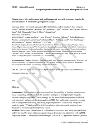
  • 5. CUAJ – Original Research Klotz et al Comparing micro-ultrasound and mpMRI for prostate cancer   5 © 2020 Canadian Urological Association patients. Sites A and I did not differentiate between GG2 and GG3 cases and were not included in assessments of GG3 or higher cancer. Significant variability in biopsy indication was evident between the enrolling centers, however the resulting sensitivity difference of MRI and micro- ultrasound was consistent (Figure 1 and Table 3). Overall, micro-ultrasound demonstrated a sensitivity of 94% (386/411) for predicting GG ≥ 2 PCa while mpMRI demonstrated a lower sensitivity of 90% (371/411) (p=0.03). Specificity was similar at 22% (138/629) for micro- ultrasound and 23% (136/629) for mpMRI (p<0.01 for non-inferiority) (Table 2). Figure 1 shows a Forest plot of sensitivity by site. This was relatively consistent between sites, whether blinded or not. The overall sensitivity of micro-ultrasound was 3.6% higher than MRI, and the p value for non-inferiority was < 0.001, and for superiority 0.03. Figure 2 shows the Forest plot of specificity. There was substantial variation between sites. Micro-Ultrasound specificity was 0.3% higher than MRI with p<0.001 for non-inferiority, and p=0.45 for superiority. All but 3 groups (all with <65 subjects) performed within the non-inferiority margin individually, suggesting robust inter-site performance. A sensitivity analysis was further conducted by removing each of the groups in order to determine whether the overall conclusion still held. P-values for non-inferiority were consistently significant for both sensitivity and specificity (<0.001-0.025 for both). Superiority p-values varied considerably for specificity as expected given the widely varying performances between sites (0.01-0.73), however sensitivity was consistently superior (<0.01 in all cases). Discussion The advent of prostate cancer imaging with mpMRI has dramatically altered the approach to prostate cancer diagnosis. Targeted biopsies result in more significant cancer and less insignificant cancer being diagnosed. However, mpMRI has a number of limitations. It is a relatively expensive technology, there is a significant educational learning curve, and involves a second visit for the fusion targeted biopsy. There is potential toxicity associated with Gadolinium. mpMRI misses about 15% of significant cancers.13,14 Many care management facilities globally face challenges in providing access to MRI for all patients in whom it is indicated. Further, MRI interpretation is complex and subject to variability. In a study of the concordance by expert GU radiologists, the kappa for peripheral zone prostate lesions was only 0.59.15 Fusion targeting introduces another potential source of error, whether performed cognitively or with an additional fusion biopsy system. Some men have a relative or absolute contraindication to MRI, including claustrophobia, prosthetic implants, pacemakers, or renal failure precluding gadolinium.16 Modifications to MRI, including the use of bi-parametric MRI, will address some of these issues, including gadolinium related toxicity and cost.17
  • 6. CUAJ – Original Research Klotz et al Comparing micro-ultrasound and mpMRI for prostate cancer   6 © 2020 Canadian Urological Association High resolution micro-ultrasound offers the benefit of a comparatively inexpensive, simple technology, with imaging and biopsy performed as a single procedure. It does not require contrast. Ultrasound skills are widely diffused in the urological and radiological community, and the learning curve for micro-ultrasound imaging appears to be short. In one study, the AUC flattened after 15 cases.18 The capital cost of the device is similar to other high-end ultrasound machines or MRI/US fusion devices, a fraction of the capital cost of MRI. The footprint is similar to conventional ultrasound. The only patients in whom the procedure is not possible are those with anal stenosis or absence post abdominal perineal resection. The sensitivity of micro-ultrasound in this registry for significant prostate cancer was superior to MRI. The specificity trended non-significantly below that of MRI. The specificity likely is strongly related to the learning curve. Acquiring the confidence to exclude certain borderline abnormalities takes more experience identifying abnormalities. It is likely that with further experience and validation, the specificity will improve. Limitations of the study: This was a ‘real world’ registry in 11 centers in 7 countries of a new technology. Thus, the results incorporate learning curves and significant variability between centers. Data was collected prospectively, but there was not a uniform protocol. Details of the methodologic variation between sites are listed in the supplementary table. Conventional 12 core systematic biopsies were not performed in most patients due to the inclusion of micro- ultrasound targets within the systematic samples and adjustment of systematic positions to reflect tissue variations observed on micro-ultrasound. Thus the performance of micro ultrasound compared to systematic biopsies could not be determined. Variation in the approach to borderline lesions was substantial. For example, site A targeted all borderline lesions comprehensively, and achieved a high sensitivity at the expense of specificity. Site C did not enroll or schedule patients for biopsy unless they had a visible lesion on MRI, potentially missing MRI negative micro-US positive cases. Despite this, the sensitivity at their site was 94% compared to MRI. The number of cores per target was not standardized. Seven of the 11 sites were unblinded to the MRI when the ultrasound was performed, introducing an important source of bias in the interpretation of the ultrasound. However, results between the blinded and unblinded sites was similar. Only men with a prior mpMRI undergoing biopsy were enrolled, and data on patients who were excluded due no prior MRI was not available for comparison. Current studies where the ultrasound annotation is performed blinded to the MRI are ongoing. This was a comparison to mpMRI, without a gold standard of surgical pathology; therefore the known inaccuracies of biopsy grading could not be avoided. The PRI-MUS system used for ultrasound grading has not been validated. Conclusions This registry, the first large scale analysis of the initial multi-centre experience with micro- ultrasound, has demonstrated comparable metrics to mpMRI with respect to sensitivity,
  • 7. CUAJ – Original Research Klotz et al Comparing micro-ultrasound and mpMRI for prostate cancer   7 © 2020 Canadian Urological Association specificity, NPV and PPV. This technology is an appealing alternative to MRI in the initial evaluation of men at risk for prostate cancer. Further studies are warranted to further validate this technology.
  • 8. CUAJ – Original Research Klotz et al Comparing micro-ultrasound and mpMRI for prostate cancer   8 © 2020 Canadian Urological Association References 1. National Cancer Institute. Cancer Stat Facts: Prostate Cancer. https://seer.cancer.gov/statfacts/html/prost.html. Accessed June 1, 2020. 2. Centers for Disease Control and Prevention. U.S. Cancer Statistics Data Visualizations Tool. https://www.cdc.gov/cancer/uscs/dataviz/index.htm. Accessed June 1, 2020. 3. National Comprehensive Cancer Network. Clinical Practice Guidelines in Oncology: Prostate Cancer. 2019;Version 4. https://www.nccn.org/professionals/physician_gls/pdf/prostate.pdf. 4. Rouvière O, Puech P, Renard-Penna R, et al. Use of prostate systematic and targeted biopsy on the basis of multiparametric MRI in biopsy-naive patients (MRI-FIRST): a prospective, multicentre, paired diagnostic study. Lancet Oncol. 2018;2045(18):1-10. doi:10.1016/S1470-2045(18)30569-2 5. Kasivisvanathan V, Rannikko AS, Borghi M, et al. MRI-Targeted or Standard Biopsy for Prostate-Cancer Diagnosis. N Engl J Med. 2018:NEJMoa1801993. doi:10.1056/NEJMoa1801993 6. Ahmed HU, El-Shater Bosaily A, Brown LC, et al. Diagnostic accuracy of multi- parametric MRI and TRUS biopsy in prostate cancer (PROMIS): a paired validating confirmatory study. Lancet. 2017;6736(16):1-8. doi:10.1016/S0140-6736(16)32401-1 7. Mottet N, Cornford P, van den Bergh RCN, et al. EAU Guidelines: Prostate Cancer. In: EAU Annual Congress. Amsterdam: EAU Guidelines Office, Arnhem, The Netherlands.; 2020. 8. NCCN Clinical Practice Guidelines in Oncology V1.2019. Prostate Cancer Early Detection Recommendations. Natl Compr Cancer Network, Inc. 2019. https://www.nccn.org/professionals/physician_gls/pdf/prostate_detection.pdf. 9. Rohrbach D, Wodlinger B, Wen J, Mamou J, Feleppa E. High-Frequency Quantitative Ultrasound for Imaging Prostate Cancer Using a Novel Micro-Ultrasound Scanner. Ultrasound Med Biol. 2018. doi:10.1016/j.ultrasmedbio.2018.02.014 10. Turkbey B, Choyke PL. Pirads 2.0: What is new? Diagnostic Interv Radiol. 2015;21(5):382-384. doi:10.5152/dir.2015.15099 11. Ghai S, Eure G, Fradet V, et al. Assessing Cancer Risk on Novel 29 MHz Micro- Ultrasound Images of the Prostate: Creation of the Micro-Ultrasound Protocol for Prostate Risk Identification. J Urol. 2016;196(2):562-569. doi:10.1016/j.juro.2015.12.093 12. Brown LD, Cai TT, DasGupta A. Interval Estimation for a Binomial Proportion. Stat Sci. 2001;16(2):101-133. doi:10.1214/ss/1009213286 13. Stabile A, Giganti F, Emberton M, Moore CM. MRI in prostate cancer diagnosis: do we need to add standard sampling? A review of the last 5 years. Prostate Cancer Prostatic Dis. 2018;21(4):473-487. doi:10.1038/s41391-018-0071-8 14. Padhani AR, Haider MA, Villers A, Barentsz JO. Multiparametric Magnetic Resonance Imaging for Prostate Cancer Detection : What We See and What We Miss. Eur Urol. 2018:0-1. doi:10.1016/j.eururo.2018.12.004 15. Rosenkrantz AB, Ginocchio LA, Cornfeld D, et al. Interobserver Reproducibility of the
  • 9. CUAJ – Original Research Klotz et al Comparing micro-ultrasound and mpMRI for prostate cancer   9 © 2020 Canadian Urological Association PI-RADS Version 2 Lexicon: A Multicenter Study of Six Experienced Prostate Radiologists. Radiology. 2016;280(3):793-804. doi:10.1148/radiol.2016152542 16. Layne KA, Dargan PI, Archer JRH, Wood DM. Gadolinium deposition and the potential for toxicological sequelae – A literature review of issues surrounding gadolinium‐based contrast agents. Br J Clin Pharmacol. 2018;84(11):2522-2534. doi:10.1111/bcp.13718 17. Porter KK, King A, Galgano SJ, Sherrer RL, Gordetsky JB, Rais-Bahrami S. Financial implications of biparametric prostate MRI. Prostate Cancer Prostatic Dis. 2020;23(1):88-93. doi:10.1038/s41391-019-0158-x 18. Hyndman M, Pavlovich C, Eure G, Fradet V, Ghai S. Prospective validation of PRI- MUSTM , the Prostate Risk Identification using Micro-Ultrasound protocol for real-time detection of prostate cancer using high-resolution micro-ultrasound imaging. In: American Urology Association. San Francisco, USA; 2018.
  • 10. CUAJ – Original Research Klotz et al Comparing micro-ultrasound and mpMRI for prostate cancer   10 © 2020 Canadian Urological Association Figures and Tables Fig 1. Forest plot demonstrating site-level sensitivity difference between micro-ultrasound and multiparametric magnetic resonance imaging. Sensitivity difference overall was +3.6% indicating superior sensitivity for micro-ultrasound (p=0.03). CI: confidence interval. Fig. 2. Forest plot demonstrating site-level specificity difference between micro-ultrasound and multiparametric magnetic resonance imaging. Specificity difference was +0.3% overall, indicating non-inferiority for micro-ultrasound (p<0.01), however, significant variability was noted between sites depending on biopsy population and user targeting habits. CI: confidence interval. Study Overall effect K J I H G F E D C B A N 1040 14 62 47 116 77 51 159 19 292 9 194 Difference (95% CI) 0.28 (-4.37-4.85) -16.13 (-45.82-10.08) 46.80 (30.24-62.10) 16.52 (-4.20-36.51) -3.54 (-18.18-10.96) -7.60 (-23.33-8.47) -13.06 (-34.17-8.54) 8.37 (-0.98-18.24) -51.34 (-81.81--11.38) 11.67 (4.06-19.48) -11.93 (-49.12-23.95) -29.52 (-40.48--18.60) Non-Inferiority (p-value) <0.01 0.01 <0.01 <0.01 <0.01 <0.01 <0.01 <0.01 0.53 <0.01 0.02 <0.01 Superiority (p-value) 0.45 0.90 <0.01 0.06 0.69 0.83 0.89 0.04 0.99 <0.01 0.76 1.00 -100 -50 0 50 100 Favours MRI Favours MicroUS
  • 11. CUAJ – Original Research Klotz et al Comparing micro-ultrasound and mpMRI for prostate cancer   11 © 2020 Canadian Urological Association Table 1. Patient demographics Overall N 1040 Age, median (IQR) 67 (61–72) PSA, median (IQR) 7 (5.1–10) DRE (positive) 208 (128 NA) Prostate volume (mL), median (IQR) 38 (28–53) Prior biopsy (positive/total) 66/352 (281 NA) Percentage equivocal imaging (PRI-MUS 3) 17% Percentage equivocal imaging (PI-RADS 3) 19% DRE: digital rectal exam; IQR: interquartile range; PSA: prostate-specific antigen. Table 2. Performance metrics comparing mpMRI and micro-ultrasound A. For detection of GG ≥2 PCa (39% of cases) Modality Sensitivity Specificity PPV NPV mpMRI 90% (371/411) 22% (136/629) 43% (371/864) 77% (136/176) Micro-ultrasound 94% (386/411) 22% (138/629) 44% (386/877) 85% (138/163) p (non- inferiority) <0.001 <0.001 <0.001 <0.001 p-value (superior) 0.03 0.45 0.32 0.04 B. For GG ≥3 (19% of cases) Modality Sensitivity Specificity PPV NPV mpMRI 94% (145/154) 17% (112/642) 21% (145/675) 93% (112/121) Micro-ultrasound 93% (144-154) 21% (136/642) 22% (144/657) 93% (136/146) p (non-inferiority) <0.001 <0.001 <0.001 <0.001 p (superior) 0.59 0.06 0.43 0.41 C. PPV by PI-RADS and PRI-MUS score 3 4 5 Unknown 4 or 5 MRI PI-RADS 14% 38% 62% 49% 40% U/S PRI-MUS 19% 39% 61% 46% 42% GG: Gleason grade; mpMRI: multiparametric magnetic resonance imaging; NPV: negative predictive value; PPV: positive predictive value; US: ultrasound.
  • 12. CUAJ – Original Research Klotz et al Comparing micro-ultrasound and mpMRI for prostate cancer   12 © 2020 Canadian Urological Association Table 3. Detailed results per site Site Multiparametric MRI Micro-ultrasound n True positive True negative False positive False negative True positive True negative False positive False negative A 70 46 64 14 84 13 97 0 194 B 3 1 5 0 3 0 6 0 9 C 97 22 163 10 97 44 141 10 292 D 9 7 1 2 10 2 6 1 19 E 76 5 76 2 74 12 69 4 159 F 20 9 20 2 22 5 24 0 51 G 24 13 38 2 25 9 42 1 77 H 29 27 53 7 31 24 56 5 116 I 19 3 25 0 17 8 20 2 47 J 21 1 40 0 19 21 20 2 62 K 3 2 8 1 4 0 10 0 14 Total 371 136 493 40 386 138 491 25 1040 While clear site-level variability is seen on targeting percentage and accuracy, only 3 sites (B, I, J) failed to achieve non-inferior sensitivity on their own. Aggregate results demonstrate superior sensitivity for micro-ultrasound (p=0.03) and non-inferior specificity (p<0.01). MRI: magnetic resonance imaging. Supplementary Table 1. Summary of registry sites with methodological variations Site identifier Clinic name Location Indication for Biopsy mpMRI specifications mpMRI targeting system Blinded or unblinded MRI A Urología Clínica, Clínica IMQ Zorrotzaurre Bilbao, Spain Clinical variables, including MRI b-value ≥1400 no ERC Cognitive fusion (micro-US- guided) Unblinded B Urology of Virginia, Eastern Virginia Medical School Virginia Beach, U.S. Clinical variables, including MRI 3T Toshiba Titan no ERC b-value 2000 Cognitive fusion (micro-US- guided) Blinded C Instituto Clinico Humanitas Rozzano, Italy Suspicious mpMRI only 1.5T and 3T Biojet robotic fusion Blinded
  • 13. CUAJ – Original Research Klotz et al Comparing micro-ultrasound and mpMRI for prostate cancer   13 © 2020 Canadian Urological Association D Glickman Urological Institute, Cleveland Clinic Cleveland, U.S. Clinical variables, including MRI 3T Siemens Skyra no ERC Uronav software fusion Unblinded E Charité Universitätsme dizin Berlin, Germany Clinical variables, including MRI 3T with pelvic phased array coil no ERC Hitachi software fusion Unblinded F Groupe Urologie Saint-Augustin Bordeaux, France Clinical variables, including MRI 1.5T and 3T Cognitive fusion (micro-US- guided) Unblinded G Sunnybrook Hospital Toronto, Canada Clinical variables, including MRI Siemens and Phillips 3T, no ERC Cognitive fusion (micro-US- guided) Unblinded H Polyclinique Reims- Bezannes Bezannes, France Clinical variables, including MRI 1.5T and 3T Cognitive fusion (micro-US- guided) Unblinded I Institut Mutualiste Montsouris Paris, France Clinical variables, including MRI 3T no ERC Cognitive fusion (micro-US- guided) Unblinded J Ordenskliniku m Linz, Austria Clinical variables, including MRI 1.5T and 3T FusionVu micro-US- guided Unblinded K Carolina Urologic Research Center Myrtle Beach, U.S. Clinical variables, including MRI 1.5T and 3T Cognitive fusion (micro-US- guided) Unblinded MRI: magnetic resonance imaging; US: ultrasound.