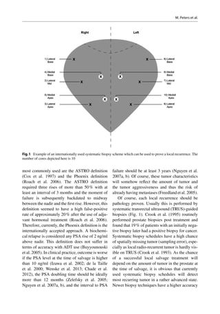
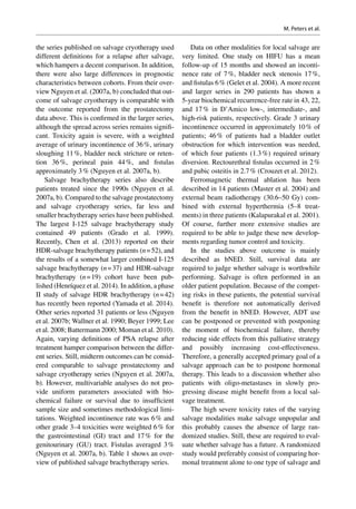
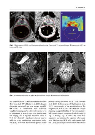
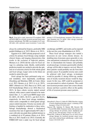
SlideShare a Scribd company logo
1 of 18
Download to read offline
Med Radiol Diagn Imaging (2016)
DOI 10.1007/174_2016_56, © Springer International Publishing Switzerland
Prostate Cancer
Max Peters, Metha Maenhout, Steven Frank,
and Marco van Vulpen
Abstract
Salvage radiotherapy for locally recurrent
prostate cancer after primary radiation is gen-
erally performed using brachytherapy. Only a
limited amount of small studies has been per-
formed so far. In these studies the rate of
severe toxicity, requiring operative reinterven-
tion, was high and cancer control outcome
was disappointing. Furthermore, it is unclear
whether salvage treatment will improve dis-
ease-specific or overall survival. For these rea-
sons, salvage brachytherapy is not popular and
usually only performed in large tertiary cen-
ters. Salvage can currently be considered in
patients with a pathology-proven local recur-
rence with an interval of at least 2–3 years
after primary treatment, together with a lim-
ited and nonaggressive tumor presentation at
time of salvage. Currently, experienced groups
recommend at least equal doses used in pri-
mary treatment, together with targeting the
entire prostate. Diagnostic developments in
magnetic resonance imaging (MRI) and posi-
tron emission tomography (PET) and biopsy
techniques such as transperineal and MRI-
targeted biopsies provide the possibility to
localize the macroscopic recurrent tumor in
the prostate. This enables a shift to focal sal-
vage techniques which can be expected to
reduce severe toxicity rates while maintaining
cancer control.
M. Peters (*) • M. Maenhout • S. Frank
M. van Vulpen
Department of Radiotherapy, University Medical
Center Utrecht, Heidelberglaan 100,
Utrecht 3584CX, The Netherlands
e-mail: M.Peters-10@umcutrecht.nl;
M.Maenhout@umcutrecht.nl; sjfrank@mdanderson.org;
M.vanvulpen@umcutrecht.nl
Contents
1 Introduction.......................................................	 000
2 Detecting a Patient with a Local
Recurrence Only...............................................	 000
3 General Results of Prostate
Cancer Salvage..................................................	000
4  Re-irradiation of Prostate Cancer...................	000
5 Focal Salvage, the Near Future........................	 000
References................................................................	000
1	 Introduction
Re-irradiation in prostate cancer after primary
curative radiotherapy or prostatectomy is gener-
ally called a salvage treatment and will further
be referred to as salvage in this entire chapter.
In addition, salvage here refers to the post-­
radiotherapy setting only. Salvage radiotherapy
post prostatectomy falls outside the scope of this
chapter.
The treatment of patients suffering from radio-­
recurrent prostate cancer is a significant clinical
problem worldwide (Ward et al. 2008). It has
been calculated that in the United States approxi-
mately 31,680 men per year will be at risk for
failure after primary radiotherapy (Ward et al.
2008). Owing to the rising incidence of prostate
cancer and the increasing use of radiotherapy, this
number is likely to increase in the future (Dutch
cancer society 2010). Other sources suggest that
up to 60 % of patients undergoing radiotherapy
as a primary treatment option may experience
a recurrence within 10 years after treatment
(Brachman et al. 2000; Agarwal et al. 2008;
Zelefsky et  al. 2007; Heidenreich et  al. 2008).
Even in the era of dose escalation (≥78 Gy) for
primary prostate cancer, biochemical recur-
rences occur in approximately 10 % of low-risk,
23 % of intermediate-risk, and 44 % of high-risk
patients after 8 years (Zumsteg et al. 2015). Even
though, following primary treatment, it is dif-
ficult to differentiate between locally recurrent
disease and distant metastases when the prostate-
specific antigen (PSA) level increases, many of
these patients will harbor organ-confined disease
(Pound et al. 2001). Some expect even over two-
thirds of patients to have locally recurrent disease
(Menard et al. 2015; Cellini et al. 2002; Pucar
et al. 2008; Arrayeh et al. 2012). Others state
most men with a rising PSA after treatment will
harbor micrometastatic disease and only a minor-
ity would have a true local recurrence only and
could potentially benefit from a salvage treat-
ment (Ward et al. 2005; Nguyen et al. 2007a, b;
Huang et al. 2007; Leibovici et al. 2012).
For patients diagnosed with recurrent pros-
tate cancer after primary radiotherapy, different
salvage treatment methods with a curative intend
exist: salvage radical prostatectomy, salvage
brachytherapy, salvage external beam radiother-
apy, salvage high-intensity focused ultrasound
(HIFU), and salvage cryosurgery (Nguyen et al.
2007a, b; Moman et al. 2009; Alongi et al. 2013;
Peters et al. 2013). These procedures are associ-
ated with high failure and high severe toxicity
rates and are therefore unpopular (Moman et al.
2009; Peters et al. 2013). This probably explains
the absence of large prospective trials in the lit-
erature. Most data are derived from retrospec-
tive evaluation of a limited number of patients,
which are often heterogeneous before salvage
treatment regarding prognostic characteris-
tics. The only palliative treatment alternative
is hormonal therapy or androgen deprivation
therapy (ADT), which is associated with often
significant cardiovascular, metabolic, and even
mental side effects (Heidenreich et al. 2014;
Nguyen et al. 2015). In the case of a biochemi-
cal recurrence after radiotherapy for prostate
cancer, hormonal therapy is the worldwide most
applied treatment, with approximately 98 % of
patients being treated in this manner (Moman
et al. 2009; Tran et al. 2014).
The high failure rates after prostate cancer sal-
vage are probably related to inaccurate patient
selection, as many of these patients will have
early distant metastases (Haider et al. 2008;
Nguyen et al. 2007a, b). The high severe toxicity
rate might be related to the fact that current sal-
vage treatments are directed at the entire prostatic
volume. This is required because of a lack of
accurate localization possibilities and blind sys-
tematic biopsies to detect a local recurrence.
Currently, new MRI and PET-imaging modalities
are adopted, which provide more accurate local-
ization information (Barentsz et al. 2012; Umbehr
et al. 2013; Fütterer et al 2015; Hamoen et al.
2015; Evangelista et al. 2013; de Rooij et al.
2015). In addition, template prostate mapping
biopsies and MRI-targeted biopsies provide addi-
tional information regarding location and the
presence of clinically significant disease
(Siddiqui et al. 2015; Valerio et al. 2015; Moore
et al. 2013). These modalities will be used in the
near future for focal salvage treatment planning
of prostate cancer recurrences (Moman et al.
M. Peters et al.
2010; Peters et al. 2014), which can be expected
to have limited severe toxicity rates.
This chapter will first discuss the possibilities
and difficulties with regard to detecting a local
recurrence and will provide current internationally
used selection criteria to identify patients for sal-
vage. Next, the results of prostate cancer salvage
will be discussed when comparing different sal-
vage methods. Further, technical details of current
radiotherapy salvage techniques will be described,
followed by new developments, most importantly
focal salvage.
2	 Detecting a Patient
with a Local Recurrence Only
As prostate cancer salvage has a high risk of
treatment failure, it is essential to perform proper
patient selection before treatment. Only patients
with a true local recurrence will be able to benefit
from any salvage treatment. Each detection tool
has several specific pitfalls in prostate cancer
recurrence assessment, and these will be dis-
cussed below. This will result in a list of interna-
tionally accepted patient selection criteria for
prostate cancer salvage.
PSA is currently used for follow-up of pros-
tate cancer treatment. The independent prognos-
tic value of PSA pre-primary treatment has been
well established. Pre-salvage, the PSA value also
seems to be a prognostic factor in many studies
using varying modalities (Chade et al. 2012;
Wenske et al. 2013; Murat et al. 2009). However,
the definition of a normal PSA value after pri-
mary treatment remains problematic and differs
between different primary treatment modalities.
The lowest PSA value after treatment is called
the nadir value. Different from the situation after
radical prostatectomy, patients following radio-
therapy will still have a prostate gland and there-
fore are not expected to achieve an undetectable
nadir. Still, post-radiation PSA values are lower
compared to the pretreatment values as irradiated
prostates show atrophy and a reduction of size
and gland tissue (Grignon and Hammond 1995).
There are potentially three sources of PSA which
contribute to the nadir: (1) remaining normal
prostate gland tissue, (2) remaining local prostate
cancer cells, and (3) subclinical metastases. Of
course, serum PSA measurements cannot differ-
entiate between any of these three PSA sources.
Therefore, changes in PSA over time are evalu-
ated. The longer the time to nadir, the more likely
it is that remaining benign prostate tissue is the
source of the residual PSA (Huang et al. 2011).
Furthermore, PSA from benign residual prostate
tissue will not show a clear rise. Early distant
metastases will also produce PSA and will even-
tually “overgrow” the PSA decline after the local
treatment, mostly in the first few years after treat-
ment. Also, these patients usually will have a
higher nadir PSA (Zietman et al. 1996). A slow
PSA rise many years after treatment will proba-
bly be due to a local recurrence as these prostate
cancer cells first would have to recover the dam-
age from the prostate cancer treatment (Zumsteg
et al. 2015). A PSA rise after treatment can fur-
ther be attributed to PSA from cancer cells or a
PSA bounce. This last phenomenon is a tempo-
rary rise of PSA followed by a PSA decline back
to nadir levels and is not associated with cancer
progression. Bounces may occur in up to 40 % of
patients (Akyol et al. 2005; Roach et al. 2006).
To date, no conclusive evidence-based explana-
tion for the bouncing behavior of PSA exists.
Precipitating factors such as ejaculation or instru-
mentation are known to cause some PSA fluctua-
tion (Das et al. 2002). Also the physiological
variability of PSA assays in healthy test subjects
was found to be considerable (Prestigiacomo and
Stamey 1996). Lastly, PSA bounces after therapy
have been associated with an increase in bio-
chemical recurrence-­free, prostate cancer-spe-
cific, and overall survival after brachytherapy
using multivariable analyses (Hinnen et al. 2012).
It is hypothesized that a delayed wave of cell
destruction due to radiation effects is responsible
for the bounce phenomenon and therefore possi-
bly leads to an increase in survival. A small frac-
tion of bounces may be caused by these factors;
however, they fail to provide a complete explana-
tion. In clinical practice a PSA rise, therefore,
needs differentiation between PSA bounce and
cancer progression. For this reason different
guidelines have been developed of which the
Prostate Cancer
most commonly used are the ASTRO definition
(Cox et al. 1997) and the Phoenix definition
(Roach et al. 2006). The ASTRO definition
required three rises of more than 50 % with at
least an interval of 3 months and the moment of
failure is subsequently backdated to midway
between the nadir and the first rise. However, this
definition seemed to have a high false-positive
rate of approximately 20 % after the use of adju-
vant hormonal treatment (Roach et al. 2006).
Therefore, currently, the Phoenix definition is the
internationally accepted approach. A biochemi-
cal relapse is considered any PSA rise of 2 ng/ml
above nadir. This definition does not suffer in
terms of accuracy with ADT use (Buyyounouski
et al. 2005). In clinical practice, outcome is worse
if the PSA level at the time of salvage is higher
than 10 ng/ml (Izawa et al. 2002; de la Taille
et  al. 2000; Wenske et  al. 2013; Chade et  al.
2012), the PSA doubling time should be ideally
more than 12 months (Zelefsky et  al. 2005;
Nguyen et al. 2007a, b), and the interval to PSA
failure should be at least 3 years (Nguyen et al.
2007a, b). Of course, these tumor characteristics
will somehow reflect the amount of tumor and
the tumor aggressiveness and thus the risk of
already having metastases (Freedland et al. 2005).
Of course, each local recurrence should be
pathology proven. Usually this is performed by
systematic transrectal ultrasound (TRUS)-guided
biopsies (Fig.  1). Crook et  al. (1995) routinely
performed prostate biopsies post treatment and
found that 19 % of patients with an initially nega-
tive biopsy later had a positive biopsy for cancer.
Systematic biopsy schedules have a high chance
of spatially missing tumor (sampling error), espe-
cially as local radio-recurrent tumor is hardly vis-
ible on TRUS (Crook et al. 1993). As the chance
of a successful local salvage treatment will
depend on the amount of tumor in the prostate at
the time of salvage, it is obvious that currently
used systematic biopsy schedules will detect
most recurring tumor in a rather advanced state.
Newer biopsy techniques have a higher accuracy
Fig.1  Example of an internationally used systematic biopsy scheme which can be used to prove a local recurrence. The
number of cores depicted here is 10
M. Peters et al.
in detecting localized recurrences and further-
more in assessing clinically significant disease.
Transperineal template prostate mapping (TTPM)
biopsies are taken with a brachytherapy grid,
usually with 5 mm spacing. With TTPM, a large
part of the prostate is sampled, usually subdi-
vided in 24 Barzell zones (Barzell and Whitmore
2003). This technique leads to the detection of
approximately 30 % more clinically significant
tumors over systematic transrectal biopsies alone
(Onik et al. 2009; Sivaraman et al. 2015). This is
mainly due to the higher amount of cores taken
but also because the anterior part of the prostate
is sampled, which is undersampled with transrec-
tal biopsies. A limitation of TTPM is the detec-
tion of more clinically insignificant tumors.
However, these lesions are often eligible for
active surveillance, which shows excellent results
regarding (cancer-specific) survival (Klotz et al.
2010, 2015). In addition, MRI-guided biopsies to
a suspicious lesion can lead to an increase in
detecting clinically significant disease while not
increasing or even decreasing the detection rate
of insignificant lesions (Siddiqui et al. 2015;
Moore et al. 2013).
Next, the evaluation of the biopsies is difficult.
In early reports, little was known about the rate of
histological clearance of irradiated tumor, and fail-
ures were determined on biopsies even 6 months
after radiotherapy (Scardino 1983). More recent
analyses have shown that early biopsies have a high
chance of being false positive. Radiation causes
postmitotic cell death. Therefore, fatally damaged
cells may even survive a limited number of cell
divisions before dying off (Mostofi et al. 1992). As
there is no visible difference between these dying
cells and viable tumor cells, it is impossible for a
pathologist to predict whether visible tumor cells
are really clonogenic and need treatment (Crook
et al. 1997). Approximately 30 % of biopsies posi-
tive in the first year after radiation treatment will
be negative for tumor in 24–30 months (Crook
et al. 1995, 2000). Crook et al. (1995) therefore
determined that the optimal time to biopsy would
be 30–36 months post radiotherapy. Of course,
even 36 months after treatment biopsies may be
false positive, although chances are reduced at that
moment. As postradiation pathology is ­difficult to
interpret, experienced uropathologists are required
to evaluate these biopsies. An extra difficulty is
the interpretation of biopsies. The currently used
Gleason system is based on the glandular pattern
of the tumor seen at relatively low magnification.
As an irradiated prostate will show a significantly
changed anatomy, especially on glandular level,
Gleason score seems less useful after radiation
treatment. Still, to evaluate the possibilities for
salvage, it will be required to have some informa-
tion on the potential aggressiveness of the visible
tumor cells. Pathologists will need to be cautious
here as one of the problems with evaluating car-
cinomas that have been treated with radiotherapy
is that the grade often appears higher (Bostwick
et al. 1982). Although post-RT prostate biopsies
are burdened with problems of timing, interpre-
tation, and sampling error, they are required as
evidence before radical local salvage for radiation
failure is considered.
Imaging to visualize the local recurrence is
regularlyperformedusingTRUS. Unfortunately,
sensitivity is very poor for the primary setting
(Crook et al. 1993; Onur et al. 2004). In radio-­
recurrent disease, accuracy may even be further
decreased due to fibrosis of the prostatic tissue.
Therefore, current clinical practice lacks a
proper imaging tool to detect a local recurrence.
Promising local imaging techniques still under
development are dynamic contrast-enhanced
magnetic resonance imaging (DCE-MRI) and
18
F or 11
C-choline positron emission tomogra-
phy (PET) (Rouviere et al. 2004; Haider et al.
2008; Moman et  al. 2010; Wang et  al. 2009;
Breeuwsma et al. 2010; Barentsz et al. 2012; de
Rooij et al. 2015; Umbehr et al. 2013). More
recent developments have provided prostate-­
specific membrane antigen (PSMA) PET-CT
using various tracers (Jadvar 2015; Rybalov
et al. 2014). However larger series evaluating
these techniques and assessment with the
pathology reference standard is needed. Recent
diagnostic meta-analyses have shown that the
use of MRI (using the prostate imaging report-
ing and data system (PI-RADS)) can help in
assessing localized disease, capsular extension,
and the exclusion of clinically significant dis-
ease with a negative predictive value up to 95 %
Prostate Cancer
(Hamoen et al. 2015; Fütterer et al. 2015; de
Rooij et al. 2015).
Imaging to exclude possible distant metasta-
ses should be performed when evaluating the
possibilities for salvage. But as a bone scan, pel-
vic computed tomography (CT), and MRI need a
significant tumor load to show metastases
(Hovels et al. 2008; Abuzallouf et al. 2004), it is
unlikely they will detect small metastases present
in patients being selected for salvage treatment,
e.g., with a PSA less than 10 ng/ml (Zagars and
Pollack 1997; Nguyen et al. 2007a, b). Next, a
pelvic lymph node dissection may be performed
to exclude metastases. As a limited pelvic node
dissection of the obturator fossa will only detect
approximately 30 % of lymph node metastases,
the value of a lymph node dissection is ques-
tioned (Heesakkers et al. 2008). For early metas-
tases detection in the future, the role of choline
PET with various tracers (Breeuwsma et al. 2010)
and magnetic nanoparticles in MRI (Barentsz
et al. 2007) needs to be further evaluated. PET-CT
imaging seems to be able to provide more accu-
rate assessment of metastatic disease and assess-
ment of prostate-confined recurrences. Recent
overviews have shown promising results regard-
ing lymph node involvement, prostate-confined
recurrences, and distant metastatic disease
(pooled sensitivity and specificity of 85.6 and
92.6 % for all three sites) (Evangelista et  al.
2013). However, the spread across series is still
large and standardization is necessary to achieve
this high diagnostic accuracy in every center.
Of course, clinical patient characteristics
before primary treatment and salvage are of
importance when evaluating the possibility of
successful salvage. Patients with a high initial
chance of developing distant metastases are
unlikely to have a local recurrence only (Nguyen
et al. 2007a, b). An initial high Gleason score,
high T-stage, high PSA value, and high PSA
kinetics (before primary treatment) are possibly
also risk factors post treatment to evaluate the
likelihood of effective salvage. Therefore, sal-
vage especially is advocated in primary low-risk
clinical features (Nguyen et  al. 2007a, b).
However, evidence in this area from multivari-
able models is generally lacking and pre-salvage
characteristics seem to have the highest predic-
tive ability for cancer control outcomes (Chade
et al. 2011, 2012; Wenske et al. 2013; Murat et al.
2009). From these larger salvage series, it seems
pre-­salvage PSA, PSADT, and pre-salvage
Gleason score are most often associated with
cancer control outcomes.
Next, the chance of having toxicity from a
local salvage treatment will be increased if a
patient had severe toxicity during or after pri-
mary radiation treatment. Also, other additional
treatments may attribute to the possible toxicity
from salvage, like a transurethral resection
(TURP), high-intensity focussed ultrasound
(HIFU) treatment, or ADT use. These treatments
will produce extra scar tissue and, therefore, will
reduce the repair capacity of normal tissue in
case of re-irradiation of the prostate. So far, how-
ever, no current salvage re-irradiation literature
exists, which models these characteristics in a
multivariable manner to the probability of devel-
oping toxicity.
Based on the above, a list of preliminary selec-
tion criteria for local salvage can be completed.
Still, it is of major importance to use such a list
reasonably, as many clinical aspects, like age,
medical history, and performance score contrib-
ute in the decision for a local salvage treatment.
The list below partially uses the criteria as
described in the currently active radiation therapy
oncology group (RTOG) trial 0526 (principle
investigator: J. Crook, MD). These selection cri-
teria were also mostly verified in a recent interna-
tional collaboration on selection for salvage
treatment (van den Bos et al. 2015).
•	 Biopsy-proven local recurrence at least 3
years after primary radiation treatment
•	 Posttreatment PSA 10 ng/ml, PSA doubling
time (PSADT) 12 months
•	 Bone scan and CT abdomen or lymph node
dissection without evidence of metastases
•	 Primary tumor characteristics preferably low
to intermediate risk
•	 Acceptable toxicity of primary radiation
treatment
•	 No other prostate treatments performed (like
TURP, HIFU, etc.)
M. Peters et al.
•	 Additional to RTOG: verification of tumor
localization with multiparametric MRI (con-
sisting at least of a 1.5 Tesla T2-weighted,
dynamic contrast-enhanced, and diffusion-
weighted imaging sequence). Exclusion of
metastatic disease using PET-CT (18
F or 11C
choline PET or Ga-68-PSMA)
3	 General Results of Prostate
Cancer Salvage
Different salvage treatment modalities are cur-
rently clinically practiced. Radical prostatec-
tomy, external beam radiotherapy, cryosurgery,
additional I125/Pd103 or Ir192 brachytherapy,
and HIFU are used (Pisters et  al. 2000; Lee
et al. 2008; Grado et al. 1999; Murat et al. 2009;
van der Poel et al. 2007; Nguyen et al. 2007a,
b; Chade et al. 2011, 2012, Paparel et al. 2009
Wenske et al. 2013, Williams et al. 2011, Spiess
et al. 2010, Chen et al. 2013, Peters et al. 2013,
Burri et al. 2010; Henríquez et al. 2014; Yamada
et al. 2014). Literature is scarce and shows dis-
cordant results in terms of treatment effect and
toxicity. Furthermore, few large series have
been published; most studies are retrospective
and contain less than 100 patients. No head-to-
head comparisons have been performed. As a
consequence, current treatment decisions often
depend on patients’ and doctors’/institutional
preferences. An overview of the literature is pre-
sented below.
Salvage radical prostatectomy has been
described in several articles and has been per-
formed in these series since the 1960s. Five-year
biochemical no evidence of disease (bNED)
ranges from 50 to 60 % (Nguyen et  al. 2007a,
b, Chade et al. 2012). In most series pre-salvage
PSA was 10 ng/ml. Only a few rather large
series have been described. Chade et al. (2011)
described the largest salvage radical prostatec-
tomy series with 404 radio-recurrent prostate
cancer patients, treated since 1985. Ward et al.
(2005) described the results of 199 patients,
treated since 1967. In addition, Paparel et  al.
(2009) described 146 patients from a single
institution. Finally, Stephenson et al. (2004)
described results of 100 patients (Stephenson
et al. 2004). The other published studies con-
tain between 6 and 51 patients (Sanderson et al.
2006; van der Poel et  al. 2007; Vaidya and
Soloway 2000). The definition of bNED after
prostatectomy was most commonly defined as
a rise in PSA of 0.2 ng/ml. It was concluded
that patients with a pre-salvage PSA of 10 ng/
ml did significantly better compared to patients
with a PSA 10 ng/ml (Ward et al. 2005; Bianco
et al. 2005). In addition, a lower pre-salvage
Gleason score was shown to improve biochemi-
cal disease-­free survival and reduced the devel-
opment of metastases in multivariable analysis
(Chade et al. 2011). Multivariable analysis was
not performed for mortality in this largest
series. However, these factors were associated
with prostate cancer-­specific survival after mul-
tivariable analysis in the series of Paparel et al.
(2009). Neoadjuvant hormonal therapy did not
seem to improve outcome (Ward et al. 2005;
van der Poel et  al. 2007). Also a cystoprosta-
tectomy did not seem to improve oncologic out-
come compared to a radical prostatectomy alone
(Ward et al. 2005). Although preoperative mor-
bidity, lasting from the previous radiotherapy
treatment, was low, urinary incontinence had a
weighted average of 41 %, bladder neck stric-
tures of 24 %, and rectal injury of approximately
5 % (Nguyen et al. 2007a, b).
Salvage cryotherapy studies have been
described with patients treated since the 1990s
(Nguyen et al. 2007a, b). Five larger series have
been published. The largest series describes 797
patients from 6 tertiary centers, of the basis of
which a pretreatment nomogram to predict bio-
chemical recurrence was created (Spiess et al.
2010). Wenske et  al. (2013) described 328
patients after either primary EBRT (n = 259), pri-
mary I-125 brachytherapy (n = 49), or primary
cryotherapy (n = 20). Furthermore, Williams
et al. described 187 patients, Izawa et al. (2002)
showed outcomes of 131 patients, and Chin et al.
(2001) described results of 125 patients. Other
studies contained 59 patients or less (Bahn et al.
2003). Most series used a double freeze-thaw
therapy (Izawa et al. 2002; Chin et al. 2001; Bahn
et al. 2003, Williams et al. 2011). Unfortunately,
Prostate Cancer
the series published on salvage cryotherapy used
different definitions for a relapse after salvage,
which hampers a decent comparison. In addition,
there were also large differences in prognostic
characteristics between cohorts. From their over-
view Nguyen et al. (2007a, b) concluded that out-
come of salvage cryotherapy is comparable with
the outcome reported from the prostatectomy
data above. This is confirmed in the larger series,
although the spread across series remains signifi-
cant. Toxicity again is severe, with a weighted
average of urinary incontinence of 36 %, urinary
sloughing 11 %, bladder neck stricture or reten-
tion 36 %, perineal pain 44 %, and fistulas
approximately 3 % (Nguyen et al. 2007a, b).
Salvage brachytherapy series also describe
patients treated since the 1990s (Nguyen et  al.
2007a, b). Compared to the salvage prostatectomy
and salvage cryotherapy series, far less and
smaller brachytherapy series have been published.
The largest I-125 salvage brachytherapy study
contained 49 patients (Grado et  al. 1999).
Recently, Chen et al. (2013) reported on their
HDR-salvage brachytherapy patients (n = 52), and
the results of a somewhat larger combined I-125
salvage brachytherapy (n = 37) and HDR-salvage
brachytherapy (n = 19) cohort have been pub-
lished (Henríquez et al. 2014). In addition, a phase
II study of salvage HDR brachytherapy (n = 42)
has recently been reported (Yamada et al. 2014).
Other series reported 31 patients or less (Nguyen
et al. 2007b; Wallner et al. 1990; Beyer 1999; Lee
et al. 2008; Battermann 2000; Moman et al. 2010).
Again, varying definitions of PSA relapse after
treatment hamper comparison between the differ-
ent series. Still, midterm outcomes can be consid-
ered comparable to salvage prostatectomy and
salvage cryotherapy series (Nguyen et al. 2007a,
b). However, multivariable analyses do not pro-
vide uniform parameters associated with bio-
chemical failure or survival due to insufficient
sample size and sometimes methodological limi-
tations. Weighted incontinence rate was 6 % and
other grade 3–4 toxicities were weighted 6 % for
the gastrointestinal (GI) tract and 17 % for the
genitourinary (GU) tract. Fistulas averaged 3 %
(Nguyen et al. 2007a, b). Table 1 shows an over-
view of published salvage brachytherapy series.
Data on other modalities for local salvage are
very limited. One study on HIFU has a mean
follow-up of 15 months and showed an inconti-
nence rate of 7 %, bladder neck stenosis 17 %,
and fistulas 6 % (Gelet et al. 2004). A more recent
and larger series in 290 patients has shown a
5-year biochemical recurrence-free rate in 43, 22,
and 17 % in D’Amico low-, intermediate-, and
high-risk patients, respectively. Grade 3 urinary
incontinence occurred in approximately 10 % of
patients; 46 % of patients had a bladder outlet
obstruction for which intervention was needed,
of which four patients (1.3 %) required urinary
diversion. Rectourethral fistulas occurred in 2 %
and pubic osteitis in 2.7 % (Crouzet et al. 2012).
Ferromagnetic thermal ablation has been
described in 14 patients (Master et al. 2004) and
external beam radiotherapy (30.6–50 Gy) com-
bined with external hyperthermia (5–8 treat-
ments) in three patients (Kalapurakal et al. 2001).
Of course, further more extensive studies are
required to be able to judge these new develop-
ments regarding tumor control and toxicity.
In the studies above outcome is mainly
described as bNED. Still, survival data are
required to judge whether salvage is worthwhile
performing. Salvage is often performed in an
older patient population. Because of the compet-
ing risks in these patients, the potential survival
benefit is therefore not automatically derived
from the benefit in bNED. However, ADT use
can be postponed or prevented with postponing
the moment of biochemical failure, thereby
reducing side effects from this palliative strategy
and possibly increasing cost-effectiveness.
Therefore, a generally accepted primary goal of a
salvage approach can be to postpone hormonal
therapy. This leads to a discussion whether also
patients with oligo-metastases in slowly pro-
gressing disease might benefit from a local sal-
vage treatment.
The high severe toxicity rates of the varying
salvage modalities make salvage unpopular and
this probably causes the absence of large ran-
domized studies. Still, these are required to eval-
uate whether salvage has a future. A randomized
study would preferably consist of comparing hor-
monal treatment alone to one type of salvage and
M. Peters et al.
Table1 Overviewofclinicaloutcomeandseveretoxicityofpublishedseriesonsalvagebrachytherapy
ReferenceiPSA(ng/ml)NHDR/seedsHT(%)FU(months)
bNED,Kaplan-­Meier
estimates%GI ≥ grade3%GU ≥ grade3
Yamadaet al.(2014)Median3.542HDR+(43)Median3669 %(5-year)08
Henríquez(2014)Median3.756HDR(n = 19),
seeds(n = 37)
+(27)Median4877 %(5-year)a,b
423
Chenet al.(2013)Median9.352HDR+(46)Median6051 %(5-year)a
02
Burriet al.(2010)Median5.637Seeds+(84)Median8654 %(10-year)38
Momanet al.(2010)Mean11.431Seeds+(16)Mean11020 %(5-year)a
06
Leeet al.(2008)Median3.821SeedsNAMedian3638 %(5-year)b
00
Nguyenet al.(2007a,b)Median7.525Seeds−Median4770 %(4-year)a
2416
Leeet al.(2007)Median5.921HDR+(52)Median1989 %(2-year)b
014
Wonget al.(2006)Median4.717Seeds+(88)Median4475 %(4-year)b
647
Loet al.(2005)NA30Seeds−Median5957 %(5-year)b
310
Koutrouveliset al.(2003)NA31Seeds+Median3087 %(3-year)b
513
Gradoet al.(1999)Median5.649Seeds+(16)Median6434 %(5-year)c
420
Beyer(1999)Median2.217Seeds+(47)Median6253 %(5-year)b
024
LoeningandTurner(1993)NA31Seeds−Median2367 %(2-year)c
00
DifferentPSAfailuredefinitionshavebeenused
Nnumberofpatients,HThormonaltherapyneoadjuvantlybeforesalvage;no,−;yes,+,%.bNEDpercentagebiochemicalnoevidenceofdiseaseincludingyearofmeasurement,
NAnotavailable,iPSAinitialPSA,HDRhighdoserate(brachytherapy),FUfollow-up,GIgastrointestinal,GUgenitourinary
a
Phoenix(nadir + 2 ng/ml)
b
ASTRO(threesuccessivePSArises50 %)
c
Otherdefinitions
Prostate Cancer
would have to prove a gain in survival. Next to
toxicity scoring, quality of life will be essential to
monitor (Nguyen et al. 2009) to judge the actual
influence of severe toxicities and compare the
difference between ADT use and salvage therapy.
4	 Re-irradiation of Prostate
Cancer
Post-radiotherapy local salvage is historically per-
formed by brachytherapy (LDR, PDR, or HDR)
or EBRT (Kimura et al. 2010). There is a prefer-
ence for brachytherapy, as it can be expected that
the surrounding area which receives a relatively
high dose will be larger after EBRT, and there-
fore EBRT can be expected to cause more severe
toxicity. This explains why there is hardly any lit-
erature on EBRT for recurrent local disease after
primary radiotherapy. In a national disease regis-
try, the Cancer of the Prostate Strategic Urologic
Research Endeavor (CaPSURE), 935 men were
described who received a salvage treatment after
primary EBRT. Of these men only eight received
salvage by EBRT (Agarwal et al. 2008). In their
article, Agarwal et al. (2008) did not notice any
survival benefit for any particular combination of
primary and salvage therapy. The inability to pro-
tectthebladderandtheanteriorrectalwallwithout
blocking a part of the prostate/tumor was largely
caused by the limitations of the EBRT technique.
Current EBRT techniques have improved enor-
mously, e.g., by daily position verification tech-
niques (van der Heide et  al. 2007) and by the
introduction of intensity-modulated radiotherapy
(IMRT). Passive-scattering proton therapy has
recently been associated with a potential increase
in GI toxicity in a large propensity score-matched
analysis (Sheets et al. 2012). Advanced forms
of proton therapy delivery with intensity-mod-
ulated proton therapy (IMPT) may provide bet-
ter salvage outcome and toxicity data, but IMPT
will need IGRT and daily position verification.
Although most articles and textbooks discourage
the use of EBRT for salvage after primary radio-
therapy, these new techniques indeed might lead
to comparable results as described above. But as
the current standard salvage ­techniques already
show hardly acceptable severe toxicity rates,
the further evaluation of EBRT for this purpose
seems not the way forward.
In salvage brachytherapy permanent seeds are
commonly used. Both I125 and Pd103 have been
described (Wallner et  al. 1990; Beyer 1999;
Grado et al. 1999; Koutrouvelis et al. 2003; Lee
et al. 2008; Battermann 2000; Nguyen et al.
2007b; Moman et al. 2010). More recently, HDR
brachytherapy is being adopted in the salvage
setting (Chen et al. 2013; Henríquez et al. 2014;
Yamada et al. 2014). Again, comparison in out-
come is hampered by differences in study popu-
lations, treatment methods, and the definition of
failure (Nguyen et al. 2007a, b). In patients who
meet the criteria described in part 2, it can be
expected to achieve a 5-year bNED rate of
approximately 50–70 % (Table  1). Five-year
overall survival rates ranging from 80 to 90 %
have been described (Beyer 1999; Lee et  al.
2008). Still, the severe toxicity rate is high, up to
approximately 10–30 % for GU and GI combined
(Moman et al. 2010; Nguyen et al. 2007b, Peters
et al. 2013). Currently, an RTOG trial (no. 0526)
is ongoing for the prospective evaluation of
brachytherapy after EBRT. The results of this
trial can be expected to give more insight in the
toxicity risk profiles of patients receiving salvage
brachytherapy. This can improve patient selec-
tion and may augment the benefit/risk ratio
(Nguyen et al. 2007a, b; Moman et al. 2010).
As all studies showed a rather large proportion
of patients with severe toxicity, specific dose con-
straints for salvage are essential. Many different
doses have been applied, but most groups used
regular doses, which are also applied for a pri-
mary treatment, e.g., 120–145  Gy in I125
implants and 100–120  Gy in Pd103. Recent
research has suggested dose constraints for
whole-gland salvage I-125 brachytherapy for the
urethra, bladder, and rectal wall (Peters et al.
2015, 2016). These constraints are, however,
based on 28 salvage patients without the use of
multivariable modeling, taking into account other
important prognostic characteristics related to
toxicity. In essence, these dose constraints there-
fore represent a univariable association between
dosimetry and late severe GU and GI toxicity for
M. Peters et al.
salvage patients. However, it was shown that
these restrictions were all set lower than in the
primary brachytherapy setting, indicating
increased sensitivity of these structures after a
primary course of radiation.
From the salvage prostatectomy data, a learn-
ing curve has been described (Ward et al. 2005),
with less severe toxicity over time. This also
seems likely for permanent seed salvage. The
technical performance of LDR salvage treatment
after primary radiotherapy should, therefore,
probably be performed in agreement with current
brachytherapy guidelines (Kovacs et al. 2005).
Still, technical difficulties remain, e.g., how to
perform post-salvage dosimetry after a primary
seed implant.
Most experience in salvage re-irradiation has
been developed in permanent seed (LDR)
brachytherapy (Battermann 2000), but HDR
might also be of use for salvage. Currently only
scarce literature exists on this topic (Table 1).
The problem with HDR is that primary treatment
often is performed in several fractions and that
catheters retract during treatment, and therefore,
it seems prone to more severe toxicity and a
poorer outcome. Still, current clinical practice is
actively working toward a single-fraction pri-
mary HDR treatment. Furthermore, current
brachytherapy groups have developed on-line
MRI guidance techniques. With these techniques
a single-fraction HDR treatment for salvage can
be developed. Current results of HDR brachy-
therapy in terms of cancer control and late toxic-
ity are promising and seem to be favorable
compared to previous LDR brachytherapy
(Table 1) (Morton et al. 2013).
5	 Focal Salvage,the Near
Future
Several studies have shown that for salvage after
primary radiotherapy, the rate of treatment-­related
toxicity is high (Nguyen et al. 2007a, b; Moman
et al. 2010, Peters et al. 2013). Multiple factors
will contribute to the fact that toxicity rates after
salvage are greater compared to primary treat-
ment, e.g., the cumulated radiotherapy dose and
the patient’s age and comorbidities. Most likely,
the area of normal tissue which receives a signifi-
cant radiation dose is too large. This probably is
caused by the fact that local salvage always is per-
formed on the entire prostate, as the spatial accu-
racy of current biopsy and imaging is poor
(Moman et al. 2010). If the location of the local
recurrence could be determined with better accu-
racy, treatment of the macroscopic tumor alone
would result in reduced normal tissue exposure.
This could be expected to reduce the rate of severe
toxicity (Moman et al. 2010).
Evidence is emerging that local recurrences
are located predominantly at the primary lesion
site (GTV) (Cellini et al. 2002; Pucar et al. 2008;
Arrayeh et al. 2012; Menard et al. 2015).
Apparently, the surviving macroscopic tumor
received an insufficient radiation dose from the
primary treatment. Minor secondary tumor
lesions (CTV) probably will have received suffi-
cient doses. This corresponds with the remarks
above that the radiation dose should not be low-
ered, and this adds to the argument of reducing
the target volume to the macroscopic lesion alone
(Moman et al. 2010). This can be called focal sal-
vage (Moman et al. 2010; Peters et al. 2014).
There are several new imaging techniques,
which may contribute to a spatially accurate
detection of a local recurrence in the prostate.
18F-fluorocholine PET-guided target volume
delineation for partial prostate re-irradiation
in locally recurrent prostate cancer has already
been described (Wang et  al. 2009). However,
PET seems to have a larger role in excluding
lymph node and distant metastases (Umbehr
et al. 2013; Evangelista et al. 2013). As resolu-
tion of MRI is better, more groups use advanced
functional MRI techniques to localize the recur-
rence in the prostate (Ahmed et al. 2012; Peters
et al. 2014; Menard et al. 2015). Dynamic
contrast-­enhanced (DCE)-MRI shows blood per-
fusion. After radiation treatment a large part of
the prostate will be fibrotic and a normal T2-­
weighted image cannot differentiate between
fibrosis and tumor. DCE-MRI will be able to
visualize a high blood perfusion, caused by neo-
vascularization of the recurring tumor, in a
fibrotic prostate bed. A sensitivity of 70–74 %
Prostate Cancer
and a specificity of 73–85 % have been described
(Rouviere et al. 2004; Haider et al. 2008). Recent
diagnostic meta-analyses have shown mp-MRI
(DCE-MRI in combination with diffusion-­
weighted imaging and/or MR spectroscopy) to be
of importance in several aspects of prostate can-
cer staging, and a negative predictive value of
95 % for clinically significant disease can be
achieved with standardized assessment (using
PIRADS). However, these studies pertain to the
primary setting (Hamoen et al. 2015; Fütterer
et al. 2015; de Rooij et al. 2015; Barentsz et al.
2012). An example of the combination of T2-­
weighted, DCE-MRI, and DWI-MRI for salvage
planning is depicted in Fig. 2. The intraoperative
MRI-based catheter reconstruction is depicted in
Fig. 3. Finally, Fig. 4 shows the same MRI
sequences and planning for a patient who under-
went focal salvage HDR after radiotherapy fail-
ure. Lastly, each suspicious lesion on MRI should
a b c
Fig.2  Multiparametric MRI used for tumor delineation. (a) Transversal T2-weighted image, (b) transversal ADC, (c)
transversal K-trans
a b
Fig.3  Catheter visualization on MRI. (a) Sagittal SPIR image, (b) transversal SPAIR image
M. Peters et al.
always be confirmed by biopsies, preferably MRI
guided (Siddiqui et al. 2015; Moore et al. 2013).
Eggener et al. (2007) already proposed a set of
criteria for primary focal therapy using multiple
clinical, biopsy, and imaging characteristics that
results in the exclusion of high-risk patients.
Moman et al. (2010) did the same for focal sal-
vage in a planning study. Ideally, multivariable
prediction models are needed to provide the opti-
mal set of selection criteria for both whole-gland
and focal salvage. Larger series in the future are
needed to attain this goal.
Focal salvage has been performed using sev-
eral modalities, most importantly cryotherapy
(Eisenberg and Shinohara 2008; de Castro Abreu
et al. 2013; Bomers et al. 2013; Li et al. 2015),
HIFU (Ahmed et al. 2012; Baco et al. 2014), and
I125 brachytherapy (Peters et al. 2014; Hsu et al.
2013). In these cohorts toxicity indeed seemed
reduced, while cancer control seemed to be main-
tained. One-, 2-, 3-, and 5-year BDFS ranges were
83–100 %, 49–100 %, 50–91 %, and 46.5–54.4 %,
respectively, for these focal salvage modalities,
which seems comparable to whole-­gland salvage
cancer control outcomes. Toxicity was mostly self-
limiting or handled by medication in these series.
Salvage brachytherapy showed a biochemical
recurrence-free rate of approximately 60 % after 3
years with only one grade three urethral stricture in
a combined set of 42 patients (Peters et al. 2014;
Hsu et al. 2013; Sasaki et al. 2013). Currently, the
FORECAST trial is under way for focal salvage
cryotherapy and HIFU, and results can be expected
in the next few years (Kanthabalan et al. 2015).
These focal salvage strategies also started a
discussion on cure versus chronic disease. As
doubling time of prostate cancer generally is very
slow and patients evaluated for salvage only have
low- to intermediate-risk features and probably
an advanced age, it remains questionable whether
salvage will be able to improve survival. Most of
these patients would probably not die from their
prostate cancer. If acceptable toxicity rates could
be achieved with focal salvage, re-treatment
would be possible if, during follow-up, another
area of locally recurrent disease would be diag-
nosed. In this way, ADT could be postponed or
even prevented in a subset of patients. This could
turn recurrent prostate cancer into a more chronic
disease and have a positive effect on the quality
of life of recurrent prostate cancer patients.
References
Abuzallouf S, Dayes I, Lukka H (2004) Baseline staging
of newly diagnosed prostate cancer: a summary of the
literature. J Urol 171:2122–2127
Agarwal PK, Sadetsky N, Konety BR, Resnick MI,
Carroll PR (2008) Treatment failure after primary and
salvage therapy for prostate cancer: likelihood, pat-
terns of care, and outcomes. Cancer 112:307–314
Ahmed HU, Cathcart P, McCartan N et al (2012) Focal
salvage therapy for localized prostate cancer recur-
rence after external beam radiotherapy: a pilot study.
Cancer 118:4148–4155
Fig. 4  From left to right: transversal T2-weighted, DWI
and DCE-MRI for a locally recurrent prostate tumor after
primary radiotherapy. This MR is an example of a 67-year-­
old male, with a prostate cancer recurrence 3 years after
primary I-125 brachytherapy treatment. PSA before sal-
vage treatment was 5.4 ng/ml. After salvage treatment,
PSA decreased to 1.0 ng/ml
Prostate Cancer
Akyol F, Ozyigit G, Selek U et al (2005) PSA bouncing
after short term androgen deprivation and
3D-conformal radiotherapy for localized prostate ade-
nocarcinoma and the relationship with the kinetics of
testosterone. Eur Urol 48:40–45
Alongi F, De Bari B, Campostrini F et al (2013) Salvage
therapy of intraprostatic failure after radical external-­
beam radiotherapy for prostate cancer: a review. Crit
Rev Oncol Hematol 88:550–563
American Society for Therapeutic Radiology and
Oncology Consensus Panel (1997) Consensus state-
ment: Guidelines for PSA following radiation therapy.
Int J Radiat Oncol Biol Phys. 37:1035–1041
Arrayeh E, Westphalen AC, Kurhanewicz J et al (2012)
Does local recurrence of prostate cancer after radia-
tion therapy occur at the site of primary tumor? Results
of a longitudinal MRI and MRSI study. Int J Radiat
Oncol Biol Phys 82:787–793
Baco E, Gelet A, Crouzet S et al (2014) Hemi salvage
high-intensity focused ultrasound (HIFU) in unilateral
radiorecurrent prostate cancer: a prospective two-­
centre study. BJU Int 114:532–540
Bahn DK, Lee F, Silverman P et al (2003) Salvage cryo-
surgery for recurrent prostate cancer after radiation
therapy: a seven-year follow-up. Clin Prostate Cancer
2:111–114
Barentsz JO, Fütterer JJ, Takahashi S (2007) Use of
ultrasmall superparamagnetic iron oxide in lymph
node MR imaging in prostate cancer patients. Eur
J Radiol 63:369–372
Barentsz JO, Richenberg J, Clements R et al (2012) ESUR
prostate MR guidelines 2012. Eur Radiol 22:746–757
Barzell WE, Whitmore WF (2003) Transperineal
template guided saturation biopsy of the prostate:
rationale, indications, and technique. Urol Times 31:
41–42
Battermann JJ (2000) Feasibility of permanent implants
for prostate cancer after previous radiotherapy in the
true pelvis. Radiother Oncol 73:297–300
Beyer DC (1999) Permanent brachytherapy as salvage
treatment for recurrent prostate cancer. Urology
54:880–883
Bianco FJ Jr, Scardino PT, Stephenson AJ et al (2005)
Long-term oncologic results of salvage radical prosta-
tectomy for locally recurrent prostate cancer after
radiotherapy. Int J Radiat Oncol Biol Phys 62:
448–453
Bomers JG, Yakar D, Overduin CG et  al (2013) MR
imaging-­guided focal cryoablation in patients with
recurrent prostate cancer. Radiology 268:451–460
Bostwick DG, Egbert BM, Fajardo LF (1982) Radiation
injury of the normal and neoplastic prostate. Am
J Surg Pathol 6:541–551
Brachman DG, Thomas T, Hilbe J, Beyer DC (2000)
Failure-free survival following brachytherapy alone or
external beam irradiation alone for T1–2 prostate
tumors in 2222 patients: results from a single practice.
Int J Radiat Oncol Biol Phys 48:111–117
Breeuwsma AJ, Pruim J, van den Bergh AC, Leliveld AM,
Nijman RJ, Dierckx RA, de Jong IJ (2010) Detection
of local, regional, and distant recurrence in patients
with psa relapse after external-beam radiotherapy
using (11)C-choline positron emission tomography.
Int J Radiat Oncol Biol Phys 77:160–164
Burri RJ, Stone NN, Unger P, Stock RG (2010) Long-term
outcome and toxicity of salvage brachytherapy for
local failure after initial radiotherapy for prostate can-
cer. Int J Radiat Oncol Biol Phys 77:1338–1344
Buyyounouski MK, Hanlon AL, Eisenberg DF, Horwitz
EM, Feigenberg SJ, Uzzo RG et al (2005) Defining
biochemical failure after radiotherapy with and with-
out androgen deprivation for prostate cancer. Int
J Radiat Oncol Biol Phys 63:1455–1462
Cellini N, Morganti AG, Mattiucci GC et  al (2002)
Analysis of intraprostatic failures in patients treated
with hormonal therapy and radiotherapy: implications
for conformal therapy planning. Int J Radiat Oncol
Biol Phys 53:595–599
Chade DC, Shariat SF, Cronin AM et al (2011) Salvage
radical prostatectomy for radiation-recurrent prostate
cancer: a multi-institutional collaboration. Eur Urol
60:205–210
Chade DC, Eastham J, Graefen M et al (2012) Cancer con-
trol and functional outcomes of salvage radical prostatec-
tomy for radiation-recurrent prostate cancer: a systematic
review of the literature. Eur Urol 61:961–971
Chen CP, Weinberg V, Shinohara K et al (2013) Salvage
HDR brachytherapy for recurrent prostate cancer after
previous definitive radiation therapy: 5-year outcomes.
Int J Radiat Oncol Biol Phys 86:324–329
Chin JL, Pautler SE, Mouraviev V et al (2001) Results of
salvage cryoablation of the prostate after radiation:
identifying predictors of treatment failure and compli-
cations. J Urol 165:1937–1941
Cox JD, Grignon DJ, Kaplan RS, Parsons JT, Schellhammer
PF (1997) Consensus statement: guidelines for PSA fol-
lowing radiation therapy. American society for thera-
peutic radiology and oncology consensus panel. Int
J Radiat Oncol Biol Phys 37:1035–1041
Crook J, Robertson S, Collin G et al (1993) Clinical rele-
vance of trans-rectal ultrasound, biopsy, and serum
prostate-specific antigen following external beam
radiotherapy for carcinoma of the prostate. Int J Radiat
Oncol Biol Phys 27:31–37
Crook JM, Perry GA, Robertson S et al (1995) Routine
prostate biopsies following radiotherapy for prostate
cancer: results for 226 patients. Urology 45:624–631
Crook J, Bahadur Y, Robertson S et al (1997) Evaluation
of radiation effect, tumor differentiation, and prostate
specific antigen staining in sequential prostate biop-
sies after external beam radiotherapy for patients with
prostate carcinoma. Cancer 27:81–89
Crook J, Malone S, Perry G et al (2000) Postradiotherapy
prostate biopsies: what do they really mean? Results for
498 patients. Int J  Radiat Oncol Biol Phys 48:
355–367
Crouzet S, Murat FJ, Pommier P et al (2012) Locally recur-
rent prostate cancer after initial radiation therapy: early
salvage high-intensity focused ultrasound improves
oncologic outcomes. Radiother Oncol 105:198–202
M. Peters et al.
Das P, Chen MH, Valentine K et al (2002) Using the mag-
nitude of PSA bounce after MRI-guided prostate
brachytherapy to distinguish recurrence, benign pre-
cipitating factors, and idiopathic bounce. Int J Radiat
Oncol Biol Phys 54:698–702
de Castro Abreu AL, Bahn D, Leslie S et  al (2013)
Salvage focal and salvage total cryoablation for locally
recurrent prostate cancer after primary radiation ther-
apy. BJU Int 112:298–307
De la Taille A, Hayek O, Benson MC et al (2000) Salvage
cryotherapy for recurrent prostate cancer after radiation
therapy: the Columbia experience. Urology
55:79–84
de Rooij M, Hamoen EH, Witjes JA, Barentsz JO, Rovers
MM (2015) Accuracy of magnetic resonance imaging
for local staging of prostate cancer: a diagnostic meta-­
analysis. Eur Urol [epub ahead of print]
Dutch Cancer Society (2010) SCK rapport kanker in
Nederland, pp  141–145. www.kwfkankerbestrijding.
nl/index.jsp?objectid=kwfredactie:6419
Eggener SE, Scardino PT et al (2007) Focal therapy for
localized prostate cancer: a critical appraisal of ratio-
nale and modalities. J Urol 178:2260–2267
Eisenberg ML, Shinohara K (2008) Partial salvage cryo-
ablation of the prostate for recurrent prostate cancer
after radiotherapy failure. Urology 72:1315–1318
Evangelista L, Zattoni F, Guttilla A et al (2013) Choline
PET or PET/CT and biochemical relapse of prostate
cancer: a systematic review and meta-analysis. Clin
Nucl Med 38:305–314
Freedland SJ, Humphreys EB, Mangold LA et al (2005)
Risk of prostate cancer-specific mortality following
biochemical recurrence after radical prostatectomy.
JAMA 294:433–439
Fütterer JJ, Briganti A, De Visschere P et al (2015) Can
clinically significant prostate cancer be detected with
multiparametric magnetic resonance imaging? A sys-
tematic review of the literature. Eur Urol 68:
1045–1053
Gelet A, Chapelon JY, Poissonnier L et al (2004) Local
recurrence of prostate cancer after external beam
radiotherapy: early experience of salvage therapy
using high-intensity focused ultrasonography. Urology
63:625–629
Grado GL, Collins JM, Kriegshauser JS et  al (1999)
Salvage brachytherapy for localized prostate cancer
after radiotherapy failure. Urology 53:2–10
Grignon DJ, Hammond EH (1995) College of American
Pathologists Conference XXVI on clinical relevance
of prognostic markers in solid tumors. Report of the
prostate cancer working group. Arch Pathol Lab Med
119:1122–1126
Haider MA, Chung P, Sweet J et al (2008) Dynamic
contrast-­enhanced magnetic resonance imaging for
localization of recurrent prostate cancer after external
beam radiotherapy. Int J Radiat Oncol Biol Phys
70:425–430
Hamoen EH, de Rooij M, Witjes JA, Barentsz JO, Rovers
MM (2015) Use of the prostate imaging reporting and
data system (PI-RADS) for prostate cancer detection
with multiparametric magnetic resonance imaging: a
diagnostic meta-analysis. Eur Urol 67:1112–1121
Heesakkers RA, Hövels AM, Jager GJ, van den Bosch
HC, Witjes JA, Raat HP, Severens JL, Adang EM, van
der Kaa CH, Fütterer JJ, Barentsz J (2008) MRI with a
lymph-node-specific contrast agent as an alternative to
CT scan and lymph-node dissection in patients with
prostate cancer: a prospective multicohort study.
Lancet Oncol 9:850–856
Heidenreich A, Aus G, Bolla M et al (2008) EAU guide-
lines on prostate cancer. Eur Urol 53:68–80
Heidenreich A, Bastian PJ, Bellmunt J et al (2014) EAU
guidelines on prostate cancer. Part II: treatment of
advanced, relapsing, and castration-resistant prostate
cancer. Eur Urol 65:467–479
Hinnen KA, Monninkhof EM, Battermann JJ et al (2012)
Prostate specific antigen bounce is related to overall
survival in prostate brachytherapy. Int J Radiat Oncol
Biol Phys 82:883–888
Henríquez I, Sancho G, Hervás A et al (2014) Salvage
brachytherapy in prostate local recurrence after radia-
tion therapy: predicting factors for control and toxic-
ity. Radiat Oncol 9:102–109
Hovels AM, Heesakkers RA, Adang EM et al (2008) The
diagnostic accuracy of CT and MRI in the staging of
pelvic lymph nodes in patients with prostate cancer: a
meta-analysis. Clin Radiol 63:387–395
Hsu CC, Hsu H, Pickett B et al (2013) Feasibility of MR
imaging/MR spectroscopy-planned focal partial sal-
vage permanent prostate implant (PPI) for localized
recurrence after initial PPI for prostate cancer. Int
J Radiat Oncol Biol Phys 85:370–377
Huang WC, Kuroiwa K, Serio AM et al (2007) The ana-
tomical and pathological characteristics of irradiated
prostate cancers may influence the oncological efficacy
of salvage ablative therapies. J Urol 177:1324–1329
Huang SP, Bao BY, Wu MT et al (2011) Impact of prostate-­
specific antigen (PSA) nadir and time to PSA nadir on
disease progression in prostate cancer treated with
androgen-deprivation therapy. Prostate 71:1189–1197
Izawa JI, Madsen LT, Scott SM et al (2002) Salvage cryo-
therapy for recurrent prostate cancer after radiother-
apy: variables affecting patient outcome. J Clin Oncol
20:2664–2671
Jadvar H (2015) PSMA PET in prostate cancer. J Nucl
Med 56:1131–1132
Kalapurakal JA, Mittal BB, Sathiaseelan V (2001)
Re-irradiation and external hyperthermia in locally
advanced, radiation recurrent, hormone refractory pros-
tate cancer: a preliminary report. Br J Radiol 74:745–751
Kanthabalan A, Shah T, Arya M et al (2015) The
FORECAST study – focal recurrent assessment and
salvage treatment for radiorecurrent prostate cancer.
Contemp Clin Trials [epub ahead of print]
Kimura M, Mouraviev V, Tsivian M, Mayes JM, Satoh T,
Polascik TJ (2010) Current salvage methods for recur-
rent prostate cancer after failure of primary radiother-
apy. BJU Int 150:191–201
Klotz L, Zhang L, Lam A, Nam R, Mamedov A, Loblaw
A (2010) Clinical results of long-term follow-up of a
Prostate Cancer
large, active surveillance cohort with localized pros-
tate cancer. J Clin Oncol 28:126–131
Klotz L, Vesprini D, Sethukavalan P et al (2015) Long-­
term follow-up of a large active surveillance cohort
of patients with prostate cancer. J Clin Oncol
33:272–277
Koutrouvelis P, Hendricks F, Lailas N et al (2003) Salvage
reimplantation in patient with local recurrent prostate
carcinoma after brachytherapy with three dimensional
computed tomography-guided permanent pararectal
implant. Technol Cancer Res Treat 2:339–344
Kovacs G, Potter R, Loch T et al (2005) GEC/ESTRO-­
EAU recommendations on temporary brachytherapy
using stepping sources for localised prostate cancer.
Radiother Oncol 74:137–148
Lee B, Shinohara K, Weinberg V et al (2007) Feasibility
of high-dose-rate brachytherapy salvage for local
prostate cancer recurrence after radiotherapy: the
University of California-San Francisco experience. Int
J Radiat Oncol Biol Phys 67:1106–1112
Lee KL, Adams MT, Motta J  (2008) Salvage prostate
brachytherapy for localized prostate cancer failure
after external beam radiation therapy. Brachytherapy
7:17–21
Leibovici D, Chiong E, Pisters LL et al (2012) Pathological
characteristics of prostate cancer recurrence after radi-
ation therapy: implications for focal salvage therapy.
J Urol 188:98–102
Li YH, Elshafei A, Agarwal G, Ruckle H, Powsang J,
Jones JS (2015) Salvage focal prostate cryoablation
for locally recurrent prostate cancer after radiother-
apy: initial results from the cryo on-line data registry.
Prostate 75:1–7
Lo K, Stock RG, Stone NN (2005) Salvage prostate
brachytherapy following radiotherapy failure. Int
J Radiat Oncol Biol Phys 63:S290–S291
Loening SA, Turner JW (1993) Use of percutaneous
transperineal 198Au seeds to treat recurrent prostate
adenocarcinoma after failure of definitive radiother-
apy. Prostate 23:283–290
Master VA, Shinohara K, Carroll PR (2004) Ferromagnetic
thermal ablation of locally recurrent prostate cancer:
prostate specific antigen results and immediate/inter-
mediate morbidities. J Urol 172:2197–2202
Menard C, Iupati D, Publicover J et al (2015) MR-guided
prostate biopsy for planning of focal salvage after
radiation therapy. Radiology 274:181–191
Moman MR, Van der Poel HG, Battermann JJ, Moerland
MA, van Vulpen M (2009) Treatment outcome and tox-
icity after salvage 125-I implantation for prostate cancer
recurrences after primary 125-I implantation and exter-
nal beam radiotherapy. Brachytherapy 9:119–125
Moman MR, van den Berg CA et al (2010) Focal salvage
guided by T2 weighted and dynamic contrast enhanced
magnetic resonance imaging for prostate cancer recur-
rences. Int J Radiat Oncol Biol Phys 76:741–746
Moore CM, Robertson NL, Arsanious N et  al (2013)
Image-guided prostate biopsy using magnetic reso-
nance imaging-derived targets: a systematic review.
Eur Urol 63:125–140
Morton GC, Hoskin PJ (2013) Brachytherapy: current sta-
tus and future strategies – can high dose rate replace
low dose rate and external beam radiotherapy? Clin
Oncol (R Coll Radiol) 25:474–482
Mostofi F, Davis C, Sesterhenn I (1992) Pathology of car-
cinoma of the prostate. Cancer 70:235–253
Murat FJ, Poissonnier L, Rabilloud M et al (2009) Mid-­
term results demonstrate salvage high-intensity
focused ultrasound (HIFU) as an effective and accept-
ably morbid salvage treatment option for locally radio-
recurrent prostate cancer. Eur Urol 55(3):640–647
Nguyen PL, Chen MH, D’Amico AV et  al (2007a)
Magnetic resonance image-guided salvage brachy-
therapy after radiation in select men who initially pre-
sented with favorable-risk prostate cancer: a
prospective phase 2 study. Cancer 110:4185–4192
Nguyen PL, D’Amico AV, Lee AK, Suh WW (2007b)
Patient selection, cancer control, and complications
after salvage local therapy for postradiation
­prostate-­specific antigen failure: a systematic review
of the literature. Cancer 110:1417–1428
Nguyen PL, Chen MH, Clark JA, Cormack RA, Loffredo
M, McMahon E, Nguyen AU, Suh W, Tempany CM,
D’Amico AV (2009) Patient-reported quality of life
after salvage brachytherapy for radio-recurrent pros-
tate cancer: a prospective phase II study. Brachytherapy
8:345–352
Nguyen PL, Alibhai SM, Basaria S et al (2015) Adverse
effects of androgen deprivation therapy and strategies
to mitigate them. Eur Urol 67:825–836
Onik G, Miessau M, Bostwick DG (2009) Three-­
dimensional prostate mapping biopsy has a potentially
significant impact on prostate cancer management.
J Clin Oncol 27:4321–4326
Onur R, Littrup PJ, Pontes JE, Bianco FJ Jr (2004)
Contemporary impact of transrectal ultrasound lesions
for prostate cancer detection. J Urol 172:512–514
Paparel P, Cronin AM, Savage C, Scardino PT, Eastham
JA (2009) Oncologic outcome and patterns of recur-
rence after salvage radical prostatectomy. Eur Urol
55:404–410
Peters M, Moman MR, van der Poel HG et  al (2013)
Patterns of outcome and toxicity after salvage prosta-
tectomy, salvage cryosurgery and salvage brachyther-
apy for prostate cancer recurrences after radiation
therapy: a multi-center experience and literature
review. World J Urol 31:403–409
Peters M, Maenhout M, van der Voort van Zyp JR et al.
(2014) Focal salvage iodine-125 brachytherapy for
prostate cancer recurrences after primary radiother-
apy: a retrospective study regarding toxicity, biochem-
ical outcome and quality of life. Radiother Oncol
112:77–82
Peters M, Van der Voort van Zyp J, Hoekstra C et al (2015)
Urethral and bladder dosimetry of total and focal sal-
vage Iodine-125 prostate brachytherapy: Late toxicity
and dose constraints. Radiother Oncol 117(2):262–269
Peters M, Hoekstra C, van der Voort van Zyp J et al (2016)
Rectal dose constraints for salvage iodine-125 prostate
brachytherapy. Brachytherapy 15:85–93
M. Peters et al.
Pisters LL, English SF, Scott SM et  al (2000) Salvage
prostatectomy with continent catheterizable urinary
reconstruction: a novel approach to recurrent prostate
cancer after radiation therapy. J Urol 163:1771–1774
Pound CR, Brawer MK, Partin AW (2001) Evaluation and
treatment of men with biochemical prostate-specific
antigen recurrence following definitive therapy for
clinically localized prostate cancer. Rev Urol 3:72–84
Prestigiacomo AF, Stamey TA (1996) Physiological varia-
tion of serum prostate specific antigen in the 4.0 to
10.0 Ng/ml range in male volunteers. J Urol
155:1977–1980
Pucar D, Sella T, Schoder H (2008) The role of imaging in
the detection of prostate cancer local recurrence after
radiation therapy and surgery. Curr Opin Urol 18:87–97
Roach M III, Hanks G, Thames H Jr, Schellhammer P,
Shipley WU, Sokol GH et  al (2006) Defining bio-
chemical failure following radiotherapy with or with-
out hormonal therapy in men with clinically localized
prostate cancer: recommendations of the RTOG-­
ASTRO Phoenix consensus conference. Int J Radiat
Oncol Biol Phys 65:965–974
Rouviere O, Valette O, Grivolat S et al (2004) Recurrent
prostate cancer after external beam radiotherapy:
value of contrast-enhanced dynamic MRI in localizing
intraprostatic tumor–correlation with biopsy findings.
Urology 63:922–927
Rybalov M, Ananias HJK, Hoving HD (2014) PSMA,
EpCAM, VEGF and GRPR as imaging targets
in locally recurrent prostate cancer after radiotherapy.
Int J Mol Sci 15:6046–6061
Sanderson KM, Penson DF, Cai J et al (2006) Salvage
radical prostatectomy: quality of life outcomes and
long-term oncological control of radiorecurrent pros-
tate cancer. J Urol 176:2025–2031
Sasaki H, Kido M, Miki K et al (2013) Salvage partial
brachytherapy for prostate cancer recurrence after pri-
mary brachytherapy. Int J Urol 21:572–577
Scardino PT (1983) The prognostic significance of biop-
sies after radiotherapy for prostatic cancer. Semin Urol
1:243–252
Sheets NC, Goldin GH, Meyer AM et al (2012) Intensity-­
modulated radiation therapy, proton therapy, or confor-
mal radiation therapy and morbidity and disease control
in localized prostate cancer. JAMA 307:1611–1620
Siddiqui MM, Rais-Bahrami S, Turkbey B et al (2015)
Comparison of MR/ultrasound fusion-guided biopsy
with ultrasound-guided biopsy for the diagnosis of
prostate cancer. JAMA 313:390–397
Sivaraman A, Sanchez-Salas R, Barret E (2015)
Transperineal template-guided mapping biopsy of the
prostate. Int J Urol 22:146–151
Spiess PE, Katz AE, Chin JL et al (2010) A pretreatment
nomogram predicting biochemical failure after sal-
vage cryotherapy for locally recurrent prostate cancer.
BJU Int 106:194–198
Stephenson AJ, Scardino PT, Bianco FJ et al (2004)
Morbidity and functional outcomes of salvage radical
prostatectomy for locally recurrent prostate cancer
after radiation therapy. J Urol 172:2239–2243
Tran H, Kwok J, Pickles T, Tyldesley S, Black PC (2014)
Underutilization of local salvage therapy after radiation
therapy for prostate cancer. Urol Oncol 32:701–706
Umbehr MH, Muntener M, Hany T, Sulser T, Bachmann
LM (2013) The role of 11C-choline and
18Ffluorocholine positron emission tomography
(PET) and PET/CT in prostate cancer: a systematic
review and meta-analysis. Eur Urol 64:106–117
Vaidya A, Soloway MS (2000) Salvage radical prostatec-
tomy for radiorecurrent prostate cancer: morbidity
revisited. J Urol 164:1998–2001
Valerio M, Anele C, Charman SC et al (2015) Transperineal
template prostate mapping biopsies: an evaluation of
different protocols in detection of clinically significant
prostate cancer. BJU Int [epub ahead of print]
Van den Bos W, Muller BG, de Bruin DM (2015) Salvage
ablative therapy in prostate cancer: international mul-
tidisciplinary consensus on trial design. Urol Oncol
33:495.e1–495.e7
Van der Heide UA, Dehnad H, Hofman P, Kotte ANTJ,
Lagendijk JJW, Van Vulpen M (2007) Analysis of
fiducial marker based position verification in the
intensity-­modulated radiotherapy of patients with
prostate cancer. Radiother Oncol 82:38–45
Van der Poel HG, Beetsma DB, van Boven H et al (2007)
Perineal salvage prostatectomy for radiation resistant
prostate cancer. Eur Urol 51:1565–1571
Wallner KE, Nori D, Morse MJ et al (1990) 125iodine
reimplantation for locally progressive prostatic carci-
noma. J Urol 144:704–706
Wang H, Vees H, Miralbell R, Wissmeyer M, Steiner C,
Ratib O, Senthamizhchelvan S, Zaidi H (2009)
18F-fluorocholine PET-guided target volume delinea-
tion techniques for partial prostate re-irradiation in local
recurrent prostate cancer. Radiother Oncol 93:220–225
Ward JF, Sebo TJ, Blute ML et al (2005) Salvage surgery
for radiorecurrent prostate cancer: contemporary out-
comes. J Urol 173:1156–1160
Ward JF, Pagliaro LC, Pisters LL et  al (2008) Salvage
therapy for radiorecurrent prostate cancer. Curr Probl
Cancer 32(6):242–271
Wenske S, Quarrier S, Katz AE (2013) Salvage cryosur-
gery of the prostate for failure after primary radiother-
apy or cryosurgery: long-term clinical, functional, and
oncologic outcomes in a large cohort at a tertiary
referral centre. Eur Urol 64:1–7
Williams AK, Martinez CH, Lu C, Ng CK, Pautler SE,
Chin JL (2011) Disease-free survival following sal-
vage cryotherapy for biopsy-proven radio-recurrent
prostate cancer. Eur Urol 60:405–410
Wong WW, Buskirk SJ, Schild SE et al (2006) Combined
prostate brachytherapy and short-term androgen depri-
vation therapy as salvage therapy for locally recurrent
prostate cancer after external beam irradiation. J Urol
176:2020–2024
Yamada Y, Kollmeier MA, Pei X et al (2014) A phase II
study of salvage high-dose-rate brachytherapy for the
treatment of locally recurrent prostate cancer after
definitive external beam radiotherapy. Brachytherapy
13:111–116
Prostate Cancer
Zagars GK, Pollack A (1997) Kinetics of serum prostate-­
specific antigen after external beam radiation for clini-
cally localized prostate cancer. Radiother Oncol
44:213–221
Zelefsky MJ, Ben-Porat L, Scher HI et al (2005) Outcome
predictors for the increasing PSA state after definitive
external-beam radiotherapy for prostate cancer. J Clin
Oncol 23:826–831
Zelefsky MJ, Kuban DA, Levy LB, Potters L, Beyer DC,
Blasko JC et al (2007) Multi-institutional analysis of
long-term outcome for stages T1–T2 prostate cancer
treated with permanent seed implantation. Int J Radiat
Oncol Biol Phys 67:327–333
Zietman AL, Tibbs MK, Dallow KC et al (1996) Use of PSA
nadir to predict subsequent biochemical outcome follow-
ing external beam radiation therapy for T1-2 adenocarci-
noma of the prostate. Radiother Oncol 40:159–162
Zumsteg ZS, Spratt DE, Romesser PB et al (2015) The
natural history and predictors of outcome following
biochemical relapse in the dose escalation
era for prostate cancer patients undergoing definitive
external beam radiotherapy. Eur Urol 67:1009–1016
M. Peters et al.

More Related Content

What's hot

Rectal cancer debate: Chemoradiation
Rectal cancer debate: ChemoradiationRectal cancer debate: Chemoradiation
Rectal cancer debate: ChemoradiationAshutosh Mukherji
 
Gastric cancer contouring panel discussion, icc 2017
Gastric cancer contouring panel discussion, icc 2017Gastric cancer contouring panel discussion, icc 2017
Gastric cancer contouring panel discussion, icc 2017Ashutosh Mukherji
 
Optimal Treatment for Clinically Node Positive Prostate Cancer -A Brief Analy...
Optimal Treatment for Clinically Node Positive Prostate Cancer -A Brief Analy...Optimal Treatment for Clinically Node Positive Prostate Cancer -A Brief Analy...
Optimal Treatment for Clinically Node Positive Prostate Cancer -A Brief Analy...Kanhu Charan
 
FAST Forward Trial breast cancer
FAST Forward Trial breast cancerFAST Forward Trial breast cancer
FAST Forward Trial breast cancerKanhu Charan
 
CRYOTHERAPY SALVAGE OF RADIATED PROSTATE
CRYOTHERAPY SALVAGE OF RADIATED PROSTATECRYOTHERAPY SALVAGE OF RADIATED PROSTATE
CRYOTHERAPY SALVAGE OF RADIATED PROSTATEGil Lederman
 
Targeted Therapy for Uveal Melanoma - Richard Carvajal, MD
Targeted Therapy for Uveal Melanoma - Richard Carvajal, MDTargeted Therapy for Uveal Melanoma - Richard Carvajal, MD
Targeted Therapy for Uveal Melanoma - Richard Carvajal, MDMelanoma Research Foundation
 
RE-IRRADIATION IN HEAD AND NECK CANCER
RE-IRRADIATION IN HEAD AND NECK CANCERRE-IRRADIATION IN HEAD AND NECK CANCER
RE-IRRADIATION IN HEAD AND NECK CANCERMUNEER khalam
 
2015 International Association for the Study of Lung Cancer (IASLC) Annual Co...
2015 International Association for the Study of Lung Cancer (IASLC) Annual Co...2015 International Association for the Study of Lung Cancer (IASLC) Annual Co...
2015 International Association for the Study of Lung Cancer (IASLC) Annual Co...drewzer
 
Pet in gynecological malignancies
Pet in gynecological malignancies Pet in gynecological malignancies
Pet in gynecological malignancies ikramdr01
 
Pancreatic Cancer
Pancreatic CancerPancreatic Cancer
Pancreatic Cancerspa718
 
BOOK ON REIRRADIATION
BOOK ON REIRRADIATIONBOOK ON REIRRADIATION
BOOK ON REIRRADIATIONKanhu Charan
 
Breast surgery for Metastatic Breast Cancer : Cochrane Analysis
Breast surgery for Metastatic Breast Cancer : Cochrane Analysis Breast surgery for Metastatic Breast Cancer : Cochrane Analysis
Breast surgery for Metastatic Breast Cancer : Cochrane Analysis Kundan Singh
 
Neoadjuvant Chemotherapy in muscle invasive bladder cancer: The Standard of ...
Neoadjuvant Chemotherapy in muscle invasive bladder cancer:The Standard of ...Neoadjuvant Chemotherapy in muscle invasive bladder cancer:The Standard of ...
Neoadjuvant Chemotherapy in muscle invasive bladder cancer: The Standard of ...Diaa A. Hameed
 
Radiotherapy for bladder cancers
Radiotherapy for bladder cancersRadiotherapy for bladder cancers
Radiotherapy for bladder cancersAshutosh Mukherji
 

What's hot (20)

Effects of lifestyle on incidence of colorectal cancer
Effects of lifestyle on incidence of colorectal cancerEffects of lifestyle on incidence of colorectal cancer
Effects of lifestyle on incidence of colorectal cancer
 
Hypofractionation in hnc
Hypofractionation in hncHypofractionation in hnc
Hypofractionation in hnc
 
Rectal cancer debate: Chemoradiation
Rectal cancer debate: ChemoradiationRectal cancer debate: Chemoradiation
Rectal cancer debate: Chemoradiation
 
IMRT in pancreas
IMRT in pancreasIMRT in pancreas
IMRT in pancreas
 
Gastric cancer contouring panel discussion, icc 2017
Gastric cancer contouring panel discussion, icc 2017Gastric cancer contouring panel discussion, icc 2017
Gastric cancer contouring panel discussion, icc 2017
 
Optimal Treatment for Clinically Node Positive Prostate Cancer -A Brief Analy...
Optimal Treatment for Clinically Node Positive Prostate Cancer -A Brief Analy...Optimal Treatment for Clinically Node Positive Prostate Cancer -A Brief Analy...
Optimal Treatment for Clinically Node Positive Prostate Cancer -A Brief Analy...
 
Prostate Cancer by Dr Ankita Purkite
Prostate Cancer by Dr Ankita PurkiteProstate Cancer by Dr Ankita Purkite
Prostate Cancer by Dr Ankita Purkite
 
FAST Forward Trial breast cancer
FAST Forward Trial breast cancerFAST Forward Trial breast cancer
FAST Forward Trial breast cancer
 
Management of Rectal Carcinoma
Management of Rectal Carcinoma Management of Rectal Carcinoma
Management of Rectal Carcinoma
 
CRYOTHERAPY SALVAGE OF RADIATED PROSTATE
CRYOTHERAPY SALVAGE OF RADIATED PROSTATECRYOTHERAPY SALVAGE OF RADIATED PROSTATE
CRYOTHERAPY SALVAGE OF RADIATED PROSTATE
 
Targeted Therapy for Uveal Melanoma - Richard Carvajal, MD
Targeted Therapy for Uveal Melanoma - Richard Carvajal, MDTargeted Therapy for Uveal Melanoma - Richard Carvajal, MD
Targeted Therapy for Uveal Melanoma - Richard Carvajal, MD
 
RE-IRRADIATION IN HEAD AND NECK CANCER
RE-IRRADIATION IN HEAD AND NECK CANCERRE-IRRADIATION IN HEAD AND NECK CANCER
RE-IRRADIATION IN HEAD AND NECK CANCER
 
2015 International Association for the Study of Lung Cancer (IASLC) Annual Co...
2015 International Association for the Study of Lung Cancer (IASLC) Annual Co...2015 International Association for the Study of Lung Cancer (IASLC) Annual Co...
2015 International Association for the Study of Lung Cancer (IASLC) Annual Co...
 
Pet in gynecological malignancies
Pet in gynecological malignancies Pet in gynecological malignancies
Pet in gynecological malignancies
 
Pancreatic Cancer
Pancreatic CancerPancreatic Cancer
Pancreatic Cancer
 
Stomach adjuvant rt
Stomach adjuvant rtStomach adjuvant rt
Stomach adjuvant rt
 
BOOK ON REIRRADIATION
BOOK ON REIRRADIATIONBOOK ON REIRRADIATION
BOOK ON REIRRADIATION
 
Breast surgery for Metastatic Breast Cancer : Cochrane Analysis
Breast surgery for Metastatic Breast Cancer : Cochrane Analysis Breast surgery for Metastatic Breast Cancer : Cochrane Analysis
Breast surgery for Metastatic Breast Cancer : Cochrane Analysis
 
Neoadjuvant Chemotherapy in muscle invasive bladder cancer: The Standard of ...
Neoadjuvant Chemotherapy in muscle invasive bladder cancer:The Standard of ...Neoadjuvant Chemotherapy in muscle invasive bladder cancer:The Standard of ...
Neoadjuvant Chemotherapy in muscle invasive bladder cancer: The Standard of ...
 
Radiotherapy for bladder cancers
Radiotherapy for bladder cancersRadiotherapy for bladder cancers
Radiotherapy for bladder cancers
 

Similar to Re-irradiation, Prostate Cancer

Digital version thesis Salvage for radiorecurrent prostate cancer, Max Peters
Digital version thesis Salvage for radiorecurrent prostate cancer, Max PetersDigital version thesis Salvage for radiorecurrent prostate cancer, Max Peters
Digital version thesis Salvage for radiorecurrent prostate cancer, Max PetersMax Peters
 
Focal Ca prostate.pdf
Focal Ca prostate.pdfFocal Ca prostate.pdf
Focal Ca prostate.pdfssusere131b1
 
Radioembolization of Hepatic Metastases with Yttrium 90 (1) (1) FINAL
Radioembolization of Hepatic Metastases with Yttrium 90 (1) (1) FINALRadioembolization of Hepatic Metastases with Yttrium 90 (1) (1) FINAL
Radioembolization of Hepatic Metastases with Yttrium 90 (1) (1) FINALBrandon Wright
 
Oligometastatic prostate cancer- radiation Therapy
Oligometastatic prostate cancer- radiation TherapyOligometastatic prostate cancer- radiation Therapy
Oligometastatic prostate cancer- radiation Therapykamali purushothaman
 
RT in early breast.pptx
RT in early breast.pptxRT in early breast.pptx
RT in early breast.pptxSheedh4
 
ca prostate by Dr. Musaib Mushtaq.ppt
ca prostate by Dr. Musaib Mushtaq.pptca prostate by Dr. Musaib Mushtaq.ppt
ca prostate by Dr. Musaib Mushtaq.pptMusaibMushtaq
 
SBRT in head and neck cancer
SBRT in  head and neck cancerSBRT in  head and neck cancer
SBRT in head and neck cancerDr Rushi Panchal
 
What’s new in prostate cancer part 2, 2021
What’s new in prostate cancer part 2, 2021What’s new in prostate cancer part 2, 2021
What’s new in prostate cancer part 2, 2021Robert J Miller MD
 
Radiotherapy in renal tumors
Radiotherapy in renal tumorsRadiotherapy in renal tumors
Radiotherapy in renal tumorsKanhu Charan
 
Controversias en cancer de pulmón oligometastásico (tratamiento sistémico vs ...
Controversias en cancer de pulmón oligometastásico (tratamiento sistémico vs ...Controversias en cancer de pulmón oligometastásico (tratamiento sistémico vs ...
Controversias en cancer de pulmón oligometastásico (tratamiento sistémico vs ...Mauricio Lema
 
Week 6 DiscussionQuestion ARisk management is a matter of id.docx
Week 6 DiscussionQuestion ARisk management is a matter of id.docxWeek 6 DiscussionQuestion ARisk management is a matter of id.docx
Week 6 DiscussionQuestion ARisk management is a matter of id.docxcockekeshia
 
Results of Stereotactic Body Radiotherapy (SBRT) for Management of Hepatic Tu...
Results of Stereotactic Body Radiotherapy (SBRT) for Management of Hepatic Tu...Results of Stereotactic Body Radiotherapy (SBRT) for Management of Hepatic Tu...
Results of Stereotactic Body Radiotherapy (SBRT) for Management of Hepatic Tu...Premier Publishers
 
Analysis of Radiation Cystitis and Radiation Proctitis Cases in Patients with...
Analysis of Radiation Cystitis and Radiation Proctitis Cases in Patients with...Analysis of Radiation Cystitis and Radiation Proctitis Cases in Patients with...
Analysis of Radiation Cystitis and Radiation Proctitis Cases in Patients with...Premier Publishers
 
Prostate cancer 2018: A brief overview
Prostate cancer 2018: A brief overviewProstate cancer 2018: A brief overview
Prostate cancer 2018: A brief overviewZeena Nackerdien
 
Prostate cancer updates 2021
Prostate cancer updates 2021Prostate cancer updates 2021
Prostate cancer updates 2021Kanhu Charan
 
ADJUVANT RADIATION IN CA GALLBLADDER
ADJUVANT RADIATION IN CA GALLBLADDERADJUVANT RADIATION IN CA GALLBLADDER
ADJUVANT RADIATION IN CA GALLBLADDERMUNEER khalam
 
Long Term Effects of Using Medicinal Mushroom Preparations in Human Colorecta...
Long Term Effects of Using Medicinal Mushroom Preparations in Human Colorecta...Long Term Effects of Using Medicinal Mushroom Preparations in Human Colorecta...
Long Term Effects of Using Medicinal Mushroom Preparations in Human Colorecta...Neven Jakopovic
 

Similar to Re-irradiation, Prostate Cancer (20)

Digital version thesis Salvage for radiorecurrent prostate cancer, Max Peters
Digital version thesis Salvage for radiorecurrent prostate cancer, Max PetersDigital version thesis Salvage for radiorecurrent prostate cancer, Max Peters
Digital version thesis Salvage for radiorecurrent prostate cancer, Max Peters
 
Focal Ca prostate.pdf
Focal Ca prostate.pdfFocal Ca prostate.pdf
Focal Ca prostate.pdf
 
Radioembolization of Hepatic Metastases with Yttrium 90 (1) (1) FINAL
Radioembolization of Hepatic Metastases with Yttrium 90 (1) (1) FINALRadioembolization of Hepatic Metastases with Yttrium 90 (1) (1) FINAL
Radioembolization of Hepatic Metastases with Yttrium 90 (1) (1) FINAL
 
Oligometastatic prostate cancer- radiation Therapy
Oligometastatic prostate cancer- radiation TherapyOligometastatic prostate cancer- radiation Therapy
Oligometastatic prostate cancer- radiation Therapy
 
RT in early breast.pptx
RT in early breast.pptxRT in early breast.pptx
RT in early breast.pptx
 
PIVOT
PIVOTPIVOT
PIVOT
 
ca prostate by Dr. Musaib Mushtaq.ppt
ca prostate by Dr. Musaib Mushtaq.pptca prostate by Dr. Musaib Mushtaq.ppt
ca prostate by Dr. Musaib Mushtaq.ppt
 
SBRT in head and neck cancer
SBRT in  head and neck cancerSBRT in  head and neck cancer
SBRT in head and neck cancer
 
What’s new in prostate cancer part 2, 2021
What’s new in prostate cancer part 2, 2021What’s new in prostate cancer part 2, 2021
What’s new in prostate cancer part 2, 2021
 
Radiotherapy in renal tumors
Radiotherapy in renal tumorsRadiotherapy in renal tumors
Radiotherapy in renal tumors
 
Cross trial
Cross trialCross trial
Cross trial
 
Controversias en cancer de pulmón oligometastásico (tratamiento sistémico vs ...
Controversias en cancer de pulmón oligometastásico (tratamiento sistémico vs ...Controversias en cancer de pulmón oligometastásico (tratamiento sistémico vs ...
Controversias en cancer de pulmón oligometastásico (tratamiento sistémico vs ...
 
Week 6 DiscussionQuestion ARisk management is a matter of id.docx
Week 6 DiscussionQuestion ARisk management is a matter of id.docxWeek 6 DiscussionQuestion ARisk management is a matter of id.docx
Week 6 DiscussionQuestion ARisk management is a matter of id.docx
 
Results of Stereotactic Body Radiotherapy (SBRT) for Management of Hepatic Tu...
Results of Stereotactic Body Radiotherapy (SBRT) for Management of Hepatic Tu...Results of Stereotactic Body Radiotherapy (SBRT) for Management of Hepatic Tu...
Results of Stereotactic Body Radiotherapy (SBRT) for Management of Hepatic Tu...
 
Analysis of Radiation Cystitis and Radiation Proctitis Cases in Patients with...
Analysis of Radiation Cystitis and Radiation Proctitis Cases in Patients with...Analysis of Radiation Cystitis and Radiation Proctitis Cases in Patients with...
Analysis of Radiation Cystitis and Radiation Proctitis Cases in Patients with...
 
Prostate cancer 2018: A brief overview
Prostate cancer 2018: A brief overviewProstate cancer 2018: A brief overview
Prostate cancer 2018: A brief overview
 
Prostate cancer updates 2021
Prostate cancer updates 2021Prostate cancer updates 2021
Prostate cancer updates 2021
 
ADJUVANT RADIATION IN CA GALLBLADDER
ADJUVANT RADIATION IN CA GALLBLADDERADJUVANT RADIATION IN CA GALLBLADDER
ADJUVANT RADIATION IN CA GALLBLADDER
 
Imrt For Anal Cancer
Imrt For Anal CancerImrt For Anal Cancer
Imrt For Anal Cancer
 
Long Term Effects of Using Medicinal Mushroom Preparations in Human Colorecta...
Long Term Effects of Using Medicinal Mushroom Preparations in Human Colorecta...Long Term Effects of Using Medicinal Mushroom Preparations in Human Colorecta...
Long Term Effects of Using Medicinal Mushroom Preparations in Human Colorecta...
 

More from Max Peters

Re-irradiation, Prostate Cancer
Re-irradiation, Prostate CancerRe-irradiation, Prostate Cancer
Re-irradiation, Prostate CancerMax Peters
 
Who can do without patients
Who can do without patientsWho can do without patients
Who can do without patientsMax Peters
 
Urethral and bladder dosimetry of total and focal salvage Iodine-125 prostate...
Urethral and bladder dosimetry of total and focal salvage Iodine-125 prostate...Urethral and bladder dosimetry of total and focal salvage Iodine-125 prostate...
Urethral and bladder dosimetry of total and focal salvage Iodine-125 prostate...Max Peters
 
Rectal dose constraints for salvage iodine-125 prostate brachytherapy.
Rectal dose constraints for salvage iodine-125 prostate brachytherapy.Rectal dose constraints for salvage iodine-125 prostate brachytherapy.
Rectal dose constraints for salvage iodine-125 prostate brachytherapy.Max Peters
 
Multivariable model development and internal validation for prostate cancer s...
Multivariable model development and internal validation for prostate cancer s...Multivariable model development and internal validation for prostate cancer s...
Multivariable model development and internal validation for prostate cancer s...Max Peters
 
Development and internal validation of a multivariable prediction model for b...
Development and internal validation of a multivariable prediction model for b...Development and internal validation of a multivariable prediction model for b...
Development and internal validation of a multivariable prediction model for b...Max Peters
 
Bedside teaching in medical education; a literature review.
Bedside teaching in medical education; a literature review.Bedside teaching in medical education; a literature review.
Bedside teaching in medical education; a literature review.Max Peters
 

More from Max Peters (7)

Re-irradiation, Prostate Cancer
Re-irradiation, Prostate CancerRe-irradiation, Prostate Cancer
Re-irradiation, Prostate Cancer
 
Who can do without patients
Who can do without patientsWho can do without patients
Who can do without patients
 
Urethral and bladder dosimetry of total and focal salvage Iodine-125 prostate...
Urethral and bladder dosimetry of total and focal salvage Iodine-125 prostate...Urethral and bladder dosimetry of total and focal salvage Iodine-125 prostate...
Urethral and bladder dosimetry of total and focal salvage Iodine-125 prostate...
 
Rectal dose constraints for salvage iodine-125 prostate brachytherapy.
Rectal dose constraints for salvage iodine-125 prostate brachytherapy.Rectal dose constraints for salvage iodine-125 prostate brachytherapy.
Rectal dose constraints for salvage iodine-125 prostate brachytherapy.
 
Multivariable model development and internal validation for prostate cancer s...
Multivariable model development and internal validation for prostate cancer s...Multivariable model development and internal validation for prostate cancer s...
Multivariable model development and internal validation for prostate cancer s...
 
Development and internal validation of a multivariable prediction model for b...
Development and internal validation of a multivariable prediction model for b...Development and internal validation of a multivariable prediction model for b...
Development and internal validation of a multivariable prediction model for b...
 
Bedside teaching in medical education; a literature review.
Bedside teaching in medical education; a literature review.Bedside teaching in medical education; a literature review.
Bedside teaching in medical education; a literature review.
 

Re-irradiation, Prostate Cancer

  • 1. Med Radiol Diagn Imaging (2016) DOI 10.1007/174_2016_56, © Springer International Publishing Switzerland Prostate Cancer Max Peters, Metha Maenhout, Steven Frank, and Marco van Vulpen Abstract Salvage radiotherapy for locally recurrent prostate cancer after primary radiation is gen- erally performed using brachytherapy. Only a limited amount of small studies has been per- formed so far. In these studies the rate of severe toxicity, requiring operative reinterven- tion, was high and cancer control outcome was disappointing. Furthermore, it is unclear whether salvage treatment will improve dis- ease-specific or overall survival. For these rea- sons, salvage brachytherapy is not popular and usually only performed in large tertiary cen- ters. Salvage can currently be considered in patients with a pathology-proven local recur- rence with an interval of at least 2–3 years after primary treatment, together with a lim- ited and nonaggressive tumor presentation at time of salvage. Currently, experienced groups recommend at least equal doses used in pri- mary treatment, together with targeting the entire prostate. Diagnostic developments in magnetic resonance imaging (MRI) and posi- tron emission tomography (PET) and biopsy techniques such as transperineal and MRI- targeted biopsies provide the possibility to localize the macroscopic recurrent tumor in the prostate. This enables a shift to focal sal- vage techniques which can be expected to reduce severe toxicity rates while maintaining cancer control. M. Peters (*) • M. Maenhout • S. Frank M. van Vulpen Department of Radiotherapy, University Medical Center Utrecht, Heidelberglaan 100, Utrecht 3584CX, The Netherlands e-mail: M.Peters-10@umcutrecht.nl; M.Maenhout@umcutrecht.nl; sjfrank@mdanderson.org; M.vanvulpen@umcutrecht.nl Contents 1 Introduction....................................................... 000 2 Detecting a Patient with a Local Recurrence Only............................................... 000 3 General Results of Prostate Cancer Salvage.................................................. 000 4  Re-irradiation of Prostate Cancer................... 000 5 Focal Salvage, the Near Future........................ 000 References................................................................ 000
  • 2. 1 Introduction Re-irradiation in prostate cancer after primary curative radiotherapy or prostatectomy is gener- ally called a salvage treatment and will further be referred to as salvage in this entire chapter. In addition, salvage here refers to the post-­ radiotherapy setting only. Salvage radiotherapy post prostatectomy falls outside the scope of this chapter. The treatment of patients suffering from radio-­ recurrent prostate cancer is a significant clinical problem worldwide (Ward et al. 2008). It has been calculated that in the United States approxi- mately 31,680 men per year will be at risk for failure after primary radiotherapy (Ward et al. 2008). Owing to the rising incidence of prostate cancer and the increasing use of radiotherapy, this number is likely to increase in the future (Dutch cancer society 2010). Other sources suggest that up to 60 % of patients undergoing radiotherapy as a primary treatment option may experience a recurrence within 10 years after treatment (Brachman et al. 2000; Agarwal et al. 2008; Zelefsky et  al. 2007; Heidenreich et  al. 2008). Even in the era of dose escalation (≥78 Gy) for primary prostate cancer, biochemical recur- rences occur in approximately 10 % of low-risk, 23 % of intermediate-risk, and 44 % of high-risk patients after 8 years (Zumsteg et al. 2015). Even though, following primary treatment, it is dif- ficult to differentiate between locally recurrent disease and distant metastases when the prostate- specific antigen (PSA) level increases, many of these patients will harbor organ-confined disease (Pound et al. 2001). Some expect even over two- thirds of patients to have locally recurrent disease (Menard et al. 2015; Cellini et al. 2002; Pucar et al. 2008; Arrayeh et al. 2012). Others state most men with a rising PSA after treatment will harbor micrometastatic disease and only a minor- ity would have a true local recurrence only and could potentially benefit from a salvage treat- ment (Ward et al. 2005; Nguyen et al. 2007a, b; Huang et al. 2007; Leibovici et al. 2012). For patients diagnosed with recurrent pros- tate cancer after primary radiotherapy, different salvage treatment methods with a curative intend exist: salvage radical prostatectomy, salvage brachytherapy, salvage external beam radiother- apy, salvage high-intensity focused ultrasound (HIFU), and salvage cryosurgery (Nguyen et al. 2007a, b; Moman et al. 2009; Alongi et al. 2013; Peters et al. 2013). These procedures are associ- ated with high failure and high severe toxicity rates and are therefore unpopular (Moman et al. 2009; Peters et al. 2013). This probably explains the absence of large prospective trials in the lit- erature. Most data are derived from retrospec- tive evaluation of a limited number of patients, which are often heterogeneous before salvage treatment regarding prognostic characteris- tics. The only palliative treatment alternative is hormonal therapy or androgen deprivation therapy (ADT), which is associated with often significant cardiovascular, metabolic, and even mental side effects (Heidenreich et al. 2014; Nguyen et al. 2015). In the case of a biochemi- cal recurrence after radiotherapy for prostate cancer, hormonal therapy is the worldwide most applied treatment, with approximately 98 % of patients being treated in this manner (Moman et al. 2009; Tran et al. 2014). The high failure rates after prostate cancer sal- vage are probably related to inaccurate patient selection, as many of these patients will have early distant metastases (Haider et al. 2008; Nguyen et al. 2007a, b). The high severe toxicity rate might be related to the fact that current sal- vage treatments are directed at the entire prostatic volume. This is required because of a lack of accurate localization possibilities and blind sys- tematic biopsies to detect a local recurrence. Currently, new MRI and PET-imaging modalities are adopted, which provide more accurate local- ization information (Barentsz et al. 2012; Umbehr et al. 2013; Fütterer et al 2015; Hamoen et al. 2015; Evangelista et al. 2013; de Rooij et al. 2015). In addition, template prostate mapping biopsies and MRI-targeted biopsies provide addi- tional information regarding location and the presence of clinically significant disease (Siddiqui et al. 2015; Valerio et al. 2015; Moore et al. 2013). These modalities will be used in the near future for focal salvage treatment planning of prostate cancer recurrences (Moman et al. M. Peters et al.
  • 3. 2010; Peters et al. 2014), which can be expected to have limited severe toxicity rates. This chapter will first discuss the possibilities and difficulties with regard to detecting a local recurrence and will provide current internationally used selection criteria to identify patients for sal- vage. Next, the results of prostate cancer salvage will be discussed when comparing different sal- vage methods. Further, technical details of current radiotherapy salvage techniques will be described, followed by new developments, most importantly focal salvage. 2 Detecting a Patient with a Local Recurrence Only As prostate cancer salvage has a high risk of treatment failure, it is essential to perform proper patient selection before treatment. Only patients with a true local recurrence will be able to benefit from any salvage treatment. Each detection tool has several specific pitfalls in prostate cancer recurrence assessment, and these will be dis- cussed below. This will result in a list of interna- tionally accepted patient selection criteria for prostate cancer salvage. PSA is currently used for follow-up of pros- tate cancer treatment. The independent prognos- tic value of PSA pre-primary treatment has been well established. Pre-salvage, the PSA value also seems to be a prognostic factor in many studies using varying modalities (Chade et al. 2012; Wenske et al. 2013; Murat et al. 2009). However, the definition of a normal PSA value after pri- mary treatment remains problematic and differs between different primary treatment modalities. The lowest PSA value after treatment is called the nadir value. Different from the situation after radical prostatectomy, patients following radio- therapy will still have a prostate gland and there- fore are not expected to achieve an undetectable nadir. Still, post-radiation PSA values are lower compared to the pretreatment values as irradiated prostates show atrophy and a reduction of size and gland tissue (Grignon and Hammond 1995). There are potentially three sources of PSA which contribute to the nadir: (1) remaining normal prostate gland tissue, (2) remaining local prostate cancer cells, and (3) subclinical metastases. Of course, serum PSA measurements cannot differ- entiate between any of these three PSA sources. Therefore, changes in PSA over time are evalu- ated. The longer the time to nadir, the more likely it is that remaining benign prostate tissue is the source of the residual PSA (Huang et al. 2011). Furthermore, PSA from benign residual prostate tissue will not show a clear rise. Early distant metastases will also produce PSA and will even- tually “overgrow” the PSA decline after the local treatment, mostly in the first few years after treat- ment. Also, these patients usually will have a higher nadir PSA (Zietman et al. 1996). A slow PSA rise many years after treatment will proba- bly be due to a local recurrence as these prostate cancer cells first would have to recover the dam- age from the prostate cancer treatment (Zumsteg et al. 2015). A PSA rise after treatment can fur- ther be attributed to PSA from cancer cells or a PSA bounce. This last phenomenon is a tempo- rary rise of PSA followed by a PSA decline back to nadir levels and is not associated with cancer progression. Bounces may occur in up to 40 % of patients (Akyol et al. 2005; Roach et al. 2006). To date, no conclusive evidence-based explana- tion for the bouncing behavior of PSA exists. Precipitating factors such as ejaculation or instru- mentation are known to cause some PSA fluctua- tion (Das et al. 2002). Also the physiological variability of PSA assays in healthy test subjects was found to be considerable (Prestigiacomo and Stamey 1996). Lastly, PSA bounces after therapy have been associated with an increase in bio- chemical recurrence-­free, prostate cancer-spe- cific, and overall survival after brachytherapy using multivariable analyses (Hinnen et al. 2012). It is hypothesized that a delayed wave of cell destruction due to radiation effects is responsible for the bounce phenomenon and therefore possi- bly leads to an increase in survival. A small frac- tion of bounces may be caused by these factors; however, they fail to provide a complete explana- tion. In clinical practice a PSA rise, therefore, needs differentiation between PSA bounce and cancer progression. For this reason different guidelines have been developed of which the Prostate Cancer
  • 4. most commonly used are the ASTRO definition (Cox et al. 1997) and the Phoenix definition (Roach et al. 2006). The ASTRO definition required three rises of more than 50 % with at least an interval of 3 months and the moment of failure is subsequently backdated to midway between the nadir and the first rise. However, this definition seemed to have a high false-positive rate of approximately 20 % after the use of adju- vant hormonal treatment (Roach et al. 2006). Therefore, currently, the Phoenix definition is the internationally accepted approach. A biochemi- cal relapse is considered any PSA rise of 2 ng/ml above nadir. This definition does not suffer in terms of accuracy with ADT use (Buyyounouski et al. 2005). In clinical practice, outcome is worse if the PSA level at the time of salvage is higher than 10 ng/ml (Izawa et al. 2002; de la Taille et  al. 2000; Wenske et  al. 2013; Chade et  al. 2012), the PSA doubling time should be ideally more than 12 months (Zelefsky et  al. 2005; Nguyen et al. 2007a, b), and the interval to PSA failure should be at least 3 years (Nguyen et al. 2007a, b). Of course, these tumor characteristics will somehow reflect the amount of tumor and the tumor aggressiveness and thus the risk of already having metastases (Freedland et al. 2005). Of course, each local recurrence should be pathology proven. Usually this is performed by systematic transrectal ultrasound (TRUS)-guided biopsies (Fig.  1). Crook et  al. (1995) routinely performed prostate biopsies post treatment and found that 19 % of patients with an initially nega- tive biopsy later had a positive biopsy for cancer. Systematic biopsy schedules have a high chance of spatially missing tumor (sampling error), espe- cially as local radio-recurrent tumor is hardly vis- ible on TRUS (Crook et al. 1993). As the chance of a successful local salvage treatment will depend on the amount of tumor in the prostate at the time of salvage, it is obvious that currently used systematic biopsy schedules will detect most recurring tumor in a rather advanced state. Newer biopsy techniques have a higher accuracy Fig.1  Example of an internationally used systematic biopsy scheme which can be used to prove a local recurrence. The number of cores depicted here is 10 M. Peters et al.
  • 5. in detecting localized recurrences and further- more in assessing clinically significant disease. Transperineal template prostate mapping (TTPM) biopsies are taken with a brachytherapy grid, usually with 5 mm spacing. With TTPM, a large part of the prostate is sampled, usually subdi- vided in 24 Barzell zones (Barzell and Whitmore 2003). This technique leads to the detection of approximately 30 % more clinically significant tumors over systematic transrectal biopsies alone (Onik et al. 2009; Sivaraman et al. 2015). This is mainly due to the higher amount of cores taken but also because the anterior part of the prostate is sampled, which is undersampled with transrec- tal biopsies. A limitation of TTPM is the detec- tion of more clinically insignificant tumors. However, these lesions are often eligible for active surveillance, which shows excellent results regarding (cancer-specific) survival (Klotz et al. 2010, 2015). In addition, MRI-guided biopsies to a suspicious lesion can lead to an increase in detecting clinically significant disease while not increasing or even decreasing the detection rate of insignificant lesions (Siddiqui et al. 2015; Moore et al. 2013). Next, the evaluation of the biopsies is difficult. In early reports, little was known about the rate of histological clearance of irradiated tumor, and fail- ures were determined on biopsies even 6 months after radiotherapy (Scardino 1983). More recent analyses have shown that early biopsies have a high chance of being false positive. Radiation causes postmitotic cell death. Therefore, fatally damaged cells may even survive a limited number of cell divisions before dying off (Mostofi et al. 1992). As there is no visible difference between these dying cells and viable tumor cells, it is impossible for a pathologist to predict whether visible tumor cells are really clonogenic and need treatment (Crook et al. 1997). Approximately 30 % of biopsies posi- tive in the first year after radiation treatment will be negative for tumor in 24–30 months (Crook et al. 1995, 2000). Crook et al. (1995) therefore determined that the optimal time to biopsy would be 30–36 months post radiotherapy. Of course, even 36 months after treatment biopsies may be false positive, although chances are reduced at that moment. As postradiation pathology is ­difficult to interpret, experienced uropathologists are required to evaluate these biopsies. An extra difficulty is the interpretation of biopsies. The currently used Gleason system is based on the glandular pattern of the tumor seen at relatively low magnification. As an irradiated prostate will show a significantly changed anatomy, especially on glandular level, Gleason score seems less useful after radiation treatment. Still, to evaluate the possibilities for salvage, it will be required to have some informa- tion on the potential aggressiveness of the visible tumor cells. Pathologists will need to be cautious here as one of the problems with evaluating car- cinomas that have been treated with radiotherapy is that the grade often appears higher (Bostwick et al. 1982). Although post-RT prostate biopsies are burdened with problems of timing, interpre- tation, and sampling error, they are required as evidence before radical local salvage for radiation failure is considered. Imaging to visualize the local recurrence is regularlyperformedusingTRUS. Unfortunately, sensitivity is very poor for the primary setting (Crook et al. 1993; Onur et al. 2004). In radio-­ recurrent disease, accuracy may even be further decreased due to fibrosis of the prostatic tissue. Therefore, current clinical practice lacks a proper imaging tool to detect a local recurrence. Promising local imaging techniques still under development are dynamic contrast-enhanced magnetic resonance imaging (DCE-MRI) and 18 F or 11 C-choline positron emission tomogra- phy (PET) (Rouviere et al. 2004; Haider et al. 2008; Moman et  al. 2010; Wang et  al. 2009; Breeuwsma et al. 2010; Barentsz et al. 2012; de Rooij et al. 2015; Umbehr et al. 2013). More recent developments have provided prostate-­ specific membrane antigen (PSMA) PET-CT using various tracers (Jadvar 2015; Rybalov et al. 2014). However larger series evaluating these techniques and assessment with the pathology reference standard is needed. Recent diagnostic meta-analyses have shown that the use of MRI (using the prostate imaging report- ing and data system (PI-RADS)) can help in assessing localized disease, capsular extension, and the exclusion of clinically significant dis- ease with a negative predictive value up to 95 % Prostate Cancer
  • 6. (Hamoen et al. 2015; Fütterer et al. 2015; de Rooij et al. 2015). Imaging to exclude possible distant metasta- ses should be performed when evaluating the possibilities for salvage. But as a bone scan, pel- vic computed tomography (CT), and MRI need a significant tumor load to show metastases (Hovels et al. 2008; Abuzallouf et al. 2004), it is unlikely they will detect small metastases present in patients being selected for salvage treatment, e.g., with a PSA less than 10 ng/ml (Zagars and Pollack 1997; Nguyen et al. 2007a, b). Next, a pelvic lymph node dissection may be performed to exclude metastases. As a limited pelvic node dissection of the obturator fossa will only detect approximately 30 % of lymph node metastases, the value of a lymph node dissection is ques- tioned (Heesakkers et al. 2008). For early metas- tases detection in the future, the role of choline PET with various tracers (Breeuwsma et al. 2010) and magnetic nanoparticles in MRI (Barentsz et al. 2007) needs to be further evaluated. PET-CT imaging seems to be able to provide more accu- rate assessment of metastatic disease and assess- ment of prostate-confined recurrences. Recent overviews have shown promising results regard- ing lymph node involvement, prostate-confined recurrences, and distant metastatic disease (pooled sensitivity and specificity of 85.6 and 92.6 % for all three sites) (Evangelista et  al. 2013). However, the spread across series is still large and standardization is necessary to achieve this high diagnostic accuracy in every center. Of course, clinical patient characteristics before primary treatment and salvage are of importance when evaluating the possibility of successful salvage. Patients with a high initial chance of developing distant metastases are unlikely to have a local recurrence only (Nguyen et al. 2007a, b). An initial high Gleason score, high T-stage, high PSA value, and high PSA kinetics (before primary treatment) are possibly also risk factors post treatment to evaluate the likelihood of effective salvage. Therefore, sal- vage especially is advocated in primary low-risk clinical features (Nguyen et  al. 2007a, b). However, evidence in this area from multivari- able models is generally lacking and pre-salvage characteristics seem to have the highest predic- tive ability for cancer control outcomes (Chade et al. 2011, 2012; Wenske et al. 2013; Murat et al. 2009). From these larger salvage series, it seems pre-­salvage PSA, PSADT, and pre-salvage Gleason score are most often associated with cancer control outcomes. Next, the chance of having toxicity from a local salvage treatment will be increased if a patient had severe toxicity during or after pri- mary radiation treatment. Also, other additional treatments may attribute to the possible toxicity from salvage, like a transurethral resection (TURP), high-intensity focussed ultrasound (HIFU) treatment, or ADT use. These treatments will produce extra scar tissue and, therefore, will reduce the repair capacity of normal tissue in case of re-irradiation of the prostate. So far, how- ever, no current salvage re-irradiation literature exists, which models these characteristics in a multivariable manner to the probability of devel- oping toxicity. Based on the above, a list of preliminary selec- tion criteria for local salvage can be completed. Still, it is of major importance to use such a list reasonably, as many clinical aspects, like age, medical history, and performance score contrib- ute in the decision for a local salvage treatment. The list below partially uses the criteria as described in the currently active radiation therapy oncology group (RTOG) trial 0526 (principle investigator: J. Crook, MD). These selection cri- teria were also mostly verified in a recent interna- tional collaboration on selection for salvage treatment (van den Bos et al. 2015). • Biopsy-proven local recurrence at least 3 years after primary radiation treatment • Posttreatment PSA 10 ng/ml, PSA doubling time (PSADT) 12 months • Bone scan and CT abdomen or lymph node dissection without evidence of metastases • Primary tumor characteristics preferably low to intermediate risk • Acceptable toxicity of primary radiation treatment • No other prostate treatments performed (like TURP, HIFU, etc.) M. Peters et al.
  • 7. • Additional to RTOG: verification of tumor localization with multiparametric MRI (con- sisting at least of a 1.5 Tesla T2-weighted, dynamic contrast-enhanced, and diffusion- weighted imaging sequence). Exclusion of metastatic disease using PET-CT (18 F or 11C choline PET or Ga-68-PSMA) 3 General Results of Prostate Cancer Salvage Different salvage treatment modalities are cur- rently clinically practiced. Radical prostatec- tomy, external beam radiotherapy, cryosurgery, additional I125/Pd103 or Ir192 brachytherapy, and HIFU are used (Pisters et  al. 2000; Lee et al. 2008; Grado et al. 1999; Murat et al. 2009; van der Poel et al. 2007; Nguyen et al. 2007a, b; Chade et al. 2011, 2012, Paparel et al. 2009 Wenske et al. 2013, Williams et al. 2011, Spiess et al. 2010, Chen et al. 2013, Peters et al. 2013, Burri et al. 2010; Henríquez et al. 2014; Yamada et al. 2014). Literature is scarce and shows dis- cordant results in terms of treatment effect and toxicity. Furthermore, few large series have been published; most studies are retrospective and contain less than 100 patients. No head-to- head comparisons have been performed. As a consequence, current treatment decisions often depend on patients’ and doctors’/institutional preferences. An overview of the literature is pre- sented below. Salvage radical prostatectomy has been described in several articles and has been per- formed in these series since the 1960s. Five-year biochemical no evidence of disease (bNED) ranges from 50 to 60 % (Nguyen et  al. 2007a, b, Chade et al. 2012). In most series pre-salvage PSA was 10 ng/ml. Only a few rather large series have been described. Chade et al. (2011) described the largest salvage radical prostatec- tomy series with 404 radio-recurrent prostate cancer patients, treated since 1985. Ward et al. (2005) described the results of 199 patients, treated since 1967. In addition, Paparel et  al. (2009) described 146 patients from a single institution. Finally, Stephenson et al. (2004) described results of 100 patients (Stephenson et al. 2004). The other published studies con- tain between 6 and 51 patients (Sanderson et al. 2006; van der Poel et  al. 2007; Vaidya and Soloway 2000). The definition of bNED after prostatectomy was most commonly defined as a rise in PSA of 0.2 ng/ml. It was concluded that patients with a pre-salvage PSA of 10 ng/ ml did significantly better compared to patients with a PSA 10 ng/ml (Ward et al. 2005; Bianco et al. 2005). In addition, a lower pre-salvage Gleason score was shown to improve biochemi- cal disease-­free survival and reduced the devel- opment of metastases in multivariable analysis (Chade et al. 2011). Multivariable analysis was not performed for mortality in this largest series. However, these factors were associated with prostate cancer-­specific survival after mul- tivariable analysis in the series of Paparel et al. (2009). Neoadjuvant hormonal therapy did not seem to improve outcome (Ward et al. 2005; van der Poel et  al. 2007). Also a cystoprosta- tectomy did not seem to improve oncologic out- come compared to a radical prostatectomy alone (Ward et al. 2005). Although preoperative mor- bidity, lasting from the previous radiotherapy treatment, was low, urinary incontinence had a weighted average of 41 %, bladder neck stric- tures of 24 %, and rectal injury of approximately 5 % (Nguyen et al. 2007a, b). Salvage cryotherapy studies have been described with patients treated since the 1990s (Nguyen et al. 2007a, b). Five larger series have been published. The largest series describes 797 patients from 6 tertiary centers, of the basis of which a pretreatment nomogram to predict bio- chemical recurrence was created (Spiess et al. 2010). Wenske et  al. (2013) described 328 patients after either primary EBRT (n = 259), pri- mary I-125 brachytherapy (n = 49), or primary cryotherapy (n = 20). Furthermore, Williams et al. described 187 patients, Izawa et al. (2002) showed outcomes of 131 patients, and Chin et al. (2001) described results of 125 patients. Other studies contained 59 patients or less (Bahn et al. 2003). Most series used a double freeze-thaw therapy (Izawa et al. 2002; Chin et al. 2001; Bahn et al. 2003, Williams et al. 2011). Unfortunately, Prostate Cancer
  • 8. the series published on salvage cryotherapy used different definitions for a relapse after salvage, which hampers a decent comparison. In addition, there were also large differences in prognostic characteristics between cohorts. From their over- view Nguyen et al. (2007a, b) concluded that out- come of salvage cryotherapy is comparable with the outcome reported from the prostatectomy data above. This is confirmed in the larger series, although the spread across series remains signifi- cant. Toxicity again is severe, with a weighted average of urinary incontinence of 36 %, urinary sloughing 11 %, bladder neck stricture or reten- tion 36 %, perineal pain 44 %, and fistulas approximately 3 % (Nguyen et al. 2007a, b). Salvage brachytherapy series also describe patients treated since the 1990s (Nguyen et  al. 2007a, b). Compared to the salvage prostatectomy and salvage cryotherapy series, far less and smaller brachytherapy series have been published. The largest I-125 salvage brachytherapy study contained 49 patients (Grado et  al. 1999). Recently, Chen et al. (2013) reported on their HDR-salvage brachytherapy patients (n = 52), and the results of a somewhat larger combined I-125 salvage brachytherapy (n = 37) and HDR-salvage brachytherapy (n = 19) cohort have been pub- lished (Henríquez et al. 2014). In addition, a phase II study of salvage HDR brachytherapy (n = 42) has recently been reported (Yamada et al. 2014). Other series reported 31 patients or less (Nguyen et al. 2007b; Wallner et al. 1990; Beyer 1999; Lee et al. 2008; Battermann 2000; Moman et al. 2010). Again, varying definitions of PSA relapse after treatment hamper comparison between the differ- ent series. Still, midterm outcomes can be consid- ered comparable to salvage prostatectomy and salvage cryotherapy series (Nguyen et al. 2007a, b). However, multivariable analyses do not pro- vide uniform parameters associated with bio- chemical failure or survival due to insufficient sample size and sometimes methodological limi- tations. Weighted incontinence rate was 6 % and other grade 3–4 toxicities were weighted 6 % for the gastrointestinal (GI) tract and 17 % for the genitourinary (GU) tract. Fistulas averaged 3 % (Nguyen et al. 2007a, b). Table 1 shows an over- view of published salvage brachytherapy series. Data on other modalities for local salvage are very limited. One study on HIFU has a mean follow-up of 15 months and showed an inconti- nence rate of 7 %, bladder neck stenosis 17 %, and fistulas 6 % (Gelet et al. 2004). A more recent and larger series in 290 patients has shown a 5-year biochemical recurrence-free rate in 43, 22, and 17 % in D’Amico low-, intermediate-, and high-risk patients, respectively. Grade 3 urinary incontinence occurred in approximately 10 % of patients; 46 % of patients had a bladder outlet obstruction for which intervention was needed, of which four patients (1.3 %) required urinary diversion. Rectourethral fistulas occurred in 2 % and pubic osteitis in 2.7 % (Crouzet et al. 2012). Ferromagnetic thermal ablation has been described in 14 patients (Master et al. 2004) and external beam radiotherapy (30.6–50 Gy) com- bined with external hyperthermia (5–8 treat- ments) in three patients (Kalapurakal et al. 2001). Of course, further more extensive studies are required to be able to judge these new develop- ments regarding tumor control and toxicity. In the studies above outcome is mainly described as bNED. Still, survival data are required to judge whether salvage is worthwhile performing. Salvage is often performed in an older patient population. Because of the compet- ing risks in these patients, the potential survival benefit is therefore not automatically derived from the benefit in bNED. However, ADT use can be postponed or prevented with postponing the moment of biochemical failure, thereby reducing side effects from this palliative strategy and possibly increasing cost-effectiveness. Therefore, a generally accepted primary goal of a salvage approach can be to postpone hormonal therapy. This leads to a discussion whether also patients with oligo-metastases in slowly pro- gressing disease might benefit from a local sal- vage treatment. The high severe toxicity rates of the varying salvage modalities make salvage unpopular and this probably causes the absence of large ran- domized studies. Still, these are required to eval- uate whether salvage has a future. A randomized study would preferably consist of comparing hor- monal treatment alone to one type of salvage and M. Peters et al.
  • 9. Table1 Overviewofclinicaloutcomeandseveretoxicityofpublishedseriesonsalvagebrachytherapy ReferenceiPSA(ng/ml)NHDR/seedsHT(%)FU(months) bNED,Kaplan-­Meier estimates%GI ≥ grade3%GU ≥ grade3 Yamadaet al.(2014)Median3.542HDR+(43)Median3669 %(5-year)08 Henríquez(2014)Median3.756HDR(n = 19), seeds(n = 37) +(27)Median4877 %(5-year)a,b 423 Chenet al.(2013)Median9.352HDR+(46)Median6051 %(5-year)a 02 Burriet al.(2010)Median5.637Seeds+(84)Median8654 %(10-year)38 Momanet al.(2010)Mean11.431Seeds+(16)Mean11020 %(5-year)a 06 Leeet al.(2008)Median3.821SeedsNAMedian3638 %(5-year)b 00 Nguyenet al.(2007a,b)Median7.525Seeds−Median4770 %(4-year)a 2416 Leeet al.(2007)Median5.921HDR+(52)Median1989 %(2-year)b 014 Wonget al.(2006)Median4.717Seeds+(88)Median4475 %(4-year)b 647 Loet al.(2005)NA30Seeds−Median5957 %(5-year)b 310 Koutrouveliset al.(2003)NA31Seeds+Median3087 %(3-year)b 513 Gradoet al.(1999)Median5.649Seeds+(16)Median6434 %(5-year)c 420 Beyer(1999)Median2.217Seeds+(47)Median6253 %(5-year)b 024 LoeningandTurner(1993)NA31Seeds−Median2367 %(2-year)c 00 DifferentPSAfailuredefinitionshavebeenused Nnumberofpatients,HThormonaltherapyneoadjuvantlybeforesalvage;no,−;yes,+,%.bNEDpercentagebiochemicalnoevidenceofdiseaseincludingyearofmeasurement, NAnotavailable,iPSAinitialPSA,HDRhighdoserate(brachytherapy),FUfollow-up,GIgastrointestinal,GUgenitourinary a Phoenix(nadir + 2 ng/ml) b ASTRO(threesuccessivePSArises50 %) c Otherdefinitions Prostate Cancer
  • 10. would have to prove a gain in survival. Next to toxicity scoring, quality of life will be essential to monitor (Nguyen et al. 2009) to judge the actual influence of severe toxicities and compare the difference between ADT use and salvage therapy. 4 Re-irradiation of Prostate Cancer Post-radiotherapy local salvage is historically per- formed by brachytherapy (LDR, PDR, or HDR) or EBRT (Kimura et al. 2010). There is a prefer- ence for brachytherapy, as it can be expected that the surrounding area which receives a relatively high dose will be larger after EBRT, and there- fore EBRT can be expected to cause more severe toxicity. This explains why there is hardly any lit- erature on EBRT for recurrent local disease after primary radiotherapy. In a national disease regis- try, the Cancer of the Prostate Strategic Urologic Research Endeavor (CaPSURE), 935 men were described who received a salvage treatment after primary EBRT. Of these men only eight received salvage by EBRT (Agarwal et al. 2008). In their article, Agarwal et al. (2008) did not notice any survival benefit for any particular combination of primary and salvage therapy. The inability to pro- tectthebladderandtheanteriorrectalwallwithout blocking a part of the prostate/tumor was largely caused by the limitations of the EBRT technique. Current EBRT techniques have improved enor- mously, e.g., by daily position verification tech- niques (van der Heide et  al. 2007) and by the introduction of intensity-modulated radiotherapy (IMRT). Passive-scattering proton therapy has recently been associated with a potential increase in GI toxicity in a large propensity score-matched analysis (Sheets et al. 2012). Advanced forms of proton therapy delivery with intensity-mod- ulated proton therapy (IMPT) may provide bet- ter salvage outcome and toxicity data, but IMPT will need IGRT and daily position verification. Although most articles and textbooks discourage the use of EBRT for salvage after primary radio- therapy, these new techniques indeed might lead to comparable results as described above. But as the current standard salvage ­techniques already show hardly acceptable severe toxicity rates, the further evaluation of EBRT for this purpose seems not the way forward. In salvage brachytherapy permanent seeds are commonly used. Both I125 and Pd103 have been described (Wallner et  al. 1990; Beyer 1999; Grado et al. 1999; Koutrouvelis et al. 2003; Lee et al. 2008; Battermann 2000; Nguyen et al. 2007b; Moman et al. 2010). More recently, HDR brachytherapy is being adopted in the salvage setting (Chen et al. 2013; Henríquez et al. 2014; Yamada et al. 2014). Again, comparison in out- come is hampered by differences in study popu- lations, treatment methods, and the definition of failure (Nguyen et al. 2007a, b). In patients who meet the criteria described in part 2, it can be expected to achieve a 5-year bNED rate of approximately 50–70 % (Table  1). Five-year overall survival rates ranging from 80 to 90 % have been described (Beyer 1999; Lee et  al. 2008). Still, the severe toxicity rate is high, up to approximately 10–30 % for GU and GI combined (Moman et al. 2010; Nguyen et al. 2007b, Peters et al. 2013). Currently, an RTOG trial (no. 0526) is ongoing for the prospective evaluation of brachytherapy after EBRT. The results of this trial can be expected to give more insight in the toxicity risk profiles of patients receiving salvage brachytherapy. This can improve patient selec- tion and may augment the benefit/risk ratio (Nguyen et al. 2007a, b; Moman et al. 2010). As all studies showed a rather large proportion of patients with severe toxicity, specific dose con- straints for salvage are essential. Many different doses have been applied, but most groups used regular doses, which are also applied for a pri- mary treatment, e.g., 120–145  Gy in I125 implants and 100–120  Gy in Pd103. Recent research has suggested dose constraints for whole-gland salvage I-125 brachytherapy for the urethra, bladder, and rectal wall (Peters et al. 2015, 2016). These constraints are, however, based on 28 salvage patients without the use of multivariable modeling, taking into account other important prognostic characteristics related to toxicity. In essence, these dose constraints there- fore represent a univariable association between dosimetry and late severe GU and GI toxicity for M. Peters et al.
  • 11. salvage patients. However, it was shown that these restrictions were all set lower than in the primary brachytherapy setting, indicating increased sensitivity of these structures after a primary course of radiation. From the salvage prostatectomy data, a learn- ing curve has been described (Ward et al. 2005), with less severe toxicity over time. This also seems likely for permanent seed salvage. The technical performance of LDR salvage treatment after primary radiotherapy should, therefore, probably be performed in agreement with current brachytherapy guidelines (Kovacs et al. 2005). Still, technical difficulties remain, e.g., how to perform post-salvage dosimetry after a primary seed implant. Most experience in salvage re-irradiation has been developed in permanent seed (LDR) brachytherapy (Battermann 2000), but HDR might also be of use for salvage. Currently only scarce literature exists on this topic (Table 1). The problem with HDR is that primary treatment often is performed in several fractions and that catheters retract during treatment, and therefore, it seems prone to more severe toxicity and a poorer outcome. Still, current clinical practice is actively working toward a single-fraction pri- mary HDR treatment. Furthermore, current brachytherapy groups have developed on-line MRI guidance techniques. With these techniques a single-fraction HDR treatment for salvage can be developed. Current results of HDR brachy- therapy in terms of cancer control and late toxic- ity are promising and seem to be favorable compared to previous LDR brachytherapy (Table 1) (Morton et al. 2013). 5 Focal Salvage,the Near Future Several studies have shown that for salvage after primary radiotherapy, the rate of treatment-­related toxicity is high (Nguyen et al. 2007a, b; Moman et al. 2010, Peters et al. 2013). Multiple factors will contribute to the fact that toxicity rates after salvage are greater compared to primary treat- ment, e.g., the cumulated radiotherapy dose and the patient’s age and comorbidities. Most likely, the area of normal tissue which receives a signifi- cant radiation dose is too large. This probably is caused by the fact that local salvage always is per- formed on the entire prostate, as the spatial accu- racy of current biopsy and imaging is poor (Moman et al. 2010). If the location of the local recurrence could be determined with better accu- racy, treatment of the macroscopic tumor alone would result in reduced normal tissue exposure. This could be expected to reduce the rate of severe toxicity (Moman et al. 2010). Evidence is emerging that local recurrences are located predominantly at the primary lesion site (GTV) (Cellini et al. 2002; Pucar et al. 2008; Arrayeh et al. 2012; Menard et al. 2015). Apparently, the surviving macroscopic tumor received an insufficient radiation dose from the primary treatment. Minor secondary tumor lesions (CTV) probably will have received suffi- cient doses. This corresponds with the remarks above that the radiation dose should not be low- ered, and this adds to the argument of reducing the target volume to the macroscopic lesion alone (Moman et al. 2010). This can be called focal sal- vage (Moman et al. 2010; Peters et al. 2014). There are several new imaging techniques, which may contribute to a spatially accurate detection of a local recurrence in the prostate. 18F-fluorocholine PET-guided target volume delineation for partial prostate re-irradiation in locally recurrent prostate cancer has already been described (Wang et  al. 2009). However, PET seems to have a larger role in excluding lymph node and distant metastases (Umbehr et al. 2013; Evangelista et al. 2013). As resolu- tion of MRI is better, more groups use advanced functional MRI techniques to localize the recur- rence in the prostate (Ahmed et al. 2012; Peters et al. 2014; Menard et al. 2015). Dynamic contrast-­enhanced (DCE)-MRI shows blood per- fusion. After radiation treatment a large part of the prostate will be fibrotic and a normal T2-­ weighted image cannot differentiate between fibrosis and tumor. DCE-MRI will be able to visualize a high blood perfusion, caused by neo- vascularization of the recurring tumor, in a fibrotic prostate bed. A sensitivity of 70–74 % Prostate Cancer
  • 12. and a specificity of 73–85 % have been described (Rouviere et al. 2004; Haider et al. 2008). Recent diagnostic meta-analyses have shown mp-MRI (DCE-MRI in combination with diffusion-­ weighted imaging and/or MR spectroscopy) to be of importance in several aspects of prostate can- cer staging, and a negative predictive value of 95 % for clinically significant disease can be achieved with standardized assessment (using PIRADS). However, these studies pertain to the primary setting (Hamoen et al. 2015; Fütterer et al. 2015; de Rooij et al. 2015; Barentsz et al. 2012). An example of the combination of T2-­ weighted, DCE-MRI, and DWI-MRI for salvage planning is depicted in Fig. 2. The intraoperative MRI-based catheter reconstruction is depicted in Fig. 3. Finally, Fig. 4 shows the same MRI sequences and planning for a patient who under- went focal salvage HDR after radiotherapy fail- ure. Lastly, each suspicious lesion on MRI should a b c Fig.2  Multiparametric MRI used for tumor delineation. (a) Transversal T2-weighted image, (b) transversal ADC, (c) transversal K-trans a b Fig.3  Catheter visualization on MRI. (a) Sagittal SPIR image, (b) transversal SPAIR image M. Peters et al.
  • 13. always be confirmed by biopsies, preferably MRI guided (Siddiqui et al. 2015; Moore et al. 2013). Eggener et al. (2007) already proposed a set of criteria for primary focal therapy using multiple clinical, biopsy, and imaging characteristics that results in the exclusion of high-risk patients. Moman et al. (2010) did the same for focal sal- vage in a planning study. Ideally, multivariable prediction models are needed to provide the opti- mal set of selection criteria for both whole-gland and focal salvage. Larger series in the future are needed to attain this goal. Focal salvage has been performed using sev- eral modalities, most importantly cryotherapy (Eisenberg and Shinohara 2008; de Castro Abreu et al. 2013; Bomers et al. 2013; Li et al. 2015), HIFU (Ahmed et al. 2012; Baco et al. 2014), and I125 brachytherapy (Peters et al. 2014; Hsu et al. 2013). In these cohorts toxicity indeed seemed reduced, while cancer control seemed to be main- tained. One-, 2-, 3-, and 5-year BDFS ranges were 83–100 %, 49–100 %, 50–91 %, and 46.5–54.4 %, respectively, for these focal salvage modalities, which seems comparable to whole-­gland salvage cancer control outcomes. Toxicity was mostly self- limiting or handled by medication in these series. Salvage brachytherapy showed a biochemical recurrence-free rate of approximately 60 % after 3 years with only one grade three urethral stricture in a combined set of 42 patients (Peters et al. 2014; Hsu et al. 2013; Sasaki et al. 2013). Currently, the FORECAST trial is under way for focal salvage cryotherapy and HIFU, and results can be expected in the next few years (Kanthabalan et al. 2015). These focal salvage strategies also started a discussion on cure versus chronic disease. As doubling time of prostate cancer generally is very slow and patients evaluated for salvage only have low- to intermediate-risk features and probably an advanced age, it remains questionable whether salvage will be able to improve survival. Most of these patients would probably not die from their prostate cancer. If acceptable toxicity rates could be achieved with focal salvage, re-treatment would be possible if, during follow-up, another area of locally recurrent disease would be diag- nosed. In this way, ADT could be postponed or even prevented in a subset of patients. This could turn recurrent prostate cancer into a more chronic disease and have a positive effect on the quality of life of recurrent prostate cancer patients. References Abuzallouf S, Dayes I, Lukka H (2004) Baseline staging of newly diagnosed prostate cancer: a summary of the literature. J Urol 171:2122–2127 Agarwal PK, Sadetsky N, Konety BR, Resnick MI, Carroll PR (2008) Treatment failure after primary and salvage therapy for prostate cancer: likelihood, pat- terns of care, and outcomes. Cancer 112:307–314 Ahmed HU, Cathcart P, McCartan N et al (2012) Focal salvage therapy for localized prostate cancer recur- rence after external beam radiotherapy: a pilot study. Cancer 118:4148–4155 Fig. 4  From left to right: transversal T2-weighted, DWI and DCE-MRI for a locally recurrent prostate tumor after primary radiotherapy. This MR is an example of a 67-year-­ old male, with a prostate cancer recurrence 3 years after primary I-125 brachytherapy treatment. PSA before sal- vage treatment was 5.4 ng/ml. After salvage treatment, PSA decreased to 1.0 ng/ml Prostate Cancer
  • 14. Akyol F, Ozyigit G, Selek U et al (2005) PSA bouncing after short term androgen deprivation and 3D-conformal radiotherapy for localized prostate ade- nocarcinoma and the relationship with the kinetics of testosterone. Eur Urol 48:40–45 Alongi F, De Bari B, Campostrini F et al (2013) Salvage therapy of intraprostatic failure after radical external-­ beam radiotherapy for prostate cancer: a review. Crit Rev Oncol Hematol 88:550–563 American Society for Therapeutic Radiology and Oncology Consensus Panel (1997) Consensus state- ment: Guidelines for PSA following radiation therapy. Int J Radiat Oncol Biol Phys. 37:1035–1041 Arrayeh E, Westphalen AC, Kurhanewicz J et al (2012) Does local recurrence of prostate cancer after radia- tion therapy occur at the site of primary tumor? Results of a longitudinal MRI and MRSI study. Int J Radiat Oncol Biol Phys 82:787–793 Baco E, Gelet A, Crouzet S et al (2014) Hemi salvage high-intensity focused ultrasound (HIFU) in unilateral radiorecurrent prostate cancer: a prospective two-­ centre study. BJU Int 114:532–540 Bahn DK, Lee F, Silverman P et al (2003) Salvage cryo- surgery for recurrent prostate cancer after radiation therapy: a seven-year follow-up. Clin Prostate Cancer 2:111–114 Barentsz JO, Fütterer JJ, Takahashi S (2007) Use of ultrasmall superparamagnetic iron oxide in lymph node MR imaging in prostate cancer patients. Eur J Radiol 63:369–372 Barentsz JO, Richenberg J, Clements R et al (2012) ESUR prostate MR guidelines 2012. Eur Radiol 22:746–757 Barzell WE, Whitmore WF (2003) Transperineal template guided saturation biopsy of the prostate: rationale, indications, and technique. Urol Times 31: 41–42 Battermann JJ (2000) Feasibility of permanent implants for prostate cancer after previous radiotherapy in the true pelvis. Radiother Oncol 73:297–300 Beyer DC (1999) Permanent brachytherapy as salvage treatment for recurrent prostate cancer. Urology 54:880–883 Bianco FJ Jr, Scardino PT, Stephenson AJ et al (2005) Long-term oncologic results of salvage radical prosta- tectomy for locally recurrent prostate cancer after radiotherapy. Int J Radiat Oncol Biol Phys 62: 448–453 Bomers JG, Yakar D, Overduin CG et  al (2013) MR imaging-­guided focal cryoablation in patients with recurrent prostate cancer. Radiology 268:451–460 Bostwick DG, Egbert BM, Fajardo LF (1982) Radiation injury of the normal and neoplastic prostate. Am J Surg Pathol 6:541–551 Brachman DG, Thomas T, Hilbe J, Beyer DC (2000) Failure-free survival following brachytherapy alone or external beam irradiation alone for T1–2 prostate tumors in 2222 patients: results from a single practice. Int J Radiat Oncol Biol Phys 48:111–117 Breeuwsma AJ, Pruim J, van den Bergh AC, Leliveld AM, Nijman RJ, Dierckx RA, de Jong IJ (2010) Detection of local, regional, and distant recurrence in patients with psa relapse after external-beam radiotherapy using (11)C-choline positron emission tomography. Int J Radiat Oncol Biol Phys 77:160–164 Burri RJ, Stone NN, Unger P, Stock RG (2010) Long-term outcome and toxicity of salvage brachytherapy for local failure after initial radiotherapy for prostate can- cer. Int J Radiat Oncol Biol Phys 77:1338–1344 Buyyounouski MK, Hanlon AL, Eisenberg DF, Horwitz EM, Feigenberg SJ, Uzzo RG et al (2005) Defining biochemical failure after radiotherapy with and with- out androgen deprivation for prostate cancer. Int J Radiat Oncol Biol Phys 63:1455–1462 Cellini N, Morganti AG, Mattiucci GC et  al (2002) Analysis of intraprostatic failures in patients treated with hormonal therapy and radiotherapy: implications for conformal therapy planning. Int J Radiat Oncol Biol Phys 53:595–599 Chade DC, Shariat SF, Cronin AM et al (2011) Salvage radical prostatectomy for radiation-recurrent prostate cancer: a multi-institutional collaboration. Eur Urol 60:205–210 Chade DC, Eastham J, Graefen M et al (2012) Cancer con- trol and functional outcomes of salvage radical prostatec- tomy for radiation-recurrent prostate cancer: a systematic review of the literature. Eur Urol 61:961–971 Chen CP, Weinberg V, Shinohara K et al (2013) Salvage HDR brachytherapy for recurrent prostate cancer after previous definitive radiation therapy: 5-year outcomes. Int J Radiat Oncol Biol Phys 86:324–329 Chin JL, Pautler SE, Mouraviev V et al (2001) Results of salvage cryoablation of the prostate after radiation: identifying predictors of treatment failure and compli- cations. J Urol 165:1937–1941 Cox JD, Grignon DJ, Kaplan RS, Parsons JT, Schellhammer PF (1997) Consensus statement: guidelines for PSA fol- lowing radiation therapy. American society for thera- peutic radiology and oncology consensus panel. Int J Radiat Oncol Biol Phys 37:1035–1041 Crook J, Robertson S, Collin G et al (1993) Clinical rele- vance of trans-rectal ultrasound, biopsy, and serum prostate-specific antigen following external beam radiotherapy for carcinoma of the prostate. Int J Radiat Oncol Biol Phys 27:31–37 Crook JM, Perry GA, Robertson S et al (1995) Routine prostate biopsies following radiotherapy for prostate cancer: results for 226 patients. Urology 45:624–631 Crook J, Bahadur Y, Robertson S et al (1997) Evaluation of radiation effect, tumor differentiation, and prostate specific antigen staining in sequential prostate biop- sies after external beam radiotherapy for patients with prostate carcinoma. Cancer 27:81–89 Crook J, Malone S, Perry G et al (2000) Postradiotherapy prostate biopsies: what do they really mean? Results for 498 patients. Int J  Radiat Oncol Biol Phys 48: 355–367 Crouzet S, Murat FJ, Pommier P et al (2012) Locally recur- rent prostate cancer after initial radiation therapy: early salvage high-intensity focused ultrasound improves oncologic outcomes. Radiother Oncol 105:198–202 M. Peters et al.
  • 15. Das P, Chen MH, Valentine K et al (2002) Using the mag- nitude of PSA bounce after MRI-guided prostate brachytherapy to distinguish recurrence, benign pre- cipitating factors, and idiopathic bounce. Int J Radiat Oncol Biol Phys 54:698–702 de Castro Abreu AL, Bahn D, Leslie S et  al (2013) Salvage focal and salvage total cryoablation for locally recurrent prostate cancer after primary radiation ther- apy. BJU Int 112:298–307 De la Taille A, Hayek O, Benson MC et al (2000) Salvage cryotherapy for recurrent prostate cancer after radiation therapy: the Columbia experience. Urology 55:79–84 de Rooij M, Hamoen EH, Witjes JA, Barentsz JO, Rovers MM (2015) Accuracy of magnetic resonance imaging for local staging of prostate cancer: a diagnostic meta-­ analysis. Eur Urol [epub ahead of print] Dutch Cancer Society (2010) SCK rapport kanker in Nederland, pp  141–145. www.kwfkankerbestrijding. nl/index.jsp?objectid=kwfredactie:6419 Eggener SE, Scardino PT et al (2007) Focal therapy for localized prostate cancer: a critical appraisal of ratio- nale and modalities. J Urol 178:2260–2267 Eisenberg ML, Shinohara K (2008) Partial salvage cryo- ablation of the prostate for recurrent prostate cancer after radiotherapy failure. Urology 72:1315–1318 Evangelista L, Zattoni F, Guttilla A et al (2013) Choline PET or PET/CT and biochemical relapse of prostate cancer: a systematic review and meta-analysis. Clin Nucl Med 38:305–314 Freedland SJ, Humphreys EB, Mangold LA et al (2005) Risk of prostate cancer-specific mortality following biochemical recurrence after radical prostatectomy. JAMA 294:433–439 Fütterer JJ, Briganti A, De Visschere P et al (2015) Can clinically significant prostate cancer be detected with multiparametric magnetic resonance imaging? A sys- tematic review of the literature. Eur Urol 68: 1045–1053 Gelet A, Chapelon JY, Poissonnier L et al (2004) Local recurrence of prostate cancer after external beam radiotherapy: early experience of salvage therapy using high-intensity focused ultrasonography. Urology 63:625–629 Grado GL, Collins JM, Kriegshauser JS et  al (1999) Salvage brachytherapy for localized prostate cancer after radiotherapy failure. Urology 53:2–10 Grignon DJ, Hammond EH (1995) College of American Pathologists Conference XXVI on clinical relevance of prognostic markers in solid tumors. Report of the prostate cancer working group. Arch Pathol Lab Med 119:1122–1126 Haider MA, Chung P, Sweet J et al (2008) Dynamic contrast-­enhanced magnetic resonance imaging for localization of recurrent prostate cancer after external beam radiotherapy. Int J Radiat Oncol Biol Phys 70:425–430 Hamoen EH, de Rooij M, Witjes JA, Barentsz JO, Rovers MM (2015) Use of the prostate imaging reporting and data system (PI-RADS) for prostate cancer detection with multiparametric magnetic resonance imaging: a diagnostic meta-analysis. Eur Urol 67:1112–1121 Heesakkers RA, Hövels AM, Jager GJ, van den Bosch HC, Witjes JA, Raat HP, Severens JL, Adang EM, van der Kaa CH, Fütterer JJ, Barentsz J (2008) MRI with a lymph-node-specific contrast agent as an alternative to CT scan and lymph-node dissection in patients with prostate cancer: a prospective multicohort study. Lancet Oncol 9:850–856 Heidenreich A, Aus G, Bolla M et al (2008) EAU guide- lines on prostate cancer. Eur Urol 53:68–80 Heidenreich A, Bastian PJ, Bellmunt J et al (2014) EAU guidelines on prostate cancer. Part II: treatment of advanced, relapsing, and castration-resistant prostate cancer. Eur Urol 65:467–479 Hinnen KA, Monninkhof EM, Battermann JJ et al (2012) Prostate specific antigen bounce is related to overall survival in prostate brachytherapy. Int J Radiat Oncol Biol Phys 82:883–888 Henríquez I, Sancho G, Hervás A et al (2014) Salvage brachytherapy in prostate local recurrence after radia- tion therapy: predicting factors for control and toxic- ity. Radiat Oncol 9:102–109 Hovels AM, Heesakkers RA, Adang EM et al (2008) The diagnostic accuracy of CT and MRI in the staging of pelvic lymph nodes in patients with prostate cancer: a meta-analysis. Clin Radiol 63:387–395 Hsu CC, Hsu H, Pickett B et al (2013) Feasibility of MR imaging/MR spectroscopy-planned focal partial sal- vage permanent prostate implant (PPI) for localized recurrence after initial PPI for prostate cancer. Int J Radiat Oncol Biol Phys 85:370–377 Huang WC, Kuroiwa K, Serio AM et al (2007) The ana- tomical and pathological characteristics of irradiated prostate cancers may influence the oncological efficacy of salvage ablative therapies. J Urol 177:1324–1329 Huang SP, Bao BY, Wu MT et al (2011) Impact of prostate-­ specific antigen (PSA) nadir and time to PSA nadir on disease progression in prostate cancer treated with androgen-deprivation therapy. Prostate 71:1189–1197 Izawa JI, Madsen LT, Scott SM et al (2002) Salvage cryo- therapy for recurrent prostate cancer after radiother- apy: variables affecting patient outcome. J Clin Oncol 20:2664–2671 Jadvar H (2015) PSMA PET in prostate cancer. J Nucl Med 56:1131–1132 Kalapurakal JA, Mittal BB, Sathiaseelan V (2001) Re-irradiation and external hyperthermia in locally advanced, radiation recurrent, hormone refractory pros- tate cancer: a preliminary report. Br J Radiol 74:745–751 Kanthabalan A, Shah T, Arya M et al (2015) The FORECAST study – focal recurrent assessment and salvage treatment for radiorecurrent prostate cancer. Contemp Clin Trials [epub ahead of print] Kimura M, Mouraviev V, Tsivian M, Mayes JM, Satoh T, Polascik TJ (2010) Current salvage methods for recur- rent prostate cancer after failure of primary radiother- apy. BJU Int 150:191–201 Klotz L, Zhang L, Lam A, Nam R, Mamedov A, Loblaw A (2010) Clinical results of long-term follow-up of a Prostate Cancer
  • 16. large, active surveillance cohort with localized pros- tate cancer. J Clin Oncol 28:126–131 Klotz L, Vesprini D, Sethukavalan P et al (2015) Long-­ term follow-up of a large active surveillance cohort of patients with prostate cancer. J Clin Oncol 33:272–277 Koutrouvelis P, Hendricks F, Lailas N et al (2003) Salvage reimplantation in patient with local recurrent prostate carcinoma after brachytherapy with three dimensional computed tomography-guided permanent pararectal implant. Technol Cancer Res Treat 2:339–344 Kovacs G, Potter R, Loch T et al (2005) GEC/ESTRO-­ EAU recommendations on temporary brachytherapy using stepping sources for localised prostate cancer. Radiother Oncol 74:137–148 Lee B, Shinohara K, Weinberg V et al (2007) Feasibility of high-dose-rate brachytherapy salvage for local prostate cancer recurrence after radiotherapy: the University of California-San Francisco experience. Int J Radiat Oncol Biol Phys 67:1106–1112 Lee KL, Adams MT, Motta J  (2008) Salvage prostate brachytherapy for localized prostate cancer failure after external beam radiation therapy. Brachytherapy 7:17–21 Leibovici D, Chiong E, Pisters LL et al (2012) Pathological characteristics of prostate cancer recurrence after radi- ation therapy: implications for focal salvage therapy. J Urol 188:98–102 Li YH, Elshafei A, Agarwal G, Ruckle H, Powsang J, Jones JS (2015) Salvage focal prostate cryoablation for locally recurrent prostate cancer after radiother- apy: initial results from the cryo on-line data registry. Prostate 75:1–7 Lo K, Stock RG, Stone NN (2005) Salvage prostate brachytherapy following radiotherapy failure. Int J Radiat Oncol Biol Phys 63:S290–S291 Loening SA, Turner JW (1993) Use of percutaneous transperineal 198Au seeds to treat recurrent prostate adenocarcinoma after failure of definitive radiother- apy. Prostate 23:283–290 Master VA, Shinohara K, Carroll PR (2004) Ferromagnetic thermal ablation of locally recurrent prostate cancer: prostate specific antigen results and immediate/inter- mediate morbidities. J Urol 172:2197–2202 Menard C, Iupati D, Publicover J et al (2015) MR-guided prostate biopsy for planning of focal salvage after radiation therapy. Radiology 274:181–191 Moman MR, Van der Poel HG, Battermann JJ, Moerland MA, van Vulpen M (2009) Treatment outcome and tox- icity after salvage 125-I implantation for prostate cancer recurrences after primary 125-I implantation and exter- nal beam radiotherapy. Brachytherapy 9:119–125 Moman MR, van den Berg CA et al (2010) Focal salvage guided by T2 weighted and dynamic contrast enhanced magnetic resonance imaging for prostate cancer recur- rences. Int J Radiat Oncol Biol Phys 76:741–746 Moore CM, Robertson NL, Arsanious N et  al (2013) Image-guided prostate biopsy using magnetic reso- nance imaging-derived targets: a systematic review. Eur Urol 63:125–140 Morton GC, Hoskin PJ (2013) Brachytherapy: current sta- tus and future strategies – can high dose rate replace low dose rate and external beam radiotherapy? Clin Oncol (R Coll Radiol) 25:474–482 Mostofi F, Davis C, Sesterhenn I (1992) Pathology of car- cinoma of the prostate. Cancer 70:235–253 Murat FJ, Poissonnier L, Rabilloud M et al (2009) Mid-­ term results demonstrate salvage high-intensity focused ultrasound (HIFU) as an effective and accept- ably morbid salvage treatment option for locally radio- recurrent prostate cancer. Eur Urol 55(3):640–647 Nguyen PL, Chen MH, D’Amico AV et  al (2007a) Magnetic resonance image-guided salvage brachy- therapy after radiation in select men who initially pre- sented with favorable-risk prostate cancer: a prospective phase 2 study. Cancer 110:4185–4192 Nguyen PL, D’Amico AV, Lee AK, Suh WW (2007b) Patient selection, cancer control, and complications after salvage local therapy for postradiation ­prostate-­specific antigen failure: a systematic review of the literature. Cancer 110:1417–1428 Nguyen PL, Chen MH, Clark JA, Cormack RA, Loffredo M, McMahon E, Nguyen AU, Suh W, Tempany CM, D’Amico AV (2009) Patient-reported quality of life after salvage brachytherapy for radio-recurrent pros- tate cancer: a prospective phase II study. Brachytherapy 8:345–352 Nguyen PL, Alibhai SM, Basaria S et al (2015) Adverse effects of androgen deprivation therapy and strategies to mitigate them. Eur Urol 67:825–836 Onik G, Miessau M, Bostwick DG (2009) Three-­ dimensional prostate mapping biopsy has a potentially significant impact on prostate cancer management. J Clin Oncol 27:4321–4326 Onur R, Littrup PJ, Pontes JE, Bianco FJ Jr (2004) Contemporary impact of transrectal ultrasound lesions for prostate cancer detection. J Urol 172:512–514 Paparel P, Cronin AM, Savage C, Scardino PT, Eastham JA (2009) Oncologic outcome and patterns of recur- rence after salvage radical prostatectomy. Eur Urol 55:404–410 Peters M, Moman MR, van der Poel HG et  al (2013) Patterns of outcome and toxicity after salvage prosta- tectomy, salvage cryosurgery and salvage brachyther- apy for prostate cancer recurrences after radiation therapy: a multi-center experience and literature review. World J Urol 31:403–409 Peters M, Maenhout M, van der Voort van Zyp JR et al. (2014) Focal salvage iodine-125 brachytherapy for prostate cancer recurrences after primary radiother- apy: a retrospective study regarding toxicity, biochem- ical outcome and quality of life. Radiother Oncol 112:77–82 Peters M, Van der Voort van Zyp J, Hoekstra C et al (2015) Urethral and bladder dosimetry of total and focal sal- vage Iodine-125 prostate brachytherapy: Late toxicity and dose constraints. Radiother Oncol 117(2):262–269 Peters M, Hoekstra C, van der Voort van Zyp J et al (2016) Rectal dose constraints for salvage iodine-125 prostate brachytherapy. Brachytherapy 15:85–93 M. Peters et al.
  • 17. Pisters LL, English SF, Scott SM et  al (2000) Salvage prostatectomy with continent catheterizable urinary reconstruction: a novel approach to recurrent prostate cancer after radiation therapy. J Urol 163:1771–1774 Pound CR, Brawer MK, Partin AW (2001) Evaluation and treatment of men with biochemical prostate-specific antigen recurrence following definitive therapy for clinically localized prostate cancer. Rev Urol 3:72–84 Prestigiacomo AF, Stamey TA (1996) Physiological varia- tion of serum prostate specific antigen in the 4.0 to 10.0 Ng/ml range in male volunteers. J Urol 155:1977–1980 Pucar D, Sella T, Schoder H (2008) The role of imaging in the detection of prostate cancer local recurrence after radiation therapy and surgery. Curr Opin Urol 18:87–97 Roach M III, Hanks G, Thames H Jr, Schellhammer P, Shipley WU, Sokol GH et  al (2006) Defining bio- chemical failure following radiotherapy with or with- out hormonal therapy in men with clinically localized prostate cancer: recommendations of the RTOG-­ ASTRO Phoenix consensus conference. Int J Radiat Oncol Biol Phys 65:965–974 Rouviere O, Valette O, Grivolat S et al (2004) Recurrent prostate cancer after external beam radiotherapy: value of contrast-enhanced dynamic MRI in localizing intraprostatic tumor–correlation with biopsy findings. Urology 63:922–927 Rybalov M, Ananias HJK, Hoving HD (2014) PSMA, EpCAM, VEGF and GRPR as imaging targets in locally recurrent prostate cancer after radiotherapy. Int J Mol Sci 15:6046–6061 Sanderson KM, Penson DF, Cai J et al (2006) Salvage radical prostatectomy: quality of life outcomes and long-term oncological control of radiorecurrent pros- tate cancer. J Urol 176:2025–2031 Sasaki H, Kido M, Miki K et al (2013) Salvage partial brachytherapy for prostate cancer recurrence after pri- mary brachytherapy. Int J Urol 21:572–577 Scardino PT (1983) The prognostic significance of biop- sies after radiotherapy for prostatic cancer. Semin Urol 1:243–252 Sheets NC, Goldin GH, Meyer AM et al (2012) Intensity-­ modulated radiation therapy, proton therapy, or confor- mal radiation therapy and morbidity and disease control in localized prostate cancer. JAMA 307:1611–1620 Siddiqui MM, Rais-Bahrami S, Turkbey B et al (2015) Comparison of MR/ultrasound fusion-guided biopsy with ultrasound-guided biopsy for the diagnosis of prostate cancer. JAMA 313:390–397 Sivaraman A, Sanchez-Salas R, Barret E (2015) Transperineal template-guided mapping biopsy of the prostate. Int J Urol 22:146–151 Spiess PE, Katz AE, Chin JL et al (2010) A pretreatment nomogram predicting biochemical failure after sal- vage cryotherapy for locally recurrent prostate cancer. BJU Int 106:194–198 Stephenson AJ, Scardino PT, Bianco FJ et al (2004) Morbidity and functional outcomes of salvage radical prostatectomy for locally recurrent prostate cancer after radiation therapy. J Urol 172:2239–2243 Tran H, Kwok J, Pickles T, Tyldesley S, Black PC (2014) Underutilization of local salvage therapy after radiation therapy for prostate cancer. Urol Oncol 32:701–706 Umbehr MH, Muntener M, Hany T, Sulser T, Bachmann LM (2013) The role of 11C-choline and 18Ffluorocholine positron emission tomography (PET) and PET/CT in prostate cancer: a systematic review and meta-analysis. Eur Urol 64:106–117 Vaidya A, Soloway MS (2000) Salvage radical prostatec- tomy for radiorecurrent prostate cancer: morbidity revisited. J Urol 164:1998–2001 Valerio M, Anele C, Charman SC et al (2015) Transperineal template prostate mapping biopsies: an evaluation of different protocols in detection of clinically significant prostate cancer. BJU Int [epub ahead of print] Van den Bos W, Muller BG, de Bruin DM (2015) Salvage ablative therapy in prostate cancer: international mul- tidisciplinary consensus on trial design. Urol Oncol 33:495.e1–495.e7 Van der Heide UA, Dehnad H, Hofman P, Kotte ANTJ, Lagendijk JJW, Van Vulpen M (2007) Analysis of fiducial marker based position verification in the intensity-­modulated radiotherapy of patients with prostate cancer. Radiother Oncol 82:38–45 Van der Poel HG, Beetsma DB, van Boven H et al (2007) Perineal salvage prostatectomy for radiation resistant prostate cancer. Eur Urol 51:1565–1571 Wallner KE, Nori D, Morse MJ et al (1990) 125iodine reimplantation for locally progressive prostatic carci- noma. J Urol 144:704–706 Wang H, Vees H, Miralbell R, Wissmeyer M, Steiner C, Ratib O, Senthamizhchelvan S, Zaidi H (2009) 18F-fluorocholine PET-guided target volume delinea- tion techniques for partial prostate re-irradiation in local recurrent prostate cancer. Radiother Oncol 93:220–225 Ward JF, Sebo TJ, Blute ML et al (2005) Salvage surgery for radiorecurrent prostate cancer: contemporary out- comes. J Urol 173:1156–1160 Ward JF, Pagliaro LC, Pisters LL et  al (2008) Salvage therapy for radiorecurrent prostate cancer. Curr Probl Cancer 32(6):242–271 Wenske S, Quarrier S, Katz AE (2013) Salvage cryosur- gery of the prostate for failure after primary radiother- apy or cryosurgery: long-term clinical, functional, and oncologic outcomes in a large cohort at a tertiary referral centre. Eur Urol 64:1–7 Williams AK, Martinez CH, Lu C, Ng CK, Pautler SE, Chin JL (2011) Disease-free survival following sal- vage cryotherapy for biopsy-proven radio-recurrent prostate cancer. Eur Urol 60:405–410 Wong WW, Buskirk SJ, Schild SE et al (2006) Combined prostate brachytherapy and short-term androgen depri- vation therapy as salvage therapy for locally recurrent prostate cancer after external beam irradiation. J Urol 176:2020–2024 Yamada Y, Kollmeier MA, Pei X et al (2014) A phase II study of salvage high-dose-rate brachytherapy for the treatment of locally recurrent prostate cancer after definitive external beam radiotherapy. Brachytherapy 13:111–116 Prostate Cancer
  • 18. Zagars GK, Pollack A (1997) Kinetics of serum prostate-­ specific antigen after external beam radiation for clini- cally localized prostate cancer. Radiother Oncol 44:213–221 Zelefsky MJ, Ben-Porat L, Scher HI et al (2005) Outcome predictors for the increasing PSA state after definitive external-beam radiotherapy for prostate cancer. J Clin Oncol 23:826–831 Zelefsky MJ, Kuban DA, Levy LB, Potters L, Beyer DC, Blasko JC et al (2007) Multi-institutional analysis of long-term outcome for stages T1–T2 prostate cancer treated with permanent seed implantation. Int J Radiat Oncol Biol Phys 67:327–333 Zietman AL, Tibbs MK, Dallow KC et al (1996) Use of PSA nadir to predict subsequent biochemical outcome follow- ing external beam radiation therapy for T1-2 adenocarci- noma of the prostate. Radiother Oncol 40:159–162 Zumsteg ZS, Spratt DE, Romesser PB et al (2015) The natural history and predictors of outcome following biochemical relapse in the dose escalation era for prostate cancer patients undergoing definitive external beam radiotherapy. Eur Urol 67:1009–1016 M. Peters et al.