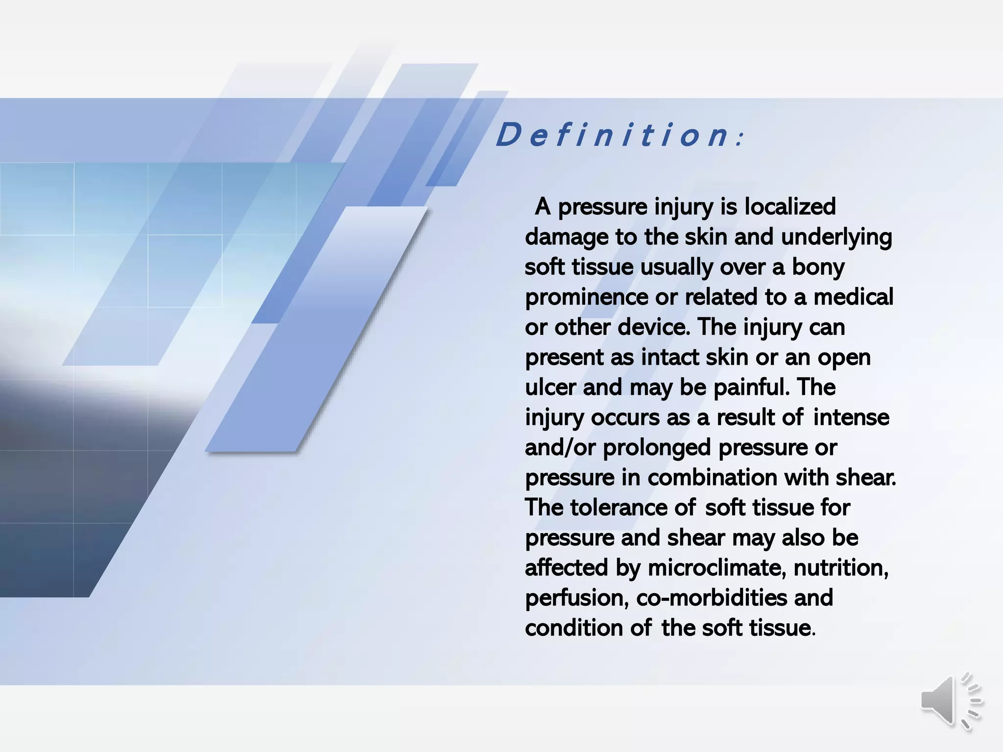
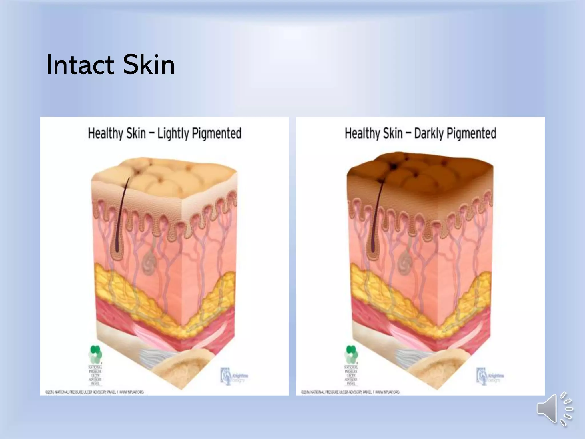
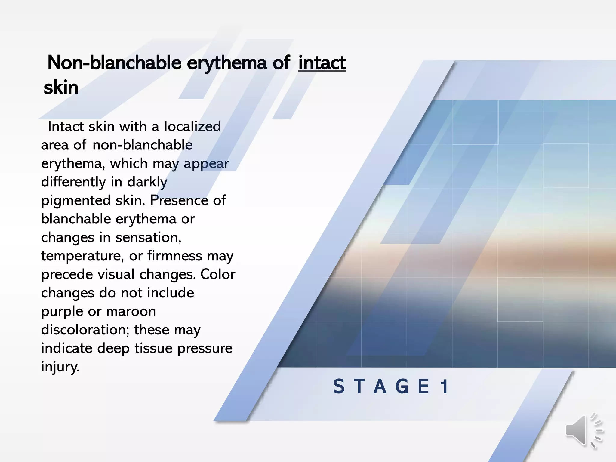
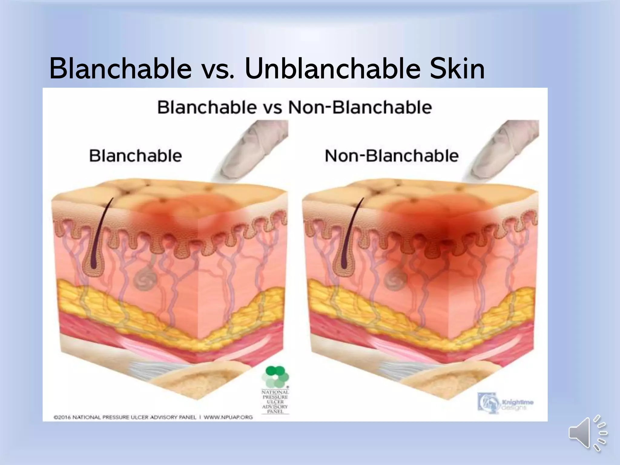
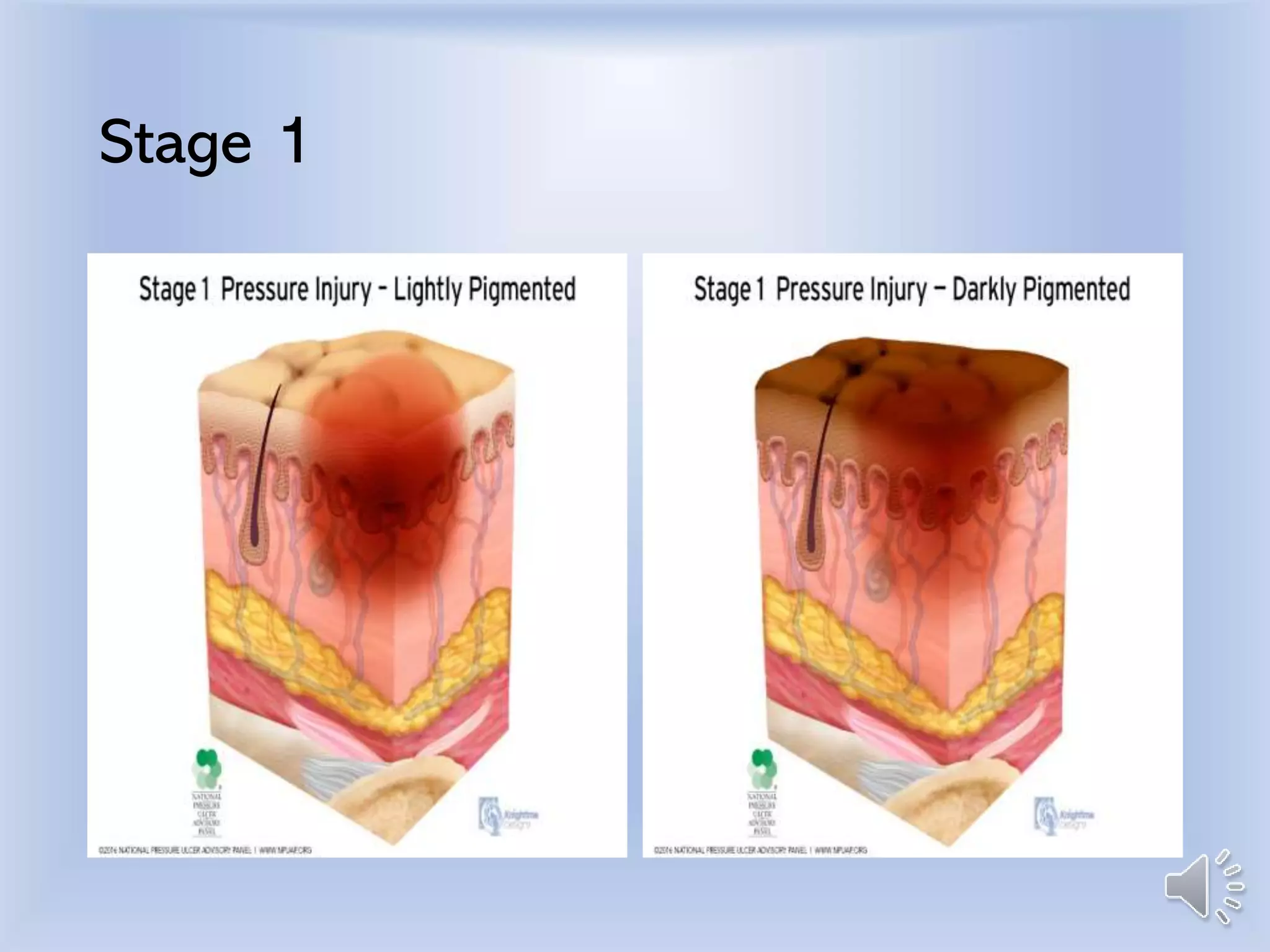
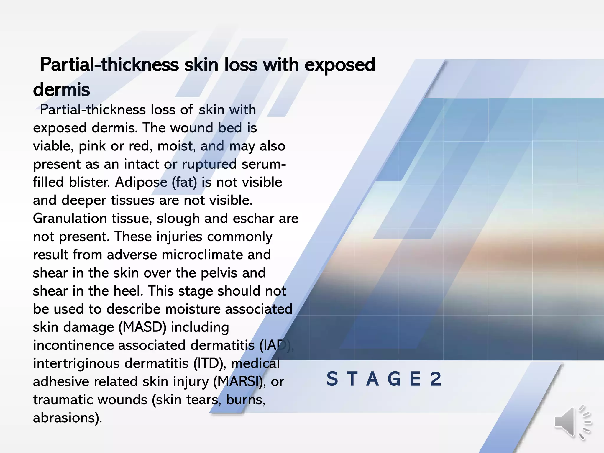
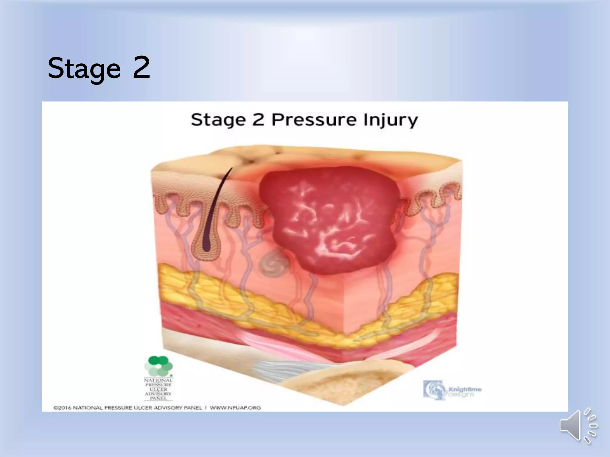
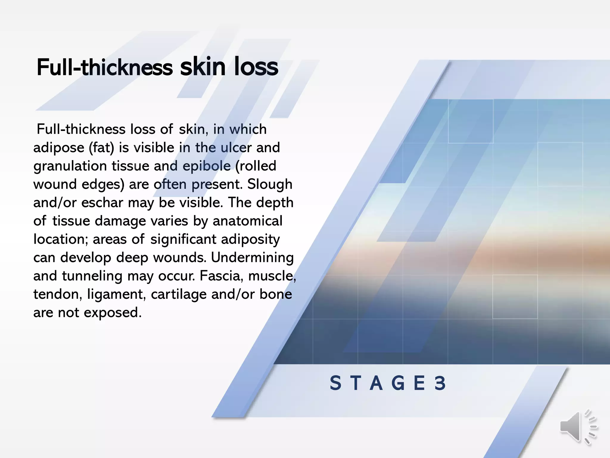
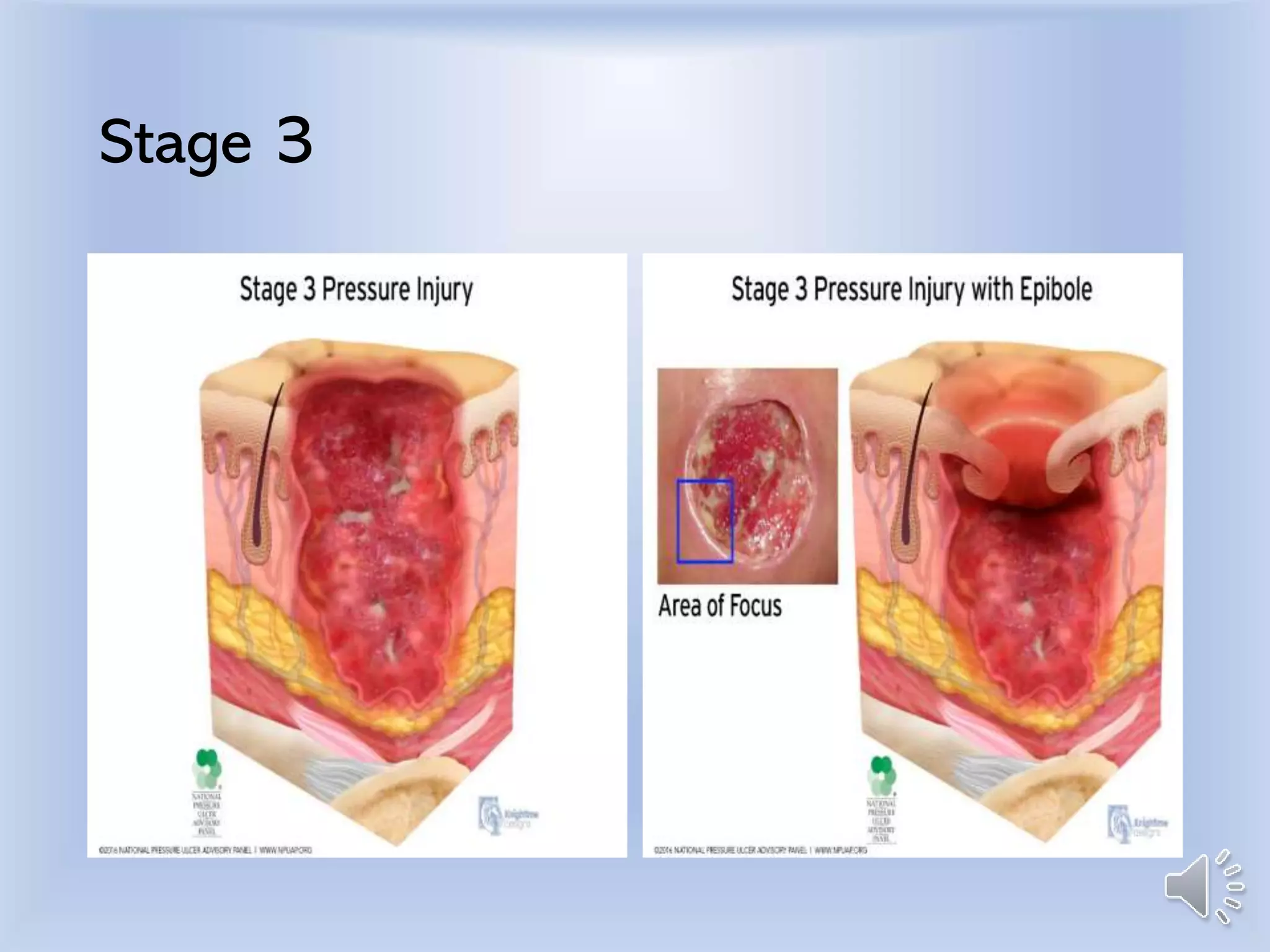
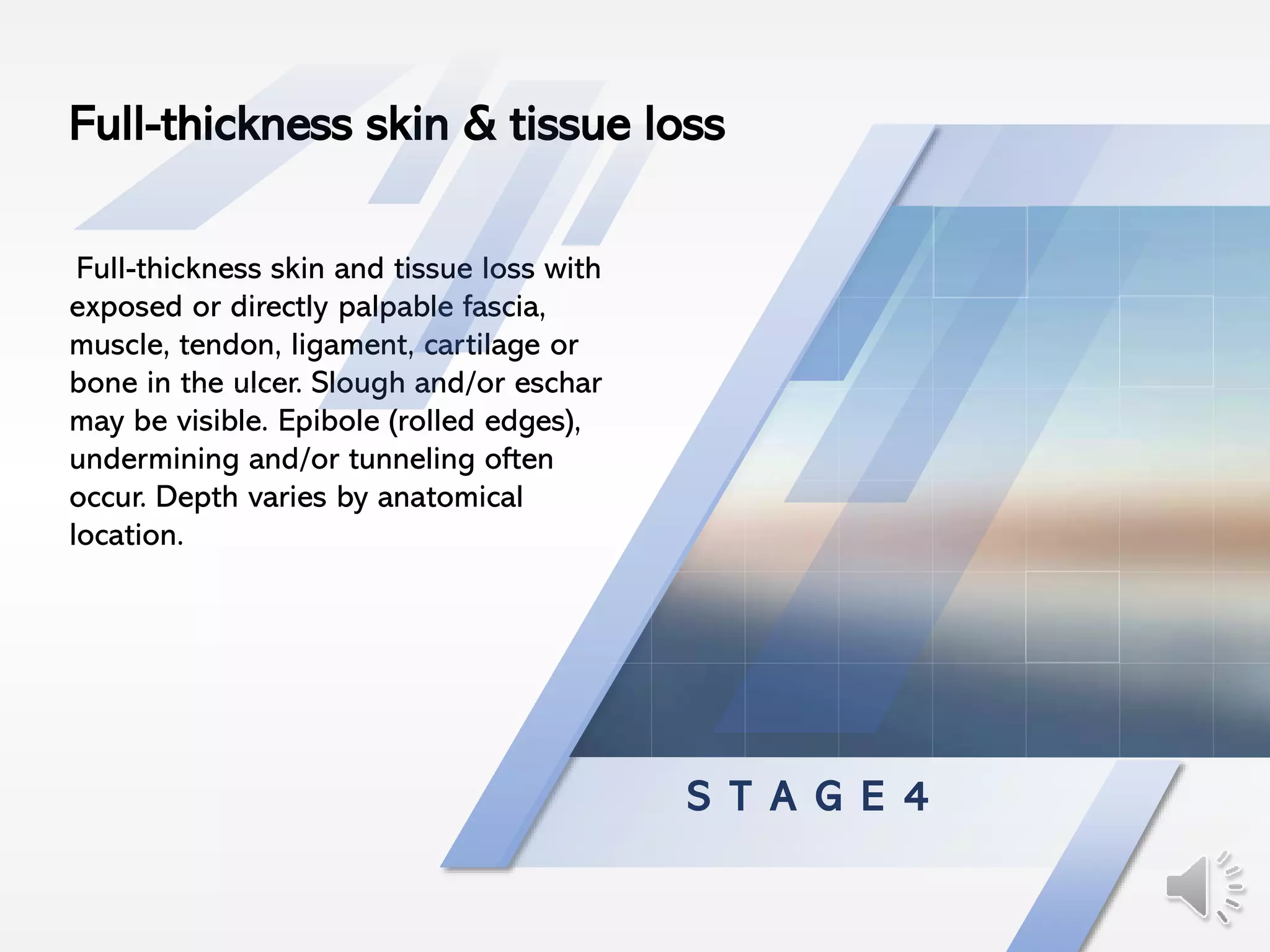
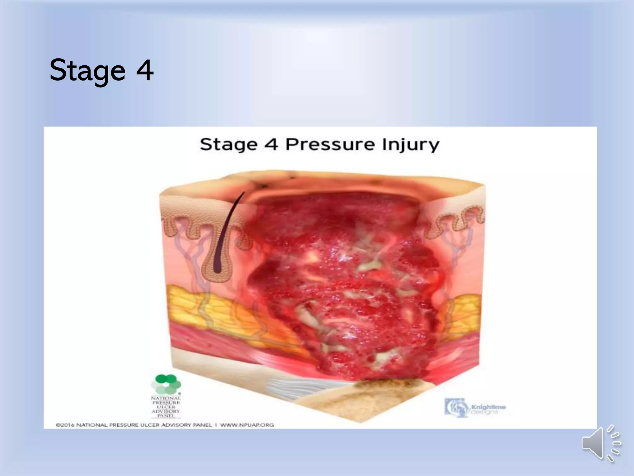
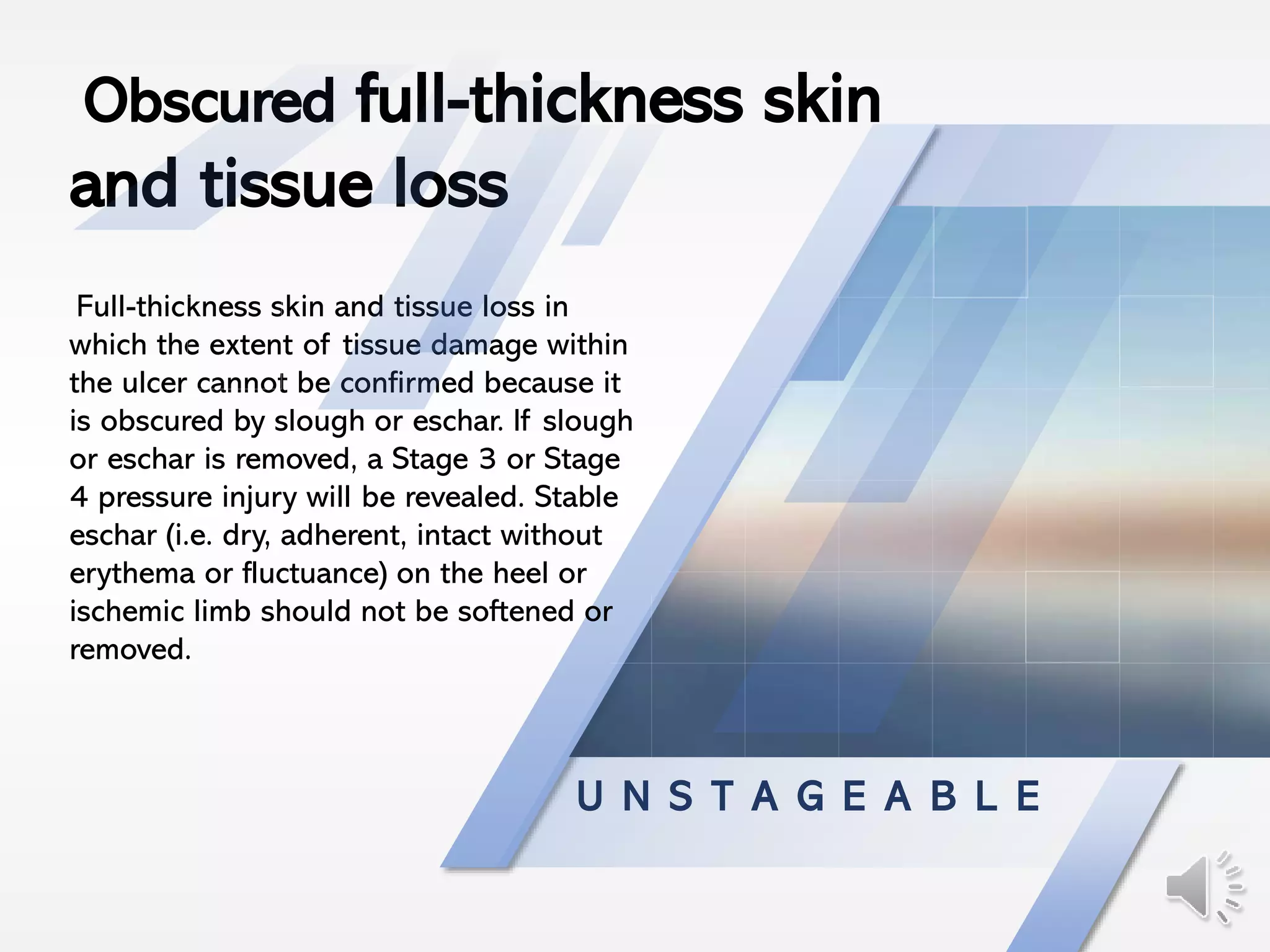
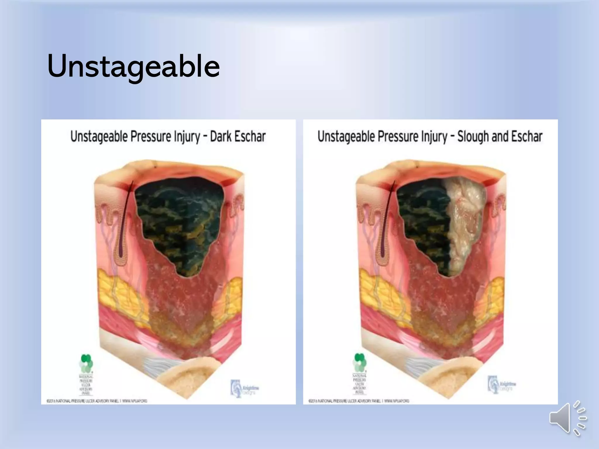
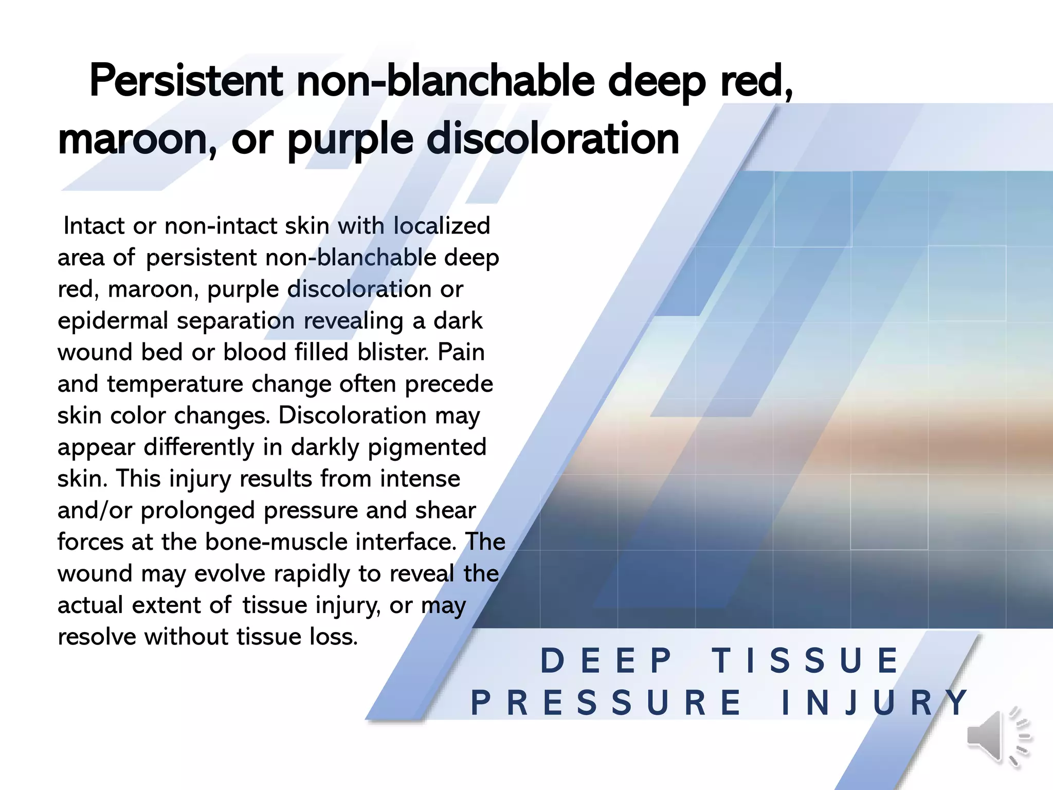
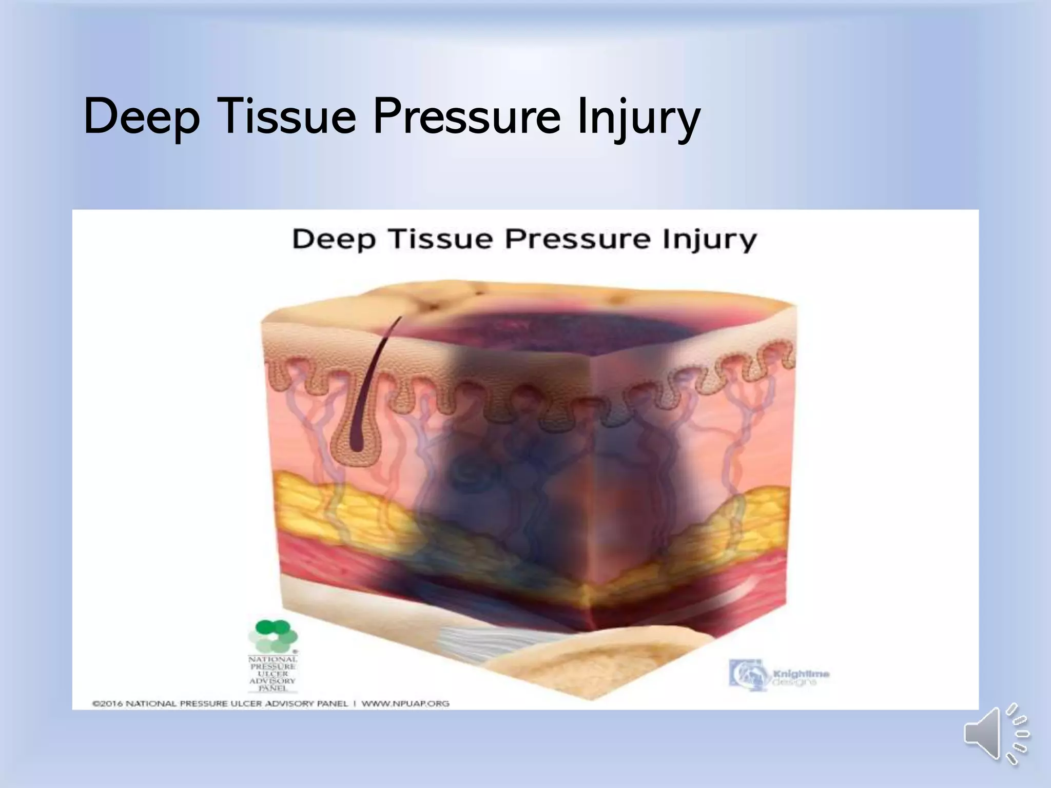
Characteristics
& Staging
M O D U L E 1
What is a Pressure
Injury?
D e f i n i t i o n :
A pressure injury is localized
damage to the skin and underlying
soft tissue usually over a bony
prominence or related to a medical
or other device. The injury can
present as intact skin or an open
ulcer and may be painful. The
injury occurs as a result of intense
and/or prolonged pressure or
pressure in combination with shear.
The tolerance of soft tissue for
pressure and shear may also be
affected by microclimate, nutrition,
perfusion, co-morbidities and
condition of the soft tissue.
Intact Skin
S T A G E 1
Intact skin with a localized
area of non-blanchable
erythema, which may appear
differently in darkly
pigmented skin. Presence of
blanchable erythema or
changes in sensation,
temperature, or firmness may
precede visual changes. Color
changes do not include
purple or maroon
discoloration; these may
indicate deep tissue pressure
injury.
Non-blanchable erythema of intact
skin
Blanchable vs. Unblanchable Skin
Stage 1
S T A G E 2
Partial-thickness loss of skin with
exposed dermis. The wound bed is
viable, pink or red, moist, and may also
present as an intact or ruptured serum-
filled blister. Adipose (fat) is not visible
and deeper tissues are not visible.
Granulation tissue, slough and eschar are
not present. These injuries commonly
result from adverse microclimate and
shear in the skin over the pelvis and
shear in the heel. This stage should not
be used to describe moisture associated
skin damage (MASD) including
incontinence associated dermatitis (IAD),
intertriginous dermatitis (ITD), medical
adhesive related skin injury (MARSI), or
traumatic wounds (skin tears, burns,
abrasions).
Partial-thickness skin loss with exposed
dermis
Stage 2
S T A G E 3
Full-thickness loss of skin, in which
adipose (fat) is visible in the ulcer and
granulation tissue and epibole (rolled
wound edges) are often present. Slough
and/or eschar may be visible. The depth
of tissue damage varies by anatomical
location; areas of significant adiposity
can develop deep wounds. Undermining
and tunneling may occur. Fascia, muscle,
tendon, ligament, cartilage and/or bone
are not exposed.
Full-thickness skin loss
Stage 3
S T A G E 4
Full-thickness skin and tissue loss with
exposed or directly palpable fascia,
muscle, tendon, ligament, cartilage or
bone in the ulcer. Slough and/or eschar
may be visible. Epibole (rolled edges),
undermining and/or tunneling often
occur. Depth varies by anatomical
location.
Full-thickness skin & tissue loss
Stage 4
U N S T A G E A B L E
Full-thickness skin and tissue loss in
which the extent of tissue damage within
the ulcer cannot be confirmed because it
is obscured by slough or eschar. If slough
or eschar is removed, a Stage 3 or Stage
4 pressure injury will be revealed. Stable
eschar (i.e. dry, adherent, intact without
erythema or fluctuance) on the heel or
ischemic limb should not be softened or
removed.
Obscured full-thickness skin
and tissue loss
Unstageable
D E E P T I S S U E
P R E S S U R E I N J U R Y
Intact or non-intact skin with localized
area of persistent non-blanchable deep
red, maroon, purple discoloration or
epidermal separation revealing a dark
wound bed or blood filled blister. Pain
and temperature change often precede
skin color changes. Discoloration may
appear differently in darkly pigmented
skin. This injury results from intense
and/or prolonged pressure and shear
forces at the bone-muscle interface. The
wound may evolve rapidly to reveal the
actual extent of tissue injury, or may
resolve without tissue loss.
Persistent non-blanchable deep red,
maroon, or purple discoloration
Deep Tissue Pressure Injury
M E D I C A L D E V I C E &
M U C O S A L I N J U R I E S
Medical device related pressure injuries
result from the use of devices designed
and applied for diagnostic or therapeutic
purposes. The resultant pressure injury
generally conforms to the pattern or
shape of the device. The injury should be
staged using the staging system.
Exception: If the injury is to a mucous
membrane ( mouth, lips, tongue, vagina)
then the wound is not staged.
Medical devices- fixators, ET tubes,
oxygen cannulas & masks, foley
catheters, fecal management
systems, ostomies.
Special Considerations
R e f e r e n c e s :
Edsberg, L. E., Black, J. M., Goldberg, M.,
McNichol, L., Moore, L., & Sieggreen, M.
(2016). Revised National Pressure Ulcer
Advisory Panel Pressure Injury Staging
System: Revised Pressure Injury Staging
System. J Wound Ostomy Continence Nurs,
43(6), 585-597.
doi:10.1097/won.0000000000000281
National Pressure Ulcer Advisory Panel.
(2018). Pressure Injury Stages. Retrieved
November 2018, from NPIAP:
https://npiap.com/page/PressureInjuryStages

Module1

  • 1.
  • 2.
    What is aPressure Injury?
  • 3.
    D e fi n i t i o n : A pressure injury is localized damage to the skin and underlying soft tissue usually over a bony prominence or related to a medical or other device. The injury can present as intact skin or an open ulcer and may be painful. The injury occurs as a result of intense and/or prolonged pressure or pressure in combination with shear. The tolerance of soft tissue for pressure and shear may also be affected by microclimate, nutrition, perfusion, co-morbidities and condition of the soft tissue.
  • 4.
  • 5.
    S T AG E 1 Intact skin with a localized area of non-blanchable erythema, which may appear differently in darkly pigmented skin. Presence of blanchable erythema or changes in sensation, temperature, or firmness may precede visual changes. Color changes do not include purple or maroon discoloration; these may indicate deep tissue pressure injury. Non-blanchable erythema of intact skin
  • 6.
  • 7.
  • 8.
    S T AG E 2 Partial-thickness loss of skin with exposed dermis. The wound bed is viable, pink or red, moist, and may also present as an intact or ruptured serum- filled blister. Adipose (fat) is not visible and deeper tissues are not visible. Granulation tissue, slough and eschar are not present. These injuries commonly result from adverse microclimate and shear in the skin over the pelvis and shear in the heel. This stage should not be used to describe moisture associated skin damage (MASD) including incontinence associated dermatitis (IAD), intertriginous dermatitis (ITD), medical adhesive related skin injury (MARSI), or traumatic wounds (skin tears, burns, abrasions). Partial-thickness skin loss with exposed dermis
  • 9.
  • 10.
    S T AG E 3 Full-thickness loss of skin, in which adipose (fat) is visible in the ulcer and granulation tissue and epibole (rolled wound edges) are often present. Slough and/or eschar may be visible. The depth of tissue damage varies by anatomical location; areas of significant adiposity can develop deep wounds. Undermining and tunneling may occur. Fascia, muscle, tendon, ligament, cartilage and/or bone are not exposed. Full-thickness skin loss
  • 11.
  • 12.
    S T AG E 4 Full-thickness skin and tissue loss with exposed or directly palpable fascia, muscle, tendon, ligament, cartilage or bone in the ulcer. Slough and/or eschar may be visible. Epibole (rolled edges), undermining and/or tunneling often occur. Depth varies by anatomical location. Full-thickness skin & tissue loss
  • 13.
  • 14.
    U N ST A G E A B L E Full-thickness skin and tissue loss in which the extent of tissue damage within the ulcer cannot be confirmed because it is obscured by slough or eschar. If slough or eschar is removed, a Stage 3 or Stage 4 pressure injury will be revealed. Stable eschar (i.e. dry, adherent, intact without erythema or fluctuance) on the heel or ischemic limb should not be softened or removed. Obscured full-thickness skin and tissue loss
  • 15.
  • 16.
    D E EP T I S S U E P R E S S U R E I N J U R Y Intact or non-intact skin with localized area of persistent non-blanchable deep red, maroon, purple discoloration or epidermal separation revealing a dark wound bed or blood filled blister. Pain and temperature change often precede skin color changes. Discoloration may appear differently in darkly pigmented skin. This injury results from intense and/or prolonged pressure and shear forces at the bone-muscle interface. The wound may evolve rapidly to reveal the actual extent of tissue injury, or may resolve without tissue loss. Persistent non-blanchable deep red, maroon, or purple discoloration
  • 17.
  • 18.
    M E DI C A L D E V I C E & M U C O S A L I N J U R I E S Medical device related pressure injuries result from the use of devices designed and applied for diagnostic or therapeutic purposes. The resultant pressure injury generally conforms to the pattern or shape of the device. The injury should be staged using the staging system. Exception: If the injury is to a mucous membrane ( mouth, lips, tongue, vagina) then the wound is not staged. Medical devices- fixators, ET tubes, oxygen cannulas & masks, foley catheters, fecal management systems, ostomies. Special Considerations
  • 19.
    R e fe r e n c e s : Edsberg, L. E., Black, J. M., Goldberg, M., McNichol, L., Moore, L., & Sieggreen, M. (2016). Revised National Pressure Ulcer Advisory Panel Pressure Injury Staging System: Revised Pressure Injury Staging System. J Wound Ostomy Continence Nurs, 43(6), 585-597. doi:10.1097/won.0000000000000281 National Pressure Ulcer Advisory Panel. (2018). Pressure Injury Stages. Retrieved November 2018, from NPIAP: https://npiap.com/page/PressureInjuryStages