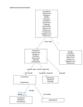
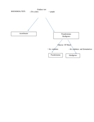
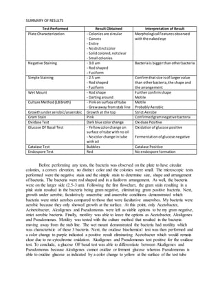
PSEUDOMONAS
Unknown Bacteria
DECEMBER 11, 2014
UOIT
Vita Koren
MORPHOLOGY CHARACTERIZING
Vita Koren
12-11-2014
Acetobacter
Acinetobacter
Alcaligenes
Bacillus
Citrobacter
Clostridium
Enterobacter
Klebsiella
Lactobacillus
Micrococcus
Pediococcus
Pseudomonas
Staphylococcus
Trichococcus
Acetobacter
Acinetobacter
Alcaligenes
Citrobacter
Enterobacter
Klebsiella
Pseudomonas
Bacillus
Clostridium
Lactobacillus
Micrococcus
Pediococcus
Staphylococcus
Trichococcus
Gram Stain
- +
Acetobacter
Acinetobacter
Alcaligenes
Pseudomonas
Growth under Aerobic/ Anaerobic
+ for Aerobic Faculative Anaerobe Anaerobic
-
Acetobacter
Alcaligenes
Pseudomonas
Acinetobacter
Enterobacter
Citrobacter
Klebsiella
Motility
+ Motile - Non-motile
BIOCHEMICAL TESTS
Acetobacter Pseudomonas
Alcaligenes
Oxidase test
- (No color) + purple
Pseudomonas
Glucose OF Basal
+ for oxidation - for oxidation and fermentation
Alcaligenes
SUMMARY OF RESULTS
Test Performed Result Obtained Interpretation of Result
Plate Characterization - Coloniesare circular
- Convex
- Entire
- Nodistinctcolor
- Solidcolored,notclear
- Small colonies
Morphological Featuresobserved
withthe nakedeye
Negative Staining - 3.0 um
- Rod shaped
- Fusiform
Bacteriais biggerthanotherbacteria
Simple Staining - 2.5 um
- Rod shaped
- Fusiform
Confirmthatsize isof largervalue
than otherbacteria,the shape and
the arrangement
Wet Mount - Rod shape
- Dartingaround
Furtherconfirmshape
Motile
Culture Method(LBBroth) - Pinkonsurface of tube
- Grew away fromstab line
Motile
ProbablyAerobic
Growth underaerobic/anaerobic Growth at the top Strict Aerobe
Gram Stain Pink Confirmedgramnegative bacteria
Oxidase Test Dark blue colorchange Oxidase Positive
Glucose OFBasal Test - Yellow colorchange on
surface of tube withno oil
- Nocolor change intube
withoil
Oxidationof glucose positive
Fermentationof glucose negative
Catalase Test Bubbles Catalase Positive
Endospore Test Red No endospore formation
Before performing any tests, the bacteria was observed on the plate to have circular
colonies, a convex elevation, no distinct color and the colonies were small. The microscopic tests
performed were the negative stain and the simple stain to determine size, shape and arrangement
of bacteria. The bacteria were rod shaped and in a fusiform arrangement. As well, the bacteria
were on the larger side (2.5-3 um). Following the first flowchart, the gram stain resulting in a
pink stain resulted in the bacteria being gram negative, eliminating gram positive bacteria. Next,
growth under aerobic, faculatively anaerobic and anaerobic conditions demonstrated which
bacteria were strict aerobes compared to those that were facultative anaerobes. My bacteria were
aerobic because they only showed growth at the surface. At this point, only Acetobacter,
Acinetobacter, Alcaligenes and Pseudomonas were left as viable options to be my gram negative,
strict aerobic bacteria. Finally, motility was able to leave the options as Acetobacter, Alcaligenes
and Pseudomonas. Motility was tested with the culture method that resulted in the bacteria
moving away from the stab line. The wet mount demonstrated the bacteria had motility which
was characteristic of these 3 bacteria. Next, the oxidase biochemical test was then performed and
a color change to purple indicated a positive result eliminating Acetobacter which would remain
clear due to no cytochrome oxidation. Alcaligenes and Pseudomonas test positive for the oxidase
test. To conclude, a glucose OF basal test was able to differentiate between Alcaligenes and
Pseudomonas because Alcaligenes cannot oxidize or ferment glucose whereas Pseudomonas is
able to oxidize glucose as indicated by a color change to yellow at the surface of the test tube
without oil. With that, the unknown was identified as Pseudomonas! Additionally, Pseudomonas
was further confirmed with a positive catalase test and a negative endospore forming test.
To further prove the determination of Pseudomonas, the citrate test can be performed and a
positive result would be obtained due to the green solution turning blue (Palleroni, 2010).
Additionally, a blood agar test can be performed and there would be beta-hemolysis of the agar
due to Pseudomonas whereas Alcaligenes would not lyse it. (Buxton, 2010).
References
Buxton, R. (2010). “Blood Agar Plates and Hemolysis: Non-Fermenting Gram-Negative Rods”
American Society for Microbiology 11 (2): 899-905
Palleroni, N. J. (2010). "The Pseudomonas Story". Environmental Microbiology 12 (6): 1377–
1383.

module 2 - final - SB5

  • 1.
  • 2.
  • 3.
    BIOCHEMICAL TESTS Acetobacter Pseudomonas Alcaligenes Oxidasetest - (No color) + purple Pseudomonas Glucose OF Basal + for oxidation - for oxidation and fermentation Alcaligenes
  • 4.
    SUMMARY OF RESULTS TestPerformed Result Obtained Interpretation of Result Plate Characterization - Coloniesare circular - Convex - Entire - Nodistinctcolor - Solidcolored,notclear - Small colonies Morphological Featuresobserved withthe nakedeye Negative Staining - 3.0 um - Rod shaped - Fusiform Bacteriais biggerthanotherbacteria Simple Staining - 2.5 um - Rod shaped - Fusiform Confirmthatsize isof largervalue than otherbacteria,the shape and the arrangement Wet Mount - Rod shape - Dartingaround Furtherconfirmshape Motile Culture Method(LBBroth) - Pinkonsurface of tube - Grew away fromstab line Motile ProbablyAerobic Growth underaerobic/anaerobic Growth at the top Strict Aerobe Gram Stain Pink Confirmedgramnegative bacteria Oxidase Test Dark blue colorchange Oxidase Positive Glucose OFBasal Test - Yellow colorchange on surface of tube withno oil - Nocolor change intube withoil Oxidationof glucose positive Fermentationof glucose negative Catalase Test Bubbles Catalase Positive Endospore Test Red No endospore formation Before performing any tests, the bacteria was observed on the plate to have circular colonies, a convex elevation, no distinct color and the colonies were small. The microscopic tests performed were the negative stain and the simple stain to determine size, shape and arrangement of bacteria. The bacteria were rod shaped and in a fusiform arrangement. As well, the bacteria were on the larger side (2.5-3 um). Following the first flowchart, the gram stain resulting in a pink stain resulted in the bacteria being gram negative, eliminating gram positive bacteria. Next, growth under aerobic, faculatively anaerobic and anaerobic conditions demonstrated which bacteria were strict aerobes compared to those that were facultative anaerobes. My bacteria were aerobic because they only showed growth at the surface. At this point, only Acetobacter, Acinetobacter, Alcaligenes and Pseudomonas were left as viable options to be my gram negative, strict aerobic bacteria. Finally, motility was able to leave the options as Acetobacter, Alcaligenes and Pseudomonas. Motility was tested with the culture method that resulted in the bacteria moving away from the stab line. The wet mount demonstrated the bacteria had motility which was characteristic of these 3 bacteria. Next, the oxidase biochemical test was then performed and a color change to purple indicated a positive result eliminating Acetobacter which would remain clear due to no cytochrome oxidation. Alcaligenes and Pseudomonas test positive for the oxidase test. To conclude, a glucose OF basal test was able to differentiate between Alcaligenes and Pseudomonas because Alcaligenes cannot oxidize or ferment glucose whereas Pseudomonas is able to oxidize glucose as indicated by a color change to yellow at the surface of the test tube
  • 5.
    without oil. Withthat, the unknown was identified as Pseudomonas! Additionally, Pseudomonas was further confirmed with a positive catalase test and a negative endospore forming test. To further prove the determination of Pseudomonas, the citrate test can be performed and a positive result would be obtained due to the green solution turning blue (Palleroni, 2010). Additionally, a blood agar test can be performed and there would be beta-hemolysis of the agar due to Pseudomonas whereas Alcaligenes would not lyse it. (Buxton, 2010). References Buxton, R. (2010). “Blood Agar Plates and Hemolysis: Non-Fermenting Gram-Negative Rods” American Society for Microbiology 11 (2): 899-905 Palleroni, N. J. (2010). "The Pseudomonas Story". Environmental Microbiology 12 (6): 1377– 1383.