Mitrochondrial enzyme by KK Sahu sir
•Download as PPTX, PDF•
0 likes•81 views

1) Introductione (2) History (3) Structure (4) list of mitochondrial enzymas a) enzymes of outer membrane b) enzymes of outer chamber c) enzymes of inner membrane d)enzymes of inner chambes (5)Mitochondrial enzymes 1 Subtypes and tissue distribution 2 Function 3 Substrate specificities 4 Clinical significance 5 Genetics (6) Enzymes of CAC (7) Enzymes of oxidative phosphorilation (8) Electron carrior and complex. (9) Respiratory chain inhibitors. (10) Conclution (11) refrences

Recommended
More Related Content
What's hot
What's hot (20)
Similar to Mitrochondrial enzyme by KK Sahu sir
Similar to Mitrochondrial enzyme by KK Sahu sir (20)
More from KAUSHAL SAHU
More from KAUSHAL SAHU (20)
Recently uploaded
Recently uploaded (20)
Mitrochondrial enzyme by KK Sahu sir
- 1. By KAUSHAL KUMAR SAHU Assistant Professor (Ad Hoc) Department of Biotechnology Govt. Digvijay Autonomous P. G. College Raj-Nandgaon ( C. G. )
- 2. Contents :- (1) Introductione (2) History (3) Structure (4) list of mitochondrial enzymas a) enzymes of outer membrane b) enzymes of outer chamber c) enzymes of inner membrane d)enzymes of inner chambes (5)Mitochondrial enzymes 1 Subtypes and tissue distribution 2 Function 3 Substrate specificities 4 Clinical significance 5 Genetics (6) Enzymes of CAC (7) Enzymes of oxidative phosphorilation (8) Electron carrior and complex. (9) Respiratory chain inhibitors. (10) Conclution (11) refrences
- 3. •introduction ; 1) Mitochondria are sometimes described as "cellular power plants“ because they generate most of the cell's supply of adenosine triphosphate (ATP), used as a source of chemical energy . 2) The word mitochondrion comes from the Greek μίτος or mitos, thread + χονδρίον or chondrion, granule. 3) the mitochondrion has its own independent genome history; •Kolliker (1850) first seen mitochondria in street muscle cells. •Flemming (1882) named fila . •Altman (1892) named Bioplast. •Benda (1898) named mitochondria. •Nass (1963) Observed DNA in mitochondria.
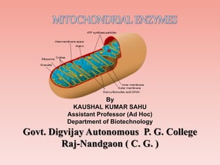
- 4. structure; A mitochondrion contains outer and inner membranes composed of phospholipid bilayers and proteins.[6] 1.outer membrane:- It contains large numbers of integral proteins called porins 2.inner membrane space:-, the concentrations of small molecules such as ions and sugars in the intermembrane space is the same as the cytosol. 3.inner membrane:-; Cardiolipin contains four fatty acids rather than two and may help to make the inner membrane impermeable. 4.Cristae:- inner mitochondrial membrane, enhancing its ability to produce ATP. These folds are studded with small round bodies known as F1 particles or oxysomes. 5.matrix:-The major functions include oxidation of pyruvate and fatty acids, and the citric acid cycle. STRUCTURE OF MITOCHONDRIA
- 5. Outer membrane enzymes *monoamine oxidase *rotenone insensitive NADH cyt-c-reductace *kynurenine hydroxylase thiokinase Outer chamber enzymes *adenylate kinase *nucieoside diphosphokinase •List of mitochondrial enzymes;
- 6. Inner membrane enzymes *respiratory chain enzymes *ATP synthetase orxidase *succinate dehydrogenase *carnitine fatty acid acyl transferase Inner chamber enzymes *isocitrate dehydrogenase *fumerase *aconitase *citrate snythetase *alfa-keto acid dehydroge *beta-oxidation enzymes *L-malate hydrogenase *L-amino levuline synthatase *L-glutamate dehyrogenase
- 7. {1}enzymesof outer membrane Monoamine oxidase :- Contents; 1. Subtypes and tissue distribution:- 2. Function:- 3. Substrate specificities (1) Serotonin, melatonin, norepinephrine, and epinephrine are mainly broken down by MAO-A. (2)Phenethylamine and benzylamine are mainly broken down by MAO-B. (3) Both forms break down dopamine, tyramine, and tryptamine equally. 4.Clinical significance 5. Genetics
- 8. Nuclieoside diphosphokinase :- . enzymes of outer chember ;- enzymes that catalyze the exchange of phosphate groups between different nucleoside diphosphates. Function:- GTP + ADP → GDP + ATP (1)Prokaryotic systems (2) Eukaryotic systems:
- 9. {3}enzymes of inner membrane {1} carnitine fatty acid acyl transferase :- Carnitine is a quaternary ammonium compound biosynthesized from the amino acids lysine and methionine. In living cells, it is required for the transport of fatty acids from the cytosol into the mitochondria during the breakdown of lipids (fats) for the generation of metabolic energy •Biochemistry Biosynthesis - Role in fatty acid metabolism – Potential uses as a pharmaceutical •Heart conditions • Kidney disease and dialysis •Effect in male infertility •As an antidote in valproic acid poisoning As a weight loss supplyment
- 10. {4}enzymes of inner chamber •Three stages of ceel respiration :- •Oxidative decarboxylation [pyruvate to acetyl co-A •CAC/ TCA/ Acetyl co-A catabolism/ kreb’s cycle •ETC/ ETS/ oxidative phosphorylation.
- 11. [a]CAC/ KREB’S CYCLE ENZYMES;
- 12. Substrates Products Enzyme Reaction type Comment Oxaloacetate + Acetyl CoA + H2O Citrate + CoA-SH Citrate synthase Aldol condensation irreversible, extends the 4C oxaloacetate to a 6C molecule Citrate cis-Aconitate + H2O Aconitase Dehydration reversible isomerisation cis-Aconitate + H2O Isocitrate Hydration Isocitrate + NAD+ Oxalosuccinate + NADH + H + Isocitrate dehydrogenase Oxidation generates NADH (equivalent of 2.5 ATP) ROLEOF CACENZYEM’S
- 13. Oxalosuccinate α-Ketoglutarate + CO2 Decarboxylation rate-limiting, irreversible stage, generates a 5C molecule α-Ketoglutarate + NAD+ + CoA-SH Succinyl-CoA + NADH + H+ + CO2 α-Ketoglutarate dehydrogenase Oxidative decarboxylation irreversible stage, generates NADH (equivalent of 2.5 ATP), regenerates the 4C chain (CoA excluded) Succinyl-CoA + GDP + Pi Succinate + CoA-SH + GTP Succinyl-CoA synthetase substrate-level phosphorylation or ADP→ATP instead of GDP→GTP,[7] generates 1 ATP or equivalent
- 14. Succinate + ubiquinone (Q) Fumarate + ubiquinol (QH2) Succinate dehydrogenase Oxidation uses FAD as a prosthetic group (FAD→FADH2 in the first step of the reaction) in the enzyme,[7] generates the equivalent of 1.5 ATP Fumarate + H2O L-Malate Fumarase H2O addition (hydration) L-Malate + NAD+ Oxaloacetate + NADH + H+ Malate dehydrogenase Oxidation reversible (in fact, equilibrium favors malate), generates NADH (equivalent of 2.5 ATP)
- 15. 1)ACONITASE;- A) Aconitase has an active [Fe4S4]2+ cluster, which may convert to an inactive [Fe3S4]+ form. ISOCITRATE DEHYDROGENASE :- Isozymes NADP+ dependent Each NADP+-dependent isozyme functions as a homodimer: NAD+ dependent The isocitrate dehydrogenase 3 isozyme is a heterotetramer that is composed of two alpha subunits, one beta subunit, and one gamma subunit.
- 16. (3) FUMERASE:- MACHANISMOF FUMARASE Clinical significance Fumarase deficiency is characterized by polyhydramnios and fetal brain abnormalitie. In the newborn period, findings include severe neurologic abnormalities, poor feeding, failure to thrive, and hypotonia.
- 17. (4) BETA –OXIDATION ENZYMES :- 1. Activation of fatty acids in the cytosol 2. Transport of fatty acids into mitochondria (carnitine shuttle) 3. Beta oxidation proper in the mitochondrial matrix. (5) L-malate dehydrogenase BETA OXIDATION CYCLE
- 19. {2} OXIDATIVE PHOSPHORILASION ENZYMES
- 20. Photosynthetic electron transport chain of the thylakoid membrane.
- 21. Electron carriers (1) FMN – 2) Coenzyme Q
- 22. 3) Heme – A prosthetic group of cytochromes. Heme contains an iron atom embedded in a porphyrin ring system. The porphyrin ring structure is planar. The iron atom of heme is usually bonded to two axial ligands, in addition to the 4 N of the porphyrin ring system.
- 23. 4) Cytochromes – 1) Are proteins with heme prosthetic groups. . 2) Some cytochromes are part of large integral membrane complexes, each consisting of several polypeptides and including multiple electron carriers. . For example, hemes a and a3 that are part of the respiratory chain complex IV are often referred to as cytochromes a and a3. Cytochrome c is instead a small, water-soluble protein, with a single heme group. 5) Iron-sulfur centers (Fe-S Electron transfer proteins may contain multiple iron-sulfur centers.
- 24. Respiratorychain 1) Most constituents of the respiratory chain are embedded in the innen Mitochondria membrane (or in the cytoplasmic membrane of aerobic bacteria 2) Electrons are transferred from NADH to O2 via multi- subunit inner membrane complexes I, III, & IV, plus coenzyme Q and cytochrome c. Within each complex, electrons pass sequentially through a series of electron carriers.
- 25. Complex Name No. of Proteins Prosthetic Groups Complex I NADH Dehydrogenase 46 FMN, 9 Fe-S centers Complex II Succinate-CoQ Reductase 5 FAD, cyt b560, 3 Fe-S centers Complex III CoQ-cyt c Reductase 11 cyt bH, cyt bL, cyt c1, Fe-SRieske Complex IV Cytochrome Oxidase 13 cyt a, cyt a3, CuA, CuB
- 26. Respiratory chain inhibitorsinclude the following: (A) Rotenone (a common rat poison) blocks electron transfer in complex I. (B) Antimycin A blocks electron transfer in complex III. (C) Cyanide and carbon monoxide inhibit complex IV. ComplexI catalyzes oxidation of NADH, with reduction of coenzyme Q: NADH + H+ + Q � NAD+ + QH2 The initial electron transfers are: NADH + H+ + FMN � NAD+ + FMNH2 FMNH2 + (Fe-S)ox � FMNH� + (Fe- S)red + H+ Succinate Dehydrogenase of the Krebs Cycle is also called complex II or Succinate-CoQ Reductase. .
- 27. Complex III accepts electrons from coenzyme QH2 that is generated by electron transfer in complexes I and II. The structure and roles of complex III are discussed in the section on oxidative phosphorylation. Cytochrome c1, a prosthetic group within complex III, reduces cytochrome c, which is the electron donor to complex IV. Cytochrome oxidase (complex IV) carries out the following irreversible reaction: O2 + 4 H+ + 4 e- � 2 H2O So the ETC cycle has been completed.
- 28. REFERENCE 1.Advanced biology-Kent michael 2.Cell and molecular Biology- Gerald Karp 3. Geoffrey M.Cooper-The cell a moleculer approach. 4. molecular and cell biology- Lodish et.al. Websites: www.kbiotech.com www.wikipedia.com