PRESENTATION TOPIC:
Isolation, Identification and Antimicrobial
Susceptibility of Microorganism
Presented By:
Arooj Fatima
BITE222104010
INTRODUCTION:
Microbes exist almost everywhere and have an important impact on both
sickness and health. In clinical microbiology, primary steps such as the isolation
and identification of the causative agents of infection, along with antimicrobial
susceptibility testing (AST), are the backbone of correct diagnosis and
successful management of infectious diseases.
Isolation: refers to the process of obtaining a pure culture of a microorganism
from a complex sample such as blood, urine, sputum, or wound swabs. This
step is crucial for eliminating contaminating flora and ensuring that the
organism responsible for infection is accurately studied.
Identification: of isolated microorganisms involves a combination of
morphological, biochemical, serological, and molecular methods.
Once identified, the susceptibility of the microorganism to various
antimicrobial agents is determined through standardized AST protocols, such as
the Kirby-Bauer disk diffusion method, broth microdilution, or E-test, as
recommended by guidelines from the Clinical and Laboratory Standards
Institute (CLSI). AST results are crucial for selecting the most effective
antibiotic therapy, especially in the context of emerging multidrug-resistant
(MDR), extensively drug-resistant (XDR), and pan-drug-resistant strains.
Material Required for the Isolation and
Identification of Micro organisms
2. Culture Media:
•Blood Agar (BA) – For hemolytic organisms
•MacConkey Agar (MAC) – Selective for Gram-negative enterics
•Mannitol Salt Agar (MSA) – For Staphylococcus species
•Chocolate Agar – Enriched medium for fastidious organisms
•Sabouraud Dextrose Agar (SDA) – For fungi and yeasts
•Selective media (e.g., XLD, TCBS, EMB, Thayer-Martin) depending on the target organi
1. Sample Collection Materials:
Sterile swabs (cotton or rayon-tipped)
Urine collection containers
Blood culture bottles (aerobic and anaerobic)
Sputum cups
Stool collection containers
Transport media (e.g., Stuart’s, Amies, Cary-Blair)
3. Incubation Equipment
Incubator (35–37°C) – For bacterial growth
CO Incubator or candle jar – For capnophilic bacteria (e.g.,
₂ Neisseria)
Fungal incubator (25–30°C) – For mold growth
4. Sterilization and Aseptic Equipment
Bunsen burner or spirit lamp
Autoclave – For sterilizing media and equipment
Laminar flow hood or biosafety cabinet
Sterile Petri dishes, pipettes, and loops
Inoculating loops and needles (metal or disposable plastic)
5. Microscopy and Staining Reagents
Gram stain kit (crystal violet, iodine, alcohol, safranin)
Acid-fast stain reagents (Ziehl-Neelsen/Kinyoun)
Lactophenol cotton blue – For fungal identification
Light microscope or phase contrast microscope
6. Biochemical Test Kits and Reagents
Oxidase and catalase reagents
Triple Sugar Iron (TSI) agar
Simmons’ Citrate, Urease, Indole tests, MR-VP broth
API strip systems or automated ID systems (e.g., VITEK, MALDI-TOF)
7. Antimicrobial Susceptibility Testing (AST) Materials
•Mueller-Hinton Agar (MHA)
•Antibiotic discs (CLSI or EUCAST approved)
•McFarland standard (0.5 turbidity) for inoculum standardization
•Zone measuring scaleor calipers
8. Personal Protective Equipment (PPE)
Lab coat, gloves, face mask/shield, safety goggles
Aseptic techniques in microbiology involve procedures that prevent contamination of
microbial cultures, laboratory equipment, and the environment. These include sterilizing
tools, using flame or laminar flow, and practicing proper hand hygiene and workspace
disinfection.
Isolation, Identification and Antimicrobial
Susceptibility of Staphylococci species:
Isolation Methods:
Media: Mannitol Salt Agar (MSA), Blood Agar, and Chromogenic Agar.
Incubation: 35-37°C for 24-48 hours.
Colony Characteristics: Golden-yellow colonies on MSA, beta-hemolysis on Blood A
Biochemical Tests: Catalase Test: Positive (bubbles form when hydrogen peroxide is
added).
Coagulase Test: Differentiates between S. aureus (positive) and other Staphylococcus
species (negative).
Other tests: DNase, urease, and carbohydrate fermentation tests.
Antimicrobial Susceptibility of Staphylococci:
Staphylococcus species, particularly Staphylococcus aureus, exhibit varied
susceptibility to antibiotics. Methicillin-resistant Staphylococcus aureus (MRSA) is a
major clinical concern due to its resistance to β-lactam antibiotics.
Commonly Tested Antibiotics:
β-lactams: Penicillin, Oxacillin, Cefoxitin (to detect MRSA)
Glycopeptides: Vancomycin, Teicoplanin
Macrolides: Erythromycin, Clindamycin Fluoroquinolones: Ciprofloxacin
Tetracyclines: Doxycycline, Minocycline
Testing Methods: Disk Diffusion (Kirby-Bauer)Minimum Inhibitory Concentration .
Isolation, Identification and Antimicrobial
Susceptibility of Streptococci species:
Hemolysis Patterns: Alpha-hemolysis (greenish discoloration)
Beta-hemolysis (complete hemolysis)
Gamma-hemolysis (no hemolysis)
CAMP Test: Positive for Group B Streptococcus (S. agalactiae)3. Optochin
Sensitivity: S. pneumoniae is sensitive to optochin
Bile Solubility: S. pneumoniae is bile soluble
Serogrouping: Lancefield grouping (A-H, K-V) using latex agglutination or other
methods.
Other Methods:
Biochemical Tests: API Strep or other biochemical panels
Molecular Methods: PCR or 16S rRNA sequencing for confirmation.
Antimicrobial susceptibility is tested using disk diffusion or MIC methods. Penicillin
remains the first-line treatment, though resistance to macrolides and tetracyclines is
increasing in some Streptococcus species.
Isolation, Identification and Antimicrobial Susceptibility of E.coli and klebsiella
species:
Isolation and Identification:
Clinical samples (urine, sputum, blood, etc.) are cultured on MacConkey agar, where E.
coli typically produces pink colonies due to lactose fermentation, while Klebsiella also
ferments lactose but forms large, mucoid colonies. Further identification is done using
biochemical tests such as the Indole test (positive for E. coli, negative for Klebsiella),
citrate utilization (positive for Klebsiella), and motility testing (motile E. coli, non-motile
Klebsiella).
Antimicrobial Susceptibility:
Susceptibility is commonly tested using the Kirby-Bauer disk diffusion method or
automated systems like VITEK. Both organisms often exhibit resistance to beta-lactam
antibiotics due to beta-lactamase enzyme production, including extended-spectrum beta-
lactamases (ESBLs). Carbapenems are typically effective, although carbapenem-resistant
strains have emerged, especially in Klebsiella pneumoniae.
Isolation, Identification and Antimicrobial
Susceptibility of Pseudomonas and Aspergillus
species:
Isolation and Identification:
1. Pseudomonas aeruginosa:
Sample sources: Clinical specimens such as sputum, wound swabs, urine, or blood.
Culture: Grown on cetrimide agar (selective medium) or MacConkey agar. Colonies
are flat with a metallic sheen and characteristic fruity odor. It may produce greenish-
blue pigment (pyocyanin).
Biochemical tests: Oxidase-positive, catalase-positive, and non-fermentative in TSI
slant. Confirmed via API 20NE or automated identification systems.
2. Aspergillus species:
Sample sources: Respiratory specimens (sputum, BAL), tissue biopsies.
Culture: Grown on Sabouraud Dextrose Agar (SDA) at 25–37°C. Colonies are fast-
growing, velvety, and often green (for A. fumigatus), yellow, or black depending on
species.
Microscopy: Lactophenol cotton blue (LPCB) staining reveals septate hyphae and
conidial heads with characteristic arrangement. Identified by morphology or PCR-
based molecular methods.
Antimicrobial/Antifungal Susceptibility:
• Pseudomonas aeruginosa:
Commonly resistant to: Penicillins, first- and second-generation cephalosporins.
Sensitive to: Piperacillin-tazobactam, ceftazidime, cefepime, carbapenems (except
imipenem in some resistant strains), and fluoroquinolones.
Resistance mechanisms: Efflux pumps, beta-lactamases (including ESBLs and
carbapenemases), porin loss.
Testing method: Disk diffusion (CLSI guidelines), E-test, or automated systems like
VITEK 2.
• Aspergillus species:
First-line drug: Voriconazole.
Alternatives: Amphotericin B, isavuconazole, or echinocandins (like caspofungin).
Resistance: Rising azole resistance (especially in A. fumigatus) due to environmental
azole use.
Testing method: CLSI broth microdilution or E-test strips on RPMI agar.
Isolation, Identification and Antimicrobial
Susceptibility of H. Influenzae species:
Isolation:
H. influenzae requires two essential growth factors:
X factor (hemin)
V factor (nicotinamide adenine dinucleotide – NAD )
⁺
These are both present in Chocolate Agar, which is the preferred medium. The organism
does not grow on standard blood agar unless satellitism is observed around organisms like
Staphylococcus aureus that provide V factor. Incubation at 35–37°C in 5–10% CO₂ for
24–48 hours is required.
Identification:
Colony morphology: Smooth, translucent, moist colonies
Gram stain: Small Gram-negative coccobacilli
Oxidase test: Positive
Catalase test: Positive
X and V factor requirement test: Growth occurs only with both factors present
Porphyrin (ALA) test: Negative for H. influenzae
Confirmatory tests: MALDI-TOF MS or PCR-based identification
Capsular typing can be done by latex agglutination or PCR for the b capsule gene.
Antimicrobial Susceptibility Testing (AST):
AST is typically done using disk diffusion, E-test, or broth microdilution methods,
following CLSI or EUCAST guidelines. Many strains produce β-lactamase,
conferring resistance to ampicillin.
Susceptible to: Cefotaxime, ceftriaxone, azithromycin, and fluoroquinolones
β-lactamase-negative strains: Often susceptible to ampicillin
β-lactamase-positive strains: Require β-lactam/β-lactamase inhibitor combinations
like amoxicillin-clavulanic acid.
Isolation, Identification and Antimicrobial
Susceptibility of Neisseria species:
Sample Collection:
N. gonorrhoeae: Urethral, cervical, rectal, and pharyngeal swabs depending on site of
infection.
N. meningitidis: Blood, cerebrospinal fluid (CSF), or nasopharyngeal swabs.
Specimens should be transported rapidly using appropriate transport media like Amies or
Stuart’s medium with charcoal to preserve viability.
Isolation:
Cultivation is done on:
Modified Thayer-Martin (MTM) agar or Chocolate agar for N. gonorrhoeae.
Chocolate agar or Blood agar for N. meningitidis.
Incubation is at 35–37°C in a 5–10% CO -enriched environment
₂ for 24–48 hours.
Identification:
Microscopy: Gram-negative kidney-shaped diplococci, often intracellular in
polymorphonuclear leukocytes (especially for N. gonorrhoeae).
Oxidase test: Positive
Catalase test: Positive
Carbohydrate utilization tests:
N. gonorrhoeae: Glucose fermentation only
N. meningitidis: Glucose and maltose fermentation
Molecular testing: NAATs (Nucleic Acid Amplification Tests) are preferred for N.
gonorrhoeae diagnosis due to their high sensitivity and specificity.
MALDI-TOF MS and PCR can confirm species-level identification.
Antimicrobial Susceptibility Testing (AST):
Testing follows CLSI or EUCAST guidelines and can include:
Disk diffusion
E-test (gradient method)
Agar dilution or broth microdilution
Current resistance patterns:
N. gonorrhoeae has shown increasing resistance to penicillin, tetracyclines, and
fluoroquinolones.
Dual therapy with ceftriaxone and azithromycin was standard, but monotherapy with
high-dose ceftriaxone is now often used due to azithromycin resistance.
N. meningitidis remains largely susceptible to penicillin, but third-generation
cephalosporins (e.g., ceftriaxone, cefotaxime) are preferred for treatment.
In conclusion, the isolation, identification, and antimicrobial susceptibility testing of
clinically important microorganisms are vital for accurate diagnosis and effective
treatment. These processes help detect pathogenic species and guide the selection of
appropriate antibiotics. Monitoring resistance patterns also aids in controlling the
spread of resistant strains. Overall, these methods are essential for improving patient
outcomes and public health.
Thanks For Listening

microorganism of isolation identification an antimicrobial

  • 1.
    PRESENTATION TOPIC: Isolation, Identificationand Antimicrobial Susceptibility of Microorganism Presented By: Arooj Fatima BITE222104010
  • 2.
    INTRODUCTION: Microbes exist almosteverywhere and have an important impact on both sickness and health. In clinical microbiology, primary steps such as the isolation and identification of the causative agents of infection, along with antimicrobial susceptibility testing (AST), are the backbone of correct diagnosis and successful management of infectious diseases. Isolation: refers to the process of obtaining a pure culture of a microorganism from a complex sample such as blood, urine, sputum, or wound swabs. This step is crucial for eliminating contaminating flora and ensuring that the organism responsible for infection is accurately studied. Identification: of isolated microorganisms involves a combination of morphological, biochemical, serological, and molecular methods. Once identified, the susceptibility of the microorganism to various antimicrobial agents is determined through standardized AST protocols, such as the Kirby-Bauer disk diffusion method, broth microdilution, or E-test, as recommended by guidelines from the Clinical and Laboratory Standards Institute (CLSI). AST results are crucial for selecting the most effective antibiotic therapy, especially in the context of emerging multidrug-resistant (MDR), extensively drug-resistant (XDR), and pan-drug-resistant strains.
  • 3.
    Material Required forthe Isolation and Identification of Micro organisms 2. Culture Media: •Blood Agar (BA) – For hemolytic organisms •MacConkey Agar (MAC) – Selective for Gram-negative enterics •Mannitol Salt Agar (MSA) – For Staphylococcus species •Chocolate Agar – Enriched medium for fastidious organisms •Sabouraud Dextrose Agar (SDA) – For fungi and yeasts •Selective media (e.g., XLD, TCBS, EMB, Thayer-Martin) depending on the target organi 1. Sample Collection Materials: Sterile swabs (cotton or rayon-tipped) Urine collection containers Blood culture bottles (aerobic and anaerobic) Sputum cups Stool collection containers Transport media (e.g., Stuart’s, Amies, Cary-Blair)
  • 4.
    3. Incubation Equipment Incubator(35–37°C) – For bacterial growth CO Incubator or candle jar – For capnophilic bacteria (e.g., ₂ Neisseria) Fungal incubator (25–30°C) – For mold growth 4. Sterilization and Aseptic Equipment Bunsen burner or spirit lamp Autoclave – For sterilizing media and equipment Laminar flow hood or biosafety cabinet Sterile Petri dishes, pipettes, and loops Inoculating loops and needles (metal or disposable plastic) 5. Microscopy and Staining Reagents Gram stain kit (crystal violet, iodine, alcohol, safranin) Acid-fast stain reagents (Ziehl-Neelsen/Kinyoun) Lactophenol cotton blue – For fungal identification Light microscope or phase contrast microscope 6. Biochemical Test Kits and Reagents Oxidase and catalase reagents Triple Sugar Iron (TSI) agar Simmons’ Citrate, Urease, Indole tests, MR-VP broth API strip systems or automated ID systems (e.g., VITEK, MALDI-TOF)
  • 5.
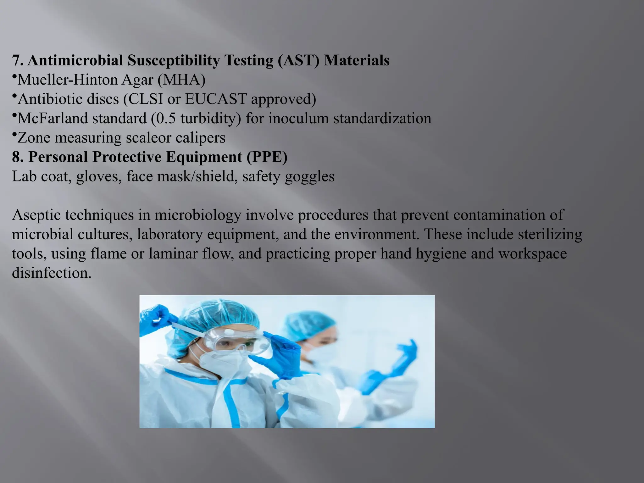
    7. Antimicrobial SusceptibilityTesting (AST) Materials •Mueller-Hinton Agar (MHA) •Antibiotic discs (CLSI or EUCAST approved) •McFarland standard (0.5 turbidity) for inoculum standardization •Zone measuring scaleor calipers 8. Personal Protective Equipment (PPE) Lab coat, gloves, face mask/shield, safety goggles Aseptic techniques in microbiology involve procedures that prevent contamination of microbial cultures, laboratory equipment, and the environment. These include sterilizing tools, using flame or laminar flow, and practicing proper hand hygiene and workspace disinfection.
  • 6.
    Isolation, Identification andAntimicrobial Susceptibility of Staphylococci species: Isolation Methods: Media: Mannitol Salt Agar (MSA), Blood Agar, and Chromogenic Agar. Incubation: 35-37°C for 24-48 hours. Colony Characteristics: Golden-yellow colonies on MSA, beta-hemolysis on Blood A Biochemical Tests: Catalase Test: Positive (bubbles form when hydrogen peroxide is added). Coagulase Test: Differentiates between S. aureus (positive) and other Staphylococcus species (negative). Other tests: DNase, urease, and carbohydrate fermentation tests. Antimicrobial Susceptibility of Staphylococci: Staphylococcus species, particularly Staphylococcus aureus, exhibit varied susceptibility to antibiotics. Methicillin-resistant Staphylococcus aureus (MRSA) is a major clinical concern due to its resistance to β-lactam antibiotics. Commonly Tested Antibiotics: β-lactams: Penicillin, Oxacillin, Cefoxitin (to detect MRSA) Glycopeptides: Vancomycin, Teicoplanin Macrolides: Erythromycin, Clindamycin Fluoroquinolones: Ciprofloxacin Tetracyclines: Doxycycline, Minocycline Testing Methods: Disk Diffusion (Kirby-Bauer)Minimum Inhibitory Concentration .
  • 7.
    Isolation, Identification andAntimicrobial Susceptibility of Streptococci species: Hemolysis Patterns: Alpha-hemolysis (greenish discoloration) Beta-hemolysis (complete hemolysis) Gamma-hemolysis (no hemolysis) CAMP Test: Positive for Group B Streptococcus (S. agalactiae)3. Optochin Sensitivity: S. pneumoniae is sensitive to optochin Bile Solubility: S. pneumoniae is bile soluble Serogrouping: Lancefield grouping (A-H, K-V) using latex agglutination or other methods. Other Methods: Biochemical Tests: API Strep or other biochemical panels Molecular Methods: PCR or 16S rRNA sequencing for confirmation. Antimicrobial susceptibility is tested using disk diffusion or MIC methods. Penicillin remains the first-line treatment, though resistance to macrolides and tetracyclines is increasing in some Streptococcus species.
  • 8.
    Isolation, Identification andAntimicrobial Susceptibility of E.coli and klebsiella species: Isolation and Identification: Clinical samples (urine, sputum, blood, etc.) are cultured on MacConkey agar, where E. coli typically produces pink colonies due to lactose fermentation, while Klebsiella also ferments lactose but forms large, mucoid colonies. Further identification is done using biochemical tests such as the Indole test (positive for E. coli, negative for Klebsiella), citrate utilization (positive for Klebsiella), and motility testing (motile E. coli, non-motile Klebsiella). Antimicrobial Susceptibility: Susceptibility is commonly tested using the Kirby-Bauer disk diffusion method or automated systems like VITEK. Both organisms often exhibit resistance to beta-lactam antibiotics due to beta-lactamase enzyme production, including extended-spectrum beta- lactamases (ESBLs). Carbapenems are typically effective, although carbapenem-resistant strains have emerged, especially in Klebsiella pneumoniae.
  • 9.
    Isolation, Identification andAntimicrobial Susceptibility of Pseudomonas and Aspergillus species: Isolation and Identification: 1. Pseudomonas aeruginosa: Sample sources: Clinical specimens such as sputum, wound swabs, urine, or blood. Culture: Grown on cetrimide agar (selective medium) or MacConkey agar. Colonies are flat with a metallic sheen and characteristic fruity odor. It may produce greenish- blue pigment (pyocyanin). Biochemical tests: Oxidase-positive, catalase-positive, and non-fermentative in TSI slant. Confirmed via API 20NE or automated identification systems. 2. Aspergillus species: Sample sources: Respiratory specimens (sputum, BAL), tissue biopsies. Culture: Grown on Sabouraud Dextrose Agar (SDA) at 25–37°C. Colonies are fast- growing, velvety, and often green (for A. fumigatus), yellow, or black depending on species. Microscopy: Lactophenol cotton blue (LPCB) staining reveals septate hyphae and conidial heads with characteristic arrangement. Identified by morphology or PCR- based molecular methods.
  • 10.
    Antimicrobial/Antifungal Susceptibility: • Pseudomonasaeruginosa: Commonly resistant to: Penicillins, first- and second-generation cephalosporins. Sensitive to: Piperacillin-tazobactam, ceftazidime, cefepime, carbapenems (except imipenem in some resistant strains), and fluoroquinolones. Resistance mechanisms: Efflux pumps, beta-lactamases (including ESBLs and carbapenemases), porin loss. Testing method: Disk diffusion (CLSI guidelines), E-test, or automated systems like VITEK 2. • Aspergillus species: First-line drug: Voriconazole. Alternatives: Amphotericin B, isavuconazole, or echinocandins (like caspofungin). Resistance: Rising azole resistance (especially in A. fumigatus) due to environmental azole use. Testing method: CLSI broth microdilution or E-test strips on RPMI agar.
  • 11.
    Isolation, Identification andAntimicrobial Susceptibility of H. Influenzae species: Isolation: H. influenzae requires two essential growth factors: X factor (hemin) V factor (nicotinamide adenine dinucleotide – NAD ) ⁺ These are both present in Chocolate Agar, which is the preferred medium. The organism does not grow on standard blood agar unless satellitism is observed around organisms like Staphylococcus aureus that provide V factor. Incubation at 35–37°C in 5–10% CO₂ for 24–48 hours is required. Identification: Colony morphology: Smooth, translucent, moist colonies Gram stain: Small Gram-negative coccobacilli Oxidase test: Positive Catalase test: Positive X and V factor requirement test: Growth occurs only with both factors present Porphyrin (ALA) test: Negative for H. influenzae Confirmatory tests: MALDI-TOF MS or PCR-based identification Capsular typing can be done by latex agglutination or PCR for the b capsule gene.
  • 12.
    Antimicrobial Susceptibility Testing(AST): AST is typically done using disk diffusion, E-test, or broth microdilution methods, following CLSI or EUCAST guidelines. Many strains produce β-lactamase, conferring resistance to ampicillin. Susceptible to: Cefotaxime, ceftriaxone, azithromycin, and fluoroquinolones β-lactamase-negative strains: Often susceptible to ampicillin β-lactamase-positive strains: Require β-lactam/β-lactamase inhibitor combinations like amoxicillin-clavulanic acid.
  • 13.
    Isolation, Identification andAntimicrobial Susceptibility of Neisseria species: Sample Collection: N. gonorrhoeae: Urethral, cervical, rectal, and pharyngeal swabs depending on site of infection. N. meningitidis: Blood, cerebrospinal fluid (CSF), or nasopharyngeal swabs. Specimens should be transported rapidly using appropriate transport media like Amies or Stuart’s medium with charcoal to preserve viability. Isolation: Cultivation is done on: Modified Thayer-Martin (MTM) agar or Chocolate agar for N. gonorrhoeae. Chocolate agar or Blood agar for N. meningitidis. Incubation is at 35–37°C in a 5–10% CO -enriched environment ₂ for 24–48 hours. Identification: Microscopy: Gram-negative kidney-shaped diplococci, often intracellular in polymorphonuclear leukocytes (especially for N. gonorrhoeae). Oxidase test: Positive Catalase test: Positive Carbohydrate utilization tests: N. gonorrhoeae: Glucose fermentation only N. meningitidis: Glucose and maltose fermentation
  • 14.
    Molecular testing: NAATs(Nucleic Acid Amplification Tests) are preferred for N. gonorrhoeae diagnosis due to their high sensitivity and specificity. MALDI-TOF MS and PCR can confirm species-level identification. Antimicrobial Susceptibility Testing (AST): Testing follows CLSI or EUCAST guidelines and can include: Disk diffusion E-test (gradient method) Agar dilution or broth microdilution Current resistance patterns: N. gonorrhoeae has shown increasing resistance to penicillin, tetracyclines, and fluoroquinolones. Dual therapy with ceftriaxone and azithromycin was standard, but monotherapy with high-dose ceftriaxone is now often used due to azithromycin resistance. N. meningitidis remains largely susceptible to penicillin, but third-generation cephalosporins (e.g., ceftriaxone, cefotaxime) are preferred for treatment.
  • 15.
    In conclusion, theisolation, identification, and antimicrobial susceptibility testing of clinically important microorganisms are vital for accurate diagnosis and effective treatment. These processes help detect pathogenic species and guide the selection of appropriate antibiotics. Monitoring resistance patterns also aids in controlling the spread of resistant strains. Overall, these methods are essential for improving patient outcomes and public health. Thanks For Listening