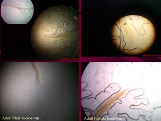
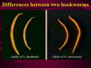
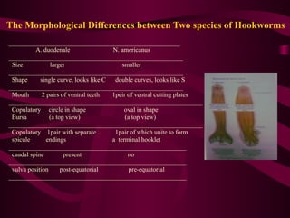
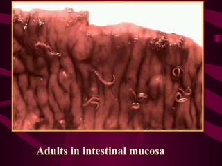
HOOKWORMS
KIMAIGA H.O
MBChB (University of Nairobi)
HOOKWORMS
• These are intestinal nematodes of the family Ancylostomatidae.
• Two main species:Ancylostoma duodenale and Necator
americanus
• A larger group of hookworms infecting animals can invade and
parasitize humans (A. ceylanicum) or can penetrate the human skin
(causing cutaneous larva migrans), but do not develop any further
(A. braziliense, A. caninum, Uncinaria stenocephala).
• Occasionally A. caninum larvae may migrate to the human
intestine, causing eosinophilic enteritis. Ancylostoma caninum
larvae have also been implicated as a cause of diffuse unilateral
subacute neuroretinitis.
ANCYLOSTOMIASIS
• It is an intestinal infection caused by Ancylostoma
duodenale and/or Necator americanus.
• It is recognized to cause intestinal blood loss,
leading to iron deficiency anemia, common in
tropical subtropical areas.
• The disease is also known as miners or brick
makers anemia, Tunnel disease, Hookworm
infection or Egyptian chlorosis.
• It presents when hookworms are in large numbers
in the intestines. Light infections are
asymptomatic.
• They penetrate the skin of man to cause cutaneous
larva migrans. They do not develop further.
• Commonly infects skin, eyes and viscera in
humans.
• It is the second most common human helminthic
infection after ascariasis.
Epidemiology
• All genders are susceptible.
• In endemic areas, high prevalence occurs among school
aged children.
• It is also prevalent pregnant women, malnourished groups
and elderly people.
• A warm, moist climate with temperatures between 250
C-
350
C with a shady, clay, sandy or loamy soil and vegetation
favour larval development.
• 22-26 is the optimal temperature for Ancylostoma
ā„ƒ
duodenale development and 31-35 is suitable for Necator
ā„ƒ
americanus
World Distribution
• Worldwide distribution.
• Hookworm is the second most common human helminthic
infection (after ascariasis).
• It is estimated that 1 billion individuals are infected with the
disease today.
• The main etiology, N. americanus, is found in the Americas,
Sub-Saharan Africa, Australia and Asia.
• A. duodenale is found in more scattered focal areas such as
the Mediterranean region, Middle East, North Africa and
southern Europe.
• Prevalent in Kenya in Coast region, Nyanza, Eastern and all
the slums.
World Distribution
Morphology
Adults:
• Adult hookworms reside in the small intestine of their hosts and
look like an odd piece thread .
• Males measure approximately 8-12 mm long and are bursate,
• The male’s posterior end is expanded to form a copulatory bursate
with two simple similar needle-like slender spicules that do not fuse
at their distal ends.
• Lateral lobes joined by small dorsal lobe
• Lateral lobes supported by fleshy rays
• Females are slightly larger than male measuring approximately 10-
15 mm long.
• Adults of both sexes have a buccal capsule containing sharp teeth.
• They are white or light pinkish when living.
Adult Female hookworm
Adult Male hookworm
Morphology of Hookworm and Strongyloides
Differences between two hookworms
Adults of A. duodenale Adults of N. americanus
The Morphological Differences between Two species of Hookworms
_____________________________________________________
A. duodenale N. americanus
______________________________________________________
Size larger smaller
______________________________________________________
Shape single curve, looks like C double curves, looks like S
______________________________________________________
Mouth 2 pairs of ventral teeth 1peir of ventral cutting plates
____________________________________________________________
Copulatory circle in shape oval in shape
Bursa (a top view) (a top view)
____________________________________________________________
Copulatory 1pair with separate 1pair of which unite to form
spicule endings a terminal hooklet
_______________________________________________________
caudal spine present no
_______________________________________________________
vulva position post-equatorial pre-equatorial
_______________________________________________________
• Scanning electron
micrograph of the mouth
capsule of Ancylostoma
duodenale.
• Has four ventral teeth, two
on each side.
• Scanning electron micrograph
of the mouth capsule of Necator
americanus.
• Has two ventral teeth semi
lunar cutting plates
• Copulatory bursa of N.
americanus(a side view)
• Ancylostoma duodenale -
copulatory bursa and
spines of male(a side
view)
Copulatory bursa of A.
duodenale (a top view)
Copulatory bursa and
spines of N. americanus (a
side view);
I J
I: Adult worm of Ancylostoma duodenale. Anterior end is depicted showing cutting
teeth.
J: Adult worm of Necator americanus. Anterior end showing mouth parts with cutting
plates.
Filariform of Necator americanus pointed tail
Hookworm larvae
20
Rhabdiform
L2
Filariform L3
Eggs
• 60-75 µm by 35-40 µm in size
• Oval in shape
• Shell is thin and colorless. Content is 2-8cells.
• The eggs of Ancylostoma and Necator cannot
be differentiated microscopically
A B
A, B: Hookworm eggs in unstained wet mounts, taken at 400Ɨ magnification.
C D
C: Hookworm egg in an unstained wet mount.
D: hookworm egg viewed under UV microscopy.
E F
E, F: Rhabditiform (L1) larvae (wet preparation). They hatch from eggs are
250-300 µm long and approximately 15-20 µm wide. They have a long buccal canal and
an inconspicuous genital primordium. Rhabditiform larvae are usually not found in stool,
but may be found there is a delay in processing the stool specimen. If larvae are seen in
stool, they must be differentiated from the L1 larvae of Stronyloides stercoralis.
G H
G, H: Filariform (L3) larvae(wet preparation).
Infective, third-stage (L3), filariform larvae are 500-600 µm long. They have a pointed
tail and a striated sheath. These L3 are found in the environment and infect the human
host by penetration of the skin.
PRINCIPAL OF COMMUNICABLE
DISEASES
Agent’s
reservoirs
Route of
exit from
reservoir
Transmissio
n
Route of entry
into host
Susceptible
host
Agent Reservoirs
• Human beings-mainly.
• Dogs
• Cats
Route of Exit from resevior
• Gastrointestinal route- through defecation.
• Respiratory route- through coughing and
sneezing of the infective larval stage(L3).
Mode of Transmission
• Indirect passive transmission-contamination
of soil.
• It occurs after 5 or more minutes of skin
contact with the soil that is infected with the
viable larvae.
Route of Entry into host
• Skin penetration-The larvae transforms and
feeds in conducive (warm, moist)
environment. At the 3rd
larval stage,
filariform larvae, it penetrates intact
skin(foot, hands and buttocks)
• It penetrates within about 5minutes of
contact.
• Eggs are passed in the stool
• Under favorable conditions (moisture, warmth, shade), larvae
hatch in 1 to 2 days and release rhabditiform (L1 ) larvae which
grow in the feces and/or the soil feeding on bacteria
• After 5 to 10 days (and two molts) they become L2, L3 filariform
larvae that are infective and can survive 3 to 4 weeks in favorable
environmental conditions.
• On contact with human skin(foot,hands and buttocks) , they
penetrate the skin using proteolytic enzymes and are carried via
the blood vessels to the right heart then penetrate pulmonary
alveoli, ascend bronchial tree to the pharynx, and are swallowed.
• The larvae reach the small intestine, where they reside and mature
into adults(L5).
Life Cycle (intestinal hookworm infection):
• Adult worms live in the small intestine and mate
• They attach to the small intestine wall.
• They suck blood and feed from the host.
• Hookworms have a anticoagulant like secretion to keep you bleeding once
they bite a blood vessel
• N.amer sucks .03mls and A.duod .15mls of blood and may stay for 2-5
yrs. This is because A.duod. Has teeth like structures while N.amer has
cutting plates.
• Most adult worms are eliminated in 1-2 years but some may take several
years.
• Some A. duodenale larvae, following penetration of the host skin, can
become dormant (in the intestine or muscle). In addition, infection by A.
duodenale may probably also occur by the oral and transmammary route.
N. americanus, however, requires a transpulmonary migration phase.
• NB: If the larvae are unable to complete their migratory cycle in humans,
they migrate just below the skin producingā€ snakeā€ like markings that is
creeping eruption/ cutaneous larva migrans
Life Cycle (intestinal hookworm infection):
Life Cycle (cutaneous larval migrans)
• Cutaneous larval migrans (also known as creeping eruption) is a
zoonotic infection with hookworm species that do not use humans
as a definitive host
• Is caused by 2 main spp related to the hookworm, known as
Ancylostoma brasiliensis & Ancylostoma canina.
• The normal definitive hosts for these species are dogs and cats.
The cycle in the definitive host is very similar to the cycle for the
human species.
1. Eggs are passed in the stool
2. Under favorable conditions (moisture, warmth, shade), larvae
hatch in 1 to 2 days. The released rhabditiform larvae grow in
the feces and/or the soil
3. After 5 to 10 days (and two molts) they become filariform
(third-stage) larvae that are infective. These infective larvae can
survive 3 to 4 weeks in favorable environmental conditions.
4. On contact with the animal host , the larvae penetrate the skin
5. They are not normal parasites of the human hence they penetrate
the skin and find themselves in a strange environment.
6. They cease progression because the human is not their definitive
host and cannot move further in their life cycle.
7. They fail to gain access to the venous system and remain in the
skin without further development.
8. They keep on migrating around the skin within the epidermis,
sometimes as much as several centimeters a day. Some larvae
may persist in deeper tissue after finishing their skin migration.
9. They cause damage to skin – skin eruptions/lesions, thus known
as Cutaneous Larva Migrans.
10. They don’t gain access to the blood system.
11. They attempt to move around without success until they are
empowered by the host’s immune system and die & disintegrate
and in some cases may calcify.
12. Commonest site is skin of feet, hands, buttocks esp. in pple who
go sunbathing.
13. Treatment is not very necessary, but the skin eruptions may be
itchy thus application of steroid creams e.g. tetramethasole.
Antipluritic (antihistamines) may suppress the itchiness (pluritis)
e.g. piriton (chlorphenamine).
14. Angiostrongilus it penetrates the skin but remains cutaneous.
Life Cycle (cutaneous larval migrans)
Susceptible Host
• The disease affects mostly school aged
children. In susceptible children, it causes
mental retardation.
• In pregnant women it causes intrauterine
growth retardation, prematurity as well as low
birth weight of the baby. It is the leading cause
of child morbidity in the developing countries.
• Also elderly people and malnaurished persons.
Pathogenesis and Clinical Manifestations
LARVAL MIGRATION
• Dermatitis, known as "ground itch" or "stool poison".The larvae
penetrating the skin cause allergic reaction, petechiae 0r papule
with itching and burning sensation. Scratching leads to
secondary infection.
• Pneumonitis (allergic reaction), Loeffier's syndrome: cough,
asthma, low fever, biood-tinged sputum or hemoptysis, chest-
pain, alveolar damage, inflammation shadows in lungs under X-
ray. These manifestations go on about 2 weeks.
• Hepatomegaly(due to venous congestion linked)
ADULTS IN SMALL INTESTINE
• Epigastric pain as that of a duodenal ulcer.
• A large worm burden results in microcytic hypochromatic
anemia (character manifestation). The symptoms are
lassitude, edema, palpitation of the heart. In severe case,
death may result from cardiac failure or physical
exhaustion.
• Allotriophagy (orpica) is due to the lack of trace element
iron .
• Amenorrhea, sterility, abortion may take place in women.
• Gastrointestinal bleeding
• Infantile hookworm disease
Hookworm Disease
• Cutaneous phase
– During larval penetration, lasts 1-2 wks;
– More common in N. americanus
– Urticarial, ground itch (ancylostome dermatitis)
– Exacerbated by pyrogenic bacteria in the larvae.
• Pulmonary phase
– Larvae break through the Bvs in the alveoli
– Bronchitis & bronchopneumonia
– Marked eosinophilia
– Haemorrhage, which is serious in massive infection.
– Symptoms like severe dry cough and pneumonitis (inflammation of
alveoli).
– NB: no voice hoarseness unlike in Ascaris pneumonitis.
• Intestinal phase
– Adult worms in the intestines.
– Worms repeatedly cut intestinal mucosa.
– Secrete anticoagulants.
– Anaemia
• Worms feed on blood (A. duodenale – 0.26ml; N.
americanus – 0.03ml)
• Loss of iron – hypochromic microcytic anaemia
• Loss of Vit B12 and folic acid deficiency – macrocytic
anaemia. It causes ilitis.
• Dimorphic anaemia.
• Severity depends on number and species of hookworms.
– Nephritic syndromes – proteinuria & loss of plasma
proteins.
Clinical Presentation
• There are no specific signs and symptoms for the infection.
This is because they present as a combination of intestinal
inflammation and progressively iron deficiency.
• Ground itch, urticaria- This is an inflammatory response
resulting from larval invasion into the skin causing intense
local itching and lesions on the legs and feet.
• Cutaneous larva migrans. These are tortuous tunnels of the
larvae between the stratum basale and stratrum corneum.
• Pneumonia- Inflammation of the lung parenchyma as result
of the larval break into the alveoli.
• Serum IgE tents to be high
• Hyperesinophilia
• Loeffluer’s syndrome- comprising of hilum lymphadenopathy,
pneumonitis and pulmonary eosinophilia.
• Coughing - Results from heavy larval migration up the trachea,
which causes tracheal irritation, coughing, wheezing and chest
pains.
• Early or late stages can be characterized by vomiting, nausea,
constipation, epigastic pains, indigestion, diarrhea and
gastrointestinal discomfort - As a result of maturation of the larva.
• Pale mucous membranes- conjuctiva, tongue, nail beds
• Nails are spoonlike
• Advanced or severe infections are characterized by anemia and
protein deficiency, including cardiac failure, emancipation and
abdominal distension with ascites, Mental and growth
retardation,delayed puberty, abortions/stillbirths, FTG poor
memory
Clinical Manifestations
• G.I manifestations
– Intermittent abdominal pains
– Epigastric tenderness (duodenal ulcers)
– Constipation
– Anorexia
– Blood-flecked/spotted faeces
• Effects of anaemia
– Pallor-eyes, lips & tongue
– Light yellow and dry skin
– Dry hair
– Enlarged liver due to
Congestive Cardiac Failure
– Heart murmurs coz of
anaemia.
– Protein loosing enteropathy
leads to a drop in plasma
protein. Oedema develops
due to exacerbation of fluid
into extracellular space.
– Oedema (usually facial but
may be generalised)
– Puffy face & swollen
eyelids
– Potbellies due to ascites
– Koilonychia (spoon nails as
a result of hypochromic
microcytic anaemia, esp
iron deficiency)
– Stunted growth and general
malaise
– Acute mental distress
– Mental dullness
– Delayed puberty
– Heavy infections – heart
failure
Abdominal distention
Adults in intestinal mucosa
Laboratory Diagnosis
• Microscopic identification of eggs in the stool. The
recommended procedure:
1. Collect a stool specimen.
2. Fix the specimen in 10% formalin.
3. Concentrate using the formalin–ethyl acetate
sedimentation technique.
4. Examine a wet mount of the sediment.
• Where concentration procedures are not available, a
direct wet mount examination of the specimen is
adequate for detecting moderate to heavy infections. For
quantitative assessments of infection, various methods
such as the Kato-Katz can be used.
• Cutaneous larval migrans is usually diagnosed clinically,
as there are no serologic tests for zoonotic hookworm
infections. Larvae may be seen in stained tissue sections,
but this procedure is usually not recommended as the
parasites are usually not found in the visible track.
• Culture of stool in test tube(oval eggs with colourless
shell, eggs have blastomeres)
• Indirect PBF RBCs-microcytic hypochromic anaemia- Hemoglobin
is lower than 120g/L in man, 110g/L in woman.
• Clinical diagnosis –pallor of mucus membranes eyes hands
• Saturated brine flotation technique
Treatment
• Expulsion of adult worm
– Albendazole (efficancy 72%)
– Mebendazole (efficancy 15%)
– Thiabendazole
– Pyrantel Pamoate
– They kill the adult worms by binding to the beta-tubulin and therefore,
preventing microtubule polymerization within the parasite.
• Dosage is the same for children as for adults
– Albendazole: 400 mg orally once
– Mebendazole: 100 mg twice a day for 3 days OR 500mg orally once.
– Pyrantel pamoate: 11mg/kg (up to a max of 1mg) orally daily for 3 days
• Larvae
– Thiabendazole (migratory or dormant larvae)
– Levamisole (less effective against N. americanus)
• WHO recommends no anti-helminthic treatment
to pregnant women at least before the end of the
first trimester.
• Mass deworming programs in schools and
children at home, twice annually.
• Deworming of pet dogs is also necessary.
• Treatment of anaemia
– Iron supplements
– Folic acid & Vit B12 in some cases
– Transfussion in severe anaemia in children
• Antispasmodics for abdominal pain
• Proteins therapy
Prevention and Control (Levels)
a) Reservoir
• Treatment of infected individuals
• Mass deworming programs in schools and
children at home, twice annually.
• Deworming of pet dogs is also necessary.
b) Portal of exit
• Proper waste disposal
c) Transmission
• Soil treatment-heat therapy, spraying of antihelminths.
d) Portal of entry
• Usage of proper footwear.
• Health education
• Mass public awareness and social education programs on modes of
transmission of the parasite
e) Host
• Treatment of the infected persons using albendazole, mebendazole,
levamisole and pyrantel pamoate.( refer to treatment of reservoir)
Prevention: putting on shoes

MICROBIOLOGY PRESENTATION ON HOOKWORMS.pptx

  • 1.
  • 2.
    HOOKWORMS • These areintestinal nematodes of the family Ancylostomatidae. • Two main species:Ancylostoma duodenale and Necator americanus • A larger group of hookworms infecting animals can invade and parasitize humans (A. ceylanicum) or can penetrate the human skin (causing cutaneous larva migrans), but do not develop any further (A. braziliense, A. caninum, Uncinaria stenocephala). • Occasionally A. caninum larvae may migrate to the human intestine, causing eosinophilic enteritis. Ancylostoma caninum larvae have also been implicated as a cause of diffuse unilateral subacute neuroretinitis.
  • 3.
    ANCYLOSTOMIASIS • It isan intestinal infection caused by Ancylostoma duodenale and/or Necator americanus. • It is recognized to cause intestinal blood loss, leading to iron deficiency anemia, common in tropical subtropical areas. • The disease is also known as miners or brick makers anemia, Tunnel disease, Hookworm infection or Egyptian chlorosis.
  • 4.
    • It presentswhen hookworms are in large numbers in the intestines. Light infections are asymptomatic. • They penetrate the skin of man to cause cutaneous larva migrans. They do not develop further. • Commonly infects skin, eyes and viscera in humans. • It is the second most common human helminthic infection after ascariasis.
  • 5.
    Epidemiology • All gendersare susceptible. • In endemic areas, high prevalence occurs among school aged children. • It is also prevalent pregnant women, malnourished groups and elderly people. • A warm, moist climate with temperatures between 250 C- 350 C with a shady, clay, sandy or loamy soil and vegetation favour larval development. • 22-26 is the optimal temperature for Ancylostoma ā„ƒ duodenale development and 31-35 is suitable for Necator ā„ƒ americanus
  • 6.
    World Distribution • Worldwidedistribution. • Hookworm is the second most common human helminthic infection (after ascariasis). • It is estimated that 1 billion individuals are infected with the disease today. • The main etiology, N. americanus, is found in the Americas, Sub-Saharan Africa, Australia and Asia. • A. duodenale is found in more scattered focal areas such as the Mediterranean region, Middle East, North Africa and southern Europe. • Prevalent in Kenya in Coast region, Nyanza, Eastern and all the slums.
  • 7.
  • 8.
    Morphology Adults: • Adult hookwormsreside in the small intestine of their hosts and look like an odd piece thread . • Males measure approximately 8-12 mm long and are bursate, • The male’s posterior end is expanded to form a copulatory bursate with two simple similar needle-like slender spicules that do not fuse at their distal ends. • Lateral lobes joined by small dorsal lobe • Lateral lobes supported by fleshy rays • Females are slightly larger than male measuring approximately 10- 15 mm long. • Adults of both sexes have a buccal capsule containing sharp teeth. • They are white or light pinkish when living.
  • 9.
  • 10.
    Morphology of Hookwormand Strongyloides
  • 11.
    Differences between twohookworms Adults of A. duodenale Adults of N. americanus
  • 12.
    The Morphological Differencesbetween Two species of Hookworms _____________________________________________________ A. duodenale N. americanus ______________________________________________________ Size larger smaller ______________________________________________________ Shape single curve, looks like C double curves, looks like S ______________________________________________________ Mouth 2 pairs of ventral teeth 1peir of ventral cutting plates ____________________________________________________________ Copulatory circle in shape oval in shape Bursa (a top view) (a top view) ____________________________________________________________ Copulatory 1pair with separate 1pair of which unite to form spicule endings a terminal hooklet _______________________________________________________ caudal spine present no _______________________________________________________ vulva position post-equatorial pre-equatorial _______________________________________________________
  • 13.
    • Scanning electron micrographof the mouth capsule of Ancylostoma duodenale. • Has four ventral teeth, two on each side. • Scanning electron micrograph of the mouth capsule of Necator americanus. • Has two ventral teeth semi lunar cutting plates
  • 15.
    • Copulatory bursaof N. americanus(a side view) • Ancylostoma duodenale - copulatory bursa and spines of male(a side view)
  • 16.
    Copulatory bursa ofA. duodenale (a top view) Copulatory bursa and spines of N. americanus (a side view);
  • 17.
    I J I: Adultworm of Ancylostoma duodenale. Anterior end is depicted showing cutting teeth. J: Adult worm of Necator americanus. Anterior end showing mouth parts with cutting plates.
  • 18.
    Filariform of Necatoramericanus pointed tail
  • 20.
  • 21.
    Eggs • 60-75 µmby 35-40 µm in size • Oval in shape • Shell is thin and colorless. Content is 2-8cells. • The eggs of Ancylostoma and Necator cannot be differentiated microscopically
  • 24.
    A B A, B:Hookworm eggs in unstained wet mounts, taken at 400Ɨ magnification.
  • 25.
    C D C: Hookwormegg in an unstained wet mount. D: hookworm egg viewed under UV microscopy.
  • 26.
    E F E, F:Rhabditiform (L1) larvae (wet preparation). They hatch from eggs are 250-300 µm long and approximately 15-20 µm wide. They have a long buccal canal and an inconspicuous genital primordium. Rhabditiform larvae are usually not found in stool, but may be found there is a delay in processing the stool specimen. If larvae are seen in stool, they must be differentiated from the L1 larvae of Stronyloides stercoralis.
  • 27.
    G H G, H:Filariform (L3) larvae(wet preparation). Infective, third-stage (L3), filariform larvae are 500-600 µm long. They have a pointed tail and a striated sheath. These L3 are found in the environment and infect the human host by penetration of the skin.
  • 28.
    PRINCIPAL OF COMMUNICABLE DISEASES Agent’s reservoirs Routeof exit from reservoir Transmissio n Route of entry into host Susceptible host
  • 29.
    Agent Reservoirs • Humanbeings-mainly. • Dogs • Cats
  • 30.
    Route of Exitfrom resevior • Gastrointestinal route- through defecation. • Respiratory route- through coughing and sneezing of the infective larval stage(L3).
  • 31.
    Mode of Transmission •Indirect passive transmission-contamination of soil. • It occurs after 5 or more minutes of skin contact with the soil that is infected with the viable larvae.
  • 32.
    Route of Entryinto host • Skin penetration-The larvae transforms and feeds in conducive (warm, moist) environment. At the 3rd larval stage, filariform larvae, it penetrates intact skin(foot, hands and buttocks) • It penetrates within about 5minutes of contact.
  • 33.
    • Eggs arepassed in the stool • Under favorable conditions (moisture, warmth, shade), larvae hatch in 1 to 2 days and release rhabditiform (L1 ) larvae which grow in the feces and/or the soil feeding on bacteria • After 5 to 10 days (and two molts) they become L2, L3 filariform larvae that are infective and can survive 3 to 4 weeks in favorable environmental conditions. • On contact with human skin(foot,hands and buttocks) , they penetrate the skin using proteolytic enzymes and are carried via the blood vessels to the right heart then penetrate pulmonary alveoli, ascend bronchial tree to the pharynx, and are swallowed. • The larvae reach the small intestine, where they reside and mature into adults(L5). Life Cycle (intestinal hookworm infection):
  • 34.
    • Adult wormslive in the small intestine and mate • They attach to the small intestine wall. • They suck blood and feed from the host. • Hookworms have a anticoagulant like secretion to keep you bleeding once they bite a blood vessel • N.amer sucks .03mls and A.duod .15mls of blood and may stay for 2-5 yrs. This is because A.duod. Has teeth like structures while N.amer has cutting plates. • Most adult worms are eliminated in 1-2 years but some may take several years. • Some A. duodenale larvae, following penetration of the host skin, can become dormant (in the intestine or muscle). In addition, infection by A. duodenale may probably also occur by the oral and transmammary route. N. americanus, however, requires a transpulmonary migration phase. • NB: If the larvae are unable to complete their migratory cycle in humans, they migrate just below the skin producingā€ snakeā€ like markings that is creeping eruption/ cutaneous larva migrans
  • 35.
    Life Cycle (intestinalhookworm infection):
  • 37.
    Life Cycle (cutaneouslarval migrans) • Cutaneous larval migrans (also known as creeping eruption) is a zoonotic infection with hookworm species that do not use humans as a definitive host • Is caused by 2 main spp related to the hookworm, known as Ancylostoma brasiliensis & Ancylostoma canina. • The normal definitive hosts for these species are dogs and cats. The cycle in the definitive host is very similar to the cycle for the human species. 1. Eggs are passed in the stool 2. Under favorable conditions (moisture, warmth, shade), larvae hatch in 1 to 2 days. The released rhabditiform larvae grow in the feces and/or the soil 3. After 5 to 10 days (and two molts) they become filariform (third-stage) larvae that are infective. These infective larvae can survive 3 to 4 weeks in favorable environmental conditions.
  • 38.
    4. On contactwith the animal host , the larvae penetrate the skin 5. They are not normal parasites of the human hence they penetrate the skin and find themselves in a strange environment. 6. They cease progression because the human is not their definitive host and cannot move further in their life cycle. 7. They fail to gain access to the venous system and remain in the skin without further development. 8. They keep on migrating around the skin within the epidermis, sometimes as much as several centimeters a day. Some larvae may persist in deeper tissue after finishing their skin migration.
  • 39.
    9. They causedamage to skin – skin eruptions/lesions, thus known as Cutaneous Larva Migrans. 10. They don’t gain access to the blood system. 11. They attempt to move around without success until they are empowered by the host’s immune system and die & disintegrate and in some cases may calcify. 12. Commonest site is skin of feet, hands, buttocks esp. in pple who go sunbathing. 13. Treatment is not very necessary, but the skin eruptions may be itchy thus application of steroid creams e.g. tetramethasole. Antipluritic (antihistamines) may suppress the itchiness (pluritis) e.g. piriton (chlorphenamine). 14. Angiostrongilus it penetrates the skin but remains cutaneous.
  • 40.
    Life Cycle (cutaneouslarval migrans)
  • 41.
    Susceptible Host • Thedisease affects mostly school aged children. In susceptible children, it causes mental retardation. • In pregnant women it causes intrauterine growth retardation, prematurity as well as low birth weight of the baby. It is the leading cause of child morbidity in the developing countries. • Also elderly people and malnaurished persons.
  • 42.
    Pathogenesis and ClinicalManifestations LARVAL MIGRATION • Dermatitis, known as "ground itch" or "stool poison".The larvae penetrating the skin cause allergic reaction, petechiae 0r papule with itching and burning sensation. Scratching leads to secondary infection. • Pneumonitis (allergic reaction), Loeffier's syndrome: cough, asthma, low fever, biood-tinged sputum or hemoptysis, chest- pain, alveolar damage, inflammation shadows in lungs under X- ray. These manifestations go on about 2 weeks. • Hepatomegaly(due to venous congestion linked)
  • 43.
    ADULTS IN SMALLINTESTINE • Epigastric pain as that of a duodenal ulcer. • A large worm burden results in microcytic hypochromatic anemia (character manifestation). The symptoms are lassitude, edema, palpitation of the heart. In severe case, death may result from cardiac failure or physical exhaustion. • Allotriophagy (orpica) is due to the lack of trace element iron . • Amenorrhea, sterility, abortion may take place in women. • Gastrointestinal bleeding • Infantile hookworm disease
  • 44.
    Hookworm Disease • Cutaneousphase – During larval penetration, lasts 1-2 wks; – More common in N. americanus – Urticarial, ground itch (ancylostome dermatitis) – Exacerbated by pyrogenic bacteria in the larvae. • Pulmonary phase – Larvae break through the Bvs in the alveoli – Bronchitis & bronchopneumonia – Marked eosinophilia – Haemorrhage, which is serious in massive infection. – Symptoms like severe dry cough and pneumonitis (inflammation of alveoli). – NB: no voice hoarseness unlike in Ascaris pneumonitis.
  • 45.
    • Intestinal phase –Adult worms in the intestines. – Worms repeatedly cut intestinal mucosa. – Secrete anticoagulants. – Anaemia • Worms feed on blood (A. duodenale – 0.26ml; N. americanus – 0.03ml) • Loss of iron – hypochromic microcytic anaemia • Loss of Vit B12 and folic acid deficiency – macrocytic anaemia. It causes ilitis. • Dimorphic anaemia. • Severity depends on number and species of hookworms. – Nephritic syndromes – proteinuria & loss of plasma proteins.
  • 47.
    Clinical Presentation • Thereare no specific signs and symptoms for the infection. This is because they present as a combination of intestinal inflammation and progressively iron deficiency. • Ground itch, urticaria- This is an inflammatory response resulting from larval invasion into the skin causing intense local itching and lesions on the legs and feet. • Cutaneous larva migrans. These are tortuous tunnels of the larvae between the stratum basale and stratrum corneum. • Pneumonia- Inflammation of the lung parenchyma as result of the larval break into the alveoli. • Serum IgE tents to be high • Hyperesinophilia
  • 48.
    • Loeffluer’s syndrome-comprising of hilum lymphadenopathy, pneumonitis and pulmonary eosinophilia. • Coughing - Results from heavy larval migration up the trachea, which causes tracheal irritation, coughing, wheezing and chest pains. • Early or late stages can be characterized by vomiting, nausea, constipation, epigastic pains, indigestion, diarrhea and gastrointestinal discomfort - As a result of maturation of the larva. • Pale mucous membranes- conjuctiva, tongue, nail beds • Nails are spoonlike • Advanced or severe infections are characterized by anemia and protein deficiency, including cardiac failure, emancipation and abdominal distension with ascites, Mental and growth retardation,delayed puberty, abortions/stillbirths, FTG poor memory
  • 49.
    Clinical Manifestations • G.Imanifestations – Intermittent abdominal pains – Epigastric tenderness (duodenal ulcers) – Constipation – Anorexia – Blood-flecked/spotted faeces
  • 50.
    • Effects ofanaemia – Pallor-eyes, lips & tongue – Light yellow and dry skin – Dry hair – Enlarged liver due to Congestive Cardiac Failure – Heart murmurs coz of anaemia. – Protein loosing enteropathy leads to a drop in plasma protein. Oedema develops due to exacerbation of fluid into extracellular space. – Oedema (usually facial but may be generalised) – Puffy face & swollen eyelids – Potbellies due to ascites – Koilonychia (spoon nails as a result of hypochromic microcytic anaemia, esp iron deficiency) – Stunted growth and general malaise – Acute mental distress – Mental dullness – Delayed puberty – Heavy infections – heart failure
  • 51.
  • 52.
  • 53.
    Laboratory Diagnosis • Microscopicidentification of eggs in the stool. The recommended procedure: 1. Collect a stool specimen. 2. Fix the specimen in 10% formalin. 3. Concentrate using the formalin–ethyl acetate sedimentation technique. 4. Examine a wet mount of the sediment. • Where concentration procedures are not available, a direct wet mount examination of the specimen is adequate for detecting moderate to heavy infections. For quantitative assessments of infection, various methods such as the Kato-Katz can be used.
  • 54.
    • Cutaneous larvalmigrans is usually diagnosed clinically, as there are no serologic tests for zoonotic hookworm infections. Larvae may be seen in stained tissue sections, but this procedure is usually not recommended as the parasites are usually not found in the visible track. • Culture of stool in test tube(oval eggs with colourless shell, eggs have blastomeres) • Indirect PBF RBCs-microcytic hypochromic anaemia- Hemoglobin is lower than 120g/L in man, 110g/L in woman. • Clinical diagnosis –pallor of mucus membranes eyes hands • Saturated brine flotation technique
  • 55.
    Treatment • Expulsion ofadult worm – Albendazole (efficancy 72%) – Mebendazole (efficancy 15%) – Thiabendazole – Pyrantel Pamoate – They kill the adult worms by binding to the beta-tubulin and therefore, preventing microtubule polymerization within the parasite. • Dosage is the same for children as for adults – Albendazole: 400 mg orally once – Mebendazole: 100 mg twice a day for 3 days OR 500mg orally once. – Pyrantel pamoate: 11mg/kg (up to a max of 1mg) orally daily for 3 days • Larvae – Thiabendazole (migratory or dormant larvae) – Levamisole (less effective against N. americanus)
  • 56.
    • WHO recommendsno anti-helminthic treatment to pregnant women at least before the end of the first trimester. • Mass deworming programs in schools and children at home, twice annually. • Deworming of pet dogs is also necessary. • Treatment of anaemia – Iron supplements – Folic acid & Vit B12 in some cases – Transfussion in severe anaemia in children • Antispasmodics for abdominal pain • Proteins therapy
  • 57.
    Prevention and Control(Levels) a) Reservoir • Treatment of infected individuals • Mass deworming programs in schools and children at home, twice annually. • Deworming of pet dogs is also necessary.
  • 58.
    b) Portal ofexit • Proper waste disposal c) Transmission • Soil treatment-heat therapy, spraying of antihelminths. d) Portal of entry • Usage of proper footwear. • Health education • Mass public awareness and social education programs on modes of transmission of the parasite e) Host • Treatment of the infected persons using albendazole, mebendazole, levamisole and pyrantel pamoate.( refer to treatment of reservoir)
  • 59.