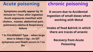
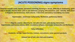
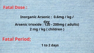
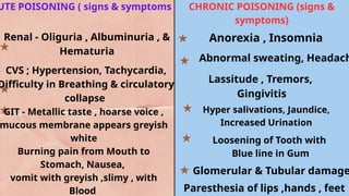
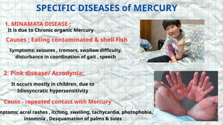
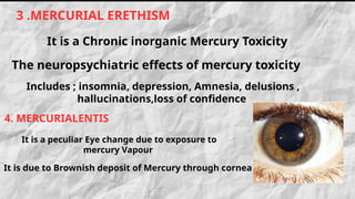
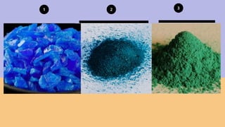
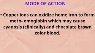
Inorganic
Metallic Irritants
1. ARSENIC
2. MERCURY
4. COPPER
3. LEAD
ARSENIC - TOXIC COMPOUNDS
1. Arsenic. Trioxide
used as weed killers , insecticides, Rat
poisons ,fruit sprays
2. Copper arsenite and
copper acetoarsenite -
3.Sodium and Potassium arsenate
- used as colouring pigment and fly paper
5.Arseniuretted hydrogen / Arsine
Used as colouring
Agent
4. Arsenic sulphide
- Colourless gas
Mode of Action
Arsenic interferes with cellular respiration by mitochondrial oxidative
phosphorylation and added with pyruvate dehydrogenase Consequently,
conversion of pyruvate to acetyl CoA is decreased, citric acid cycle activity is
decreased and production of ( cellular ATP is decreased).
• It inhibits cellular glucose uptake, gluconeogenesis, fatty acid oxidation and
further production of acetyl CoA
Locally, it causes irritation of the mucous membranes, and remotely, depression of
the nervous system.
• Arsenic is a carcinogenic substance since lung, skin and bladder (transitional cell)
carcinoma has been observed in populations with multiple exposures.
Acute poisoning chronic poisoning
Symptoms usually appear by 10
minute to 1 hour after ingestion
Acute exposures manifest with
cholera , nausea, abdominal pain,
pulmonary edema & Respiratory
failure
* In FULMINANT Type - when large
dose is taken (>3g) , no GIT
symptoms and Death occurs in 1 to
3hr
It occurs due to Accidental
ingestion of small doses when
working with Metal
intake of food &drink in which
there are traces of arsenic
Recovery from Acute
Poisoning
(ACUTE POISONING) signs symptoms
Sweetish metallic taste, nausea, persistent vomiting, burning in throat, difficulty in swallowing, garlic
odor in breath, intense thirst, pain in and abdomen, purging with tenesmus, pain . Initially, defecation
is frequent and involuntary, dark-colored, it becomes and watery resembling rice-water.
Hypotension , ventricular tachycardia, fibrillation, pulmonary edema
Oliguria, uremia, albuminuria, red cells and casts, pain during micturition.
Hypotension, pulmonary edema, ARDS, circulatory collapse, ventricular tachycardia and fibrillation.
Fatty infiltration.
Headache, vertigo, hyperthermia, tremors, convulsions, coma, general paralysis.
.
Delayed loss of hair, skin rash and eruptions
Skin :
CNS :
Hepatic :
CVS:
GIT :
Renal :
Chronic Poison signs and symptoms
. GIT disturbances
ii. Catarrhal changes
iii. Skin rashes
iv. Nervous disturbances
Fatal Dose :
Inorganic Arsenic : 0.6mg / kg /
day
Arsenic trioxide : 120 - 200mg ( adults)
2 mg / kg ( children )
Fatal Period;
1 to 2 days
CIRCUMSTANCES OF ARSENIC POISON
1. Contaminated groundwater/ well water
2. Industrial exposure (mining, smelting,)
Pesticide or herbicide use (agricultural or gardening)
4. Ingestion of contaminated food (rice, seafood, fruits)
5. Exposure to arsenic-treated wood or lumber
6. Medical procedures (cancer treatment, dermatology)
7. Contaminated soil or dust (near industrial sites)
8. Old or poorly maintained buildings (paint, insulation)
9. Imported consumer products (, jewelry, cosmetics)
Remove the patient from the source of exposure and gastric
decontamination should be done, if there is evidence of arsenic in GIT.
Vigorous scrubbing with soap and water will remove arsenic from skin.
Administer BAL in usual doses.
Vitamin B complex and IV sodium thiosulfate are useful; hemodialysis is
not indicated.
Mineral supplements and antioxidant therapy are beneficial.
. Symptomatic treatment.
outside food should be forbidden
TREATMENT
Stomach: It may be normal or may show a
chronic gastritis. Some rugae may show
patchy inflammatory redness or focal
ulceration.
ii. Small intestine: Reddish with thickened
mucosa.
Liver: Hepatomegaly, fatty degeneration or
even necrosis with non-cirrhotic portal
fibrosis.
iv. Kidneys: Tubular necrosis.
v. Heart: Myocardial necrosis may be seen.
• Bone marrow histopathology will show
hypoplasia
Emaciation,
pigmentation,
keratosis,
alopecia, white
streaks nails,
jaundice,
wasting of muscles,
and ulceration of
sasal septum.
POSTMORTEM FINDINGS :
External ;
Internal :
1. People living near industrial sites
or contaminated areas
2. Agricultural workers or farmers
3. Individuals with compromised
immune systems
4. Pregnant women and fetuses
5. Children under 6 years old
HIgh Risk
Groups
Colurdess, odorless, white crystalline powder, but has a nauseous
metallic taste. It is the most toxic inorganic salt, and common cause of
acute poisoning
(calomel): Heavy, amorphous, white and tasteless powder
I(cinnabar or vermilion): It is not absorbed tamsugh sin, and is as such
non-poisonous (red crystalline powder)
B. Organic salts include mettyi mercury (most toxic) dimethyl mercury,
ethyl mercury and phenyl mercury.
MERCURIC CHLORIDE
MERCUROUS CHLORIDE
MERCURIC SULPHIDE
MERCURIC OXIDE, and IODIDE
2. MERCURY
Mode of Action
◆ Mercury binds with sulfhydryl groups resulting
in enzyme inhibition and pathological alteration of
cellular membranes.
• Elemental mercury and methyl mercury are toxic
to the CNS. Metallic mercury vapor is also a
pulmonary irritant.
Inorganic mercury salts are corrosive to the skin,
eyes and GIT, and nephrotoxic.
Absorption and Excretion
• It is absorbed through the GIT and respiratory tract.
◆ After absorption, mercury gets deposited in all tissues,
particularly in the liver, kidneys, spleen and bones. When
inhaled, the maximum concentration occurs in the brain.
• Mainly excreted through the kidneys (urine), liver (bile)
and colonic mucous membrane (feces). It passes rapidly
to the fetus through placental
ACUTE POISONING CHRONIC POISONING
results from:
Continuous accidental absorption by
workers.
◆ Excessive therapeutic use.
◆ Recovery from a large dose.
◆ If an ointment is used as an external
application for a long time.
In the US, exposure to organic mercury is
primarily through ingestion of
contaminated fish (seafood).
Mercurialism is poisoning
resulting from the ingestion or
inhalation of mercury /its
compound ,It can be acute or
chronic.
The target organ for inhaled
mercury vapor is mainly the
brain, mercurous & mercuric salts
damage the GUT lining and
kidneys, while methyl mercury is
widely distributed throughout the
UTE POISONING ( signs & symptoms CHRONIC POISONING (signs &
symptoms)
Anorexia , Insomnia
Abnormal sweating, Headach
Lassitude , Tremors,
Gingivitis
Hyper salivations, Jaundice,
Increased Urination
Loosening of Tooth with
Blue line in Gum
Glomerular & Tubular damage
Paresthesia of lips ,hands , feet
Renal - Oliguria , Albuminuria , &
Hematuria
CVS ; Hypertension, Tachycardia,
Difficulty in Breathing & circulatory
collapse
GIT - Metallic taste , hoarse voice ,
mucous membrane appears greyish
white
Burning pain from Mouth to
Stomach, Nausea,
vomit with greyish ,slimy , with
Blood
SPECIFIC DISEASES of MERCURY
1. MINAMATA DISEASE ;
It is due to Chronic organic Mercury
Causes ; Eating contaminated & shell Fish
Symptoms: seizures , tremors, swallow difficulty,
disturbance in coordination of gait , speech
2. Pink disease/ Acrodynia;
It occurs mostly in children, due to
Idiosyncratic hypersensitivity
Cause - repeated contact with Mercury
mptoms; acral rashes , itching, swelling, tachycardia, photophobia,
insomnia , Desquamation of palms & soles
Includes ; insomnia, depression, Amnesia, delusions ,
hallucinations,loss of confidence
It is due to Brownish deposit of Mercury through cornea
3 .MERCURIAL ERETHISM
It is a Chronic inorganic Mercury Toxicity
The neuropsychiatric effects of mercury toxicity
4. MERCURIALENTIS
It is a peculiar Eye change due to exposure to
mercury Vapour
FATAL DOSE :
◆ 1-4 g of mercuric chloride (30-50 mg/kg)
◆ 10-60 mg/kg of methyl mercury
◆ 10 mg/m³ of mercury vapor.
FATAL PERIOD ; 3 to 5 days
TREATMENT
Remove the patient from the source of exposure.
ii. N-acetyl penicillamine is the chelator of choice.
How- ever, DMPS may improve the neurological
features.
iii. Oral hygiene.
iv. Demulcent drinks.
v. Saline purgatives.
POSTMORTEM FINDINGS
Emaciated body with pale skin. Erosions of oral mucosa, gum o
lower jaw may show bluish gray lines of pigment deposition,
along with loosening of teeth.
• Brain: In organic mercury poisoning, the brain is
predominantly affected. Constriction of visual
EXTERNAL
INTERNAL
mercury Poisoning may cause Cerebral infarctions ,
pneumonia, Renal cortical necrosis
LEAD
Lead (shisha) is ubiquitous in our environment but has no physiologic
role in biological systems.
• Acute toxicity is related to occupational exposure and is quite
uncommon, while chronic toxicity is much more common (commonest
heavy metal causing toxicity).
Central nervous system (CNS) and neuromuscular manifestations
result from intense exposure, while gastrointestinal tract (GIT)
features result from exposure over longer periods.
The nervous system appears to be the most sensitive and chief target
for lead induced toxicity
TOXIC COMPOUNDS :
LEAD ACETATE ( sugar of lead) -
LEAD TETRAOXIDE ( vermilion) -
TETRAETHYL LEAD -
LEAD SULFIDE ( surma )-
LEAD CARBONATE -
earlier used as astringent and sedative
for sprains
Used as Sindoor
Antiknock for petrol
Applied on the eyes
Manufacture of Paints
Lead combines with sulfhydryl groups and interferes with
mitochondrial oxidative phosphorylation, ATPases,
calcium-dependent messengers, and enhances oxidation
and cell apoptosis.
This causes defective heme synthesis, proximal renal
tubular and osteoblast dysfunction.
ii. In the CNS, it has deleterious effects on the nerve cells
and myelin sheaths, and also causes cerebral edema.
MODE of ACTION ;
ABSORPTION & EXCRETION :
• Lead is absorbed through the GIT, respiratory tract (dust and fumes) and skin (lead
tetraoxide).
• . GIT lead absorption is increased by iron deficiency and low dietary calcium, and
decreased by coingestion with food.
It is a cumulative poison. In chronic exposure, it deposits in tissues, mostly in the
bones (90%), liver and kidneys.
• It is mainly excreted through the urine (70%), but rate of excretion is low; smaller
amounts are eliminated via feces, and scant amounts via the hair, nails and sweat.
ACUTE (Signs & Symptoms) :
GIT: Metallic taste, dry throat,
thirst, vomiting, nausea,
burning abdominal pain (colic)
and blood-stained diarrhea
leading to circulatory collapse.
• CNS: Headache, lethargy,
arthralgia, myalgia, anorexia,
insomnia, paresthesia,
depression, coma and death
TREATMENT
1. Gastric lavage with
1% solu of
sodium/magnesium
sulphate
2 . Whole bowel
irrigation
3. Demulcents &
repeated cathartics
4.Hemodialysis
CHRONIC LEAD POISONING
* . It was also called colica pictorum (the colic of painters), painter's
colic or Devonshire colic.
* Lead is a cumulative poison, remains accumulated in bones as
phosphate and carbonate.
* High calcium level favors storage, while calcium deficiency causes
lead to be released into the blood stream
* Other factors promoting release of stored lead: acidosis, fever,
sweating, consumption of alcohol and exposure to sunlight.
Fatal Dose ; 1. Lead carbonate : 40g
2. Lead Acetate - 20g
Fatal
Period ;
1 to 2 days
Laboratory Diagnosis ;
1. Porphyrinuria due to coproporphyrin
2. Blood lead level >70 - 100ug /dL.
Protoporphyrin - >35 ug/ dL
3. Urine lead level >0.15 - 0.3 mg/L
POSTMORTEM FINDINGS
A blue line may be seen on the gums in patients with poor oral
hygiene, but it is not a constant feature.
. Paralyzed muscles show fatty degeneration.
Heart: It may be hypertrophied and there may be atherosclerosis of
aorta.
Stomach and intestines: It may show ulcerative or hemorrhagic
changes with contraction and thickening.
Liver and kidneys: Contracted and hard.
Brain: Pale (almost white), and swollen with flattening of gyri
TREATMENT
Remove the patient from the source of exposure Potassium or sodium iodide 1-2 g
TDS orally.
. Sodium bicarbonate 20-30 g in 4 or 5 divided dose orally
. MgSO, or sodium sulfate 8-12 g orally.
CaNa EDTA IV in usual doses. Chelation therapy is Indicated for adults with blood
lead >70 µg/dL and for Children with encephalopathy or blood lead >45 µg/dL
. BAL: Chelator of choice in case of renal impairment. DMSA is given in mild to
moderate toxicity in a dose of 10 mg/kg orally every 8 h for 5 days, then every 12 h
for 2 weeks.
. N-acetylcysteine (NAC) has shown a significant reduction in the blood lead levels.
Dietary management's , Ammonium chloride 1g
3 to 4 times given daily
COPPER
COPPER TOXIC COMPOUNDS
COPPER SULFATE
( Blue vitriol, Nila tutia
it occurs in large crystals, used in plant
Fungicide, algicide , leather agent
* it was being used as precipitator in heavy
metal poisoning & Treat Gastric and Topical
exposure to Phosphorus
COPPER SUBACETATE
( verdigris )
it occurs as Bluish green masses ,
used in field of Arts & external medicine
COPPER CARBONATE
It is blue green used as Fungicide
OCCUPATIONAL EXPOSURE
Mining and smelting
Manufacturing (pesticides,
herbicides)
Construction and
demolition
Agricultural work
Wood treatment and
preservation
Proximity to industrial sites
or waste disposal areas
Climate (temperature,
humidity) affecting arsenic
release
Geological location
(arsenic-rich soil or rock)
ENVIRONMENTAL FACTORS
Use chemical
formulae to represent
compounds.
1 2 3
MODE OF ACTION
• Copper ions can oxidize heme iron to form
meth- emoglobin which may cause
cyanosis (clinically) and chocolate brown
color blood.
ABSORPTION & EXCRETION
• After ingestion, maximum absorption of copper
occurs In the stomach and jejunum.
Absorbed copper is initially bound to albumin &
transported from GIT to liver where it transferred to
ceruloplasmin.
* Copper is eliminated mostly through the feces after
excretion into the bile.
Urinary excretion of copper is low in humans.
Adults have urinary excretion of 25 µg/24 hours (h).
ACUTE INGESTION ACUTE INHALATION
Hemolysis and hemoglobinuria are
present in severe cases. Individuals
with G-6-PD deficiency may be at
increased risk of hematologic effects
of copper.
• Multi organ dysfunction syndrome
may occur.
• Early death is attributed to
hypotension and shock, late death to
hepatic and/or renal failure.
Acute inhalation of large doses of
copper dusts or fumes can cause
metal fume fever, nausea, gastric pain
and diarrhea.
• Upper respiratory tract irritation
may result in sore throat and cough.
• Conjunctivitis, palpebral edema and
sinus irritation may occur.
• Nasal mucous membrane may show
atrophy with perforation.
CHRONIC COPPER POISONING
Chronic copper toxicity may occur from eating acidic
foods cooked in uncoated copper cookware, / from
exposure to excess copper in drinking water or food
contaminated with verdigris, or other sources
SIGNS & SYMPTOMS
Green or purple line on the gums, a constant metallic
taste, nausea, dyspepsia, vomiting and diarrhea with
colicky pain & headache.
Laryngitis and bronchitis.
General signs emaciation, anemia. malaise and debility.
. atrophy of muscles.
copper dust may cause inflammation of the conjunctiva and
ulceration of the cornea.
Skin becomes jaundiced. Urine and perspiration become green
Bronze diabetes may be present.
FATAL DOSE
Copper subacetate - 15g
Copper sulfate : 10 to 20g
FATAL PERIOD -
18 to 24hr , it may extend to 1 to 3 days
* Skin may be yellow due to jaundice.
* Greenish-blue froth from the mouth and nostrils.
. Mucous membrane of the mouth and tongue may have bluish or greenish-blue
tinge.
* Internally, bluish or greenish-blue discoloration is present in the mucous
membrane of the esophagus, stomach and intestines. Caustic burns of esophagus
superficial and deep ulcers in the stomach and small intestine may be seen.
Stomach: Gastric mucosa is congested with desquamation and hemorrhages at
places
* Small intestine: Mucosa (upper part) may show necrosis.
*. Liver: Soft and fatty.
*Kidneys: It may show acute proximal tubular necrosis. Hemoglobin casts may b
seen
Postmortem Findings :
No need to use emetics as vomiting occurs in 5-10 min after ingestion. Moreover, emetics
should be avoided to prevent re-exposure of the esophagus to the corrosive agent.
ii. Gastric lavage with 1% potassium ferrocyanide, which acts as antidote by forming
cupric ferrocyanide (insoluble). If not available, plain water can be used.
iii. Demulcents: Egg white or milk (form insoluble Deuminate of copper). Sucralfate may
help to relieve the symptoms of mucosal injury.
iv. Castor oil is given to remove poison from the intestines V. Patients with
methemoglobinemia should be given methylene blue (dose is 1-2 mg/kg of 1% solution IV
over 5 min).
vi. Chelating agents: D-penicillamine given in usual doses is very effective. The hydrophilic
dithiol chelators DMSA and DMPS are more efficient and suitable alternatives. EDTA or BAL
in usual doses are other alternatives.
vii. Allay pain by injecting morphine, and use diuretics, if urine is suppressed.
viii. Hypotension is treated with fluids, dopamine andh text
Treatment
Metallic Poisons -common metals with dosage

Metallic Poisons -common metals with dosage

  • 1.
  • 2.
  • 3.
    ARSENIC - TOXICCOMPOUNDS 1. Arsenic. Trioxide used as weed killers , insecticides, Rat poisons ,fruit sprays 2. Copper arsenite and copper acetoarsenite - 3.Sodium and Potassium arsenate - used as colouring pigment and fly paper 5.Arseniuretted hydrogen / Arsine Used as colouring Agent 4. Arsenic sulphide - Colourless gas
  • 4.
    Mode of Action Arsenicinterferes with cellular respiration by mitochondrial oxidative phosphorylation and added with pyruvate dehydrogenase Consequently, conversion of pyruvate to acetyl CoA is decreased, citric acid cycle activity is decreased and production of ( cellular ATP is decreased). • It inhibits cellular glucose uptake, gluconeogenesis, fatty acid oxidation and further production of acetyl CoA Locally, it causes irritation of the mucous membranes, and remotely, depression of the nervous system. • Arsenic is a carcinogenic substance since lung, skin and bladder (transitional cell) carcinoma has been observed in populations with multiple exposures.
  • 5.
    Acute poisoning chronicpoisoning Symptoms usually appear by 10 minute to 1 hour after ingestion Acute exposures manifest with cholera , nausea, abdominal pain, pulmonary edema & Respiratory failure * In FULMINANT Type - when large dose is taken (>3g) , no GIT symptoms and Death occurs in 1 to 3hr It occurs due to Accidental ingestion of small doses when working with Metal intake of food &drink in which there are traces of arsenic Recovery from Acute Poisoning
  • 6.
    (ACUTE POISONING) signssymptoms Sweetish metallic taste, nausea, persistent vomiting, burning in throat, difficulty in swallowing, garlic odor in breath, intense thirst, pain in and abdomen, purging with tenesmus, pain . Initially, defecation is frequent and involuntary, dark-colored, it becomes and watery resembling rice-water. Hypotension , ventricular tachycardia, fibrillation, pulmonary edema Oliguria, uremia, albuminuria, red cells and casts, pain during micturition. Hypotension, pulmonary edema, ARDS, circulatory collapse, ventricular tachycardia and fibrillation. Fatty infiltration. Headache, vertigo, hyperthermia, tremors, convulsions, coma, general paralysis. . Delayed loss of hair, skin rash and eruptions Skin : CNS : Hepatic : CVS: GIT : Renal :
  • 7.
    Chronic Poison signsand symptoms . GIT disturbances ii. Catarrhal changes iii. Skin rashes iv. Nervous disturbances
  • 8.
    Fatal Dose : InorganicArsenic : 0.6mg / kg / day Arsenic trioxide : 120 - 200mg ( adults) 2 mg / kg ( children ) Fatal Period; 1 to 2 days
  • 9.
    CIRCUMSTANCES OF ARSENICPOISON 1. Contaminated groundwater/ well water 2. Industrial exposure (mining, smelting,) Pesticide or herbicide use (agricultural or gardening) 4. Ingestion of contaminated food (rice, seafood, fruits) 5. Exposure to arsenic-treated wood or lumber 6. Medical procedures (cancer treatment, dermatology) 7. Contaminated soil or dust (near industrial sites) 8. Old or poorly maintained buildings (paint, insulation) 9. Imported consumer products (, jewelry, cosmetics)
  • 10.
    Remove the patientfrom the source of exposure and gastric decontamination should be done, if there is evidence of arsenic in GIT. Vigorous scrubbing with soap and water will remove arsenic from skin. Administer BAL in usual doses. Vitamin B complex and IV sodium thiosulfate are useful; hemodialysis is not indicated. Mineral supplements and antioxidant therapy are beneficial. . Symptomatic treatment. outside food should be forbidden TREATMENT
  • 11.
    Stomach: It maybe normal or may show a chronic gastritis. Some rugae may show patchy inflammatory redness or focal ulceration. ii. Small intestine: Reddish with thickened mucosa. Liver: Hepatomegaly, fatty degeneration or even necrosis with non-cirrhotic portal fibrosis. iv. Kidneys: Tubular necrosis. v. Heart: Myocardial necrosis may be seen. • Bone marrow histopathology will show hypoplasia Emaciation, pigmentation, keratosis, alopecia, white streaks nails, jaundice, wasting of muscles, and ulceration of sasal septum. POSTMORTEM FINDINGS : External ; Internal :
  • 12.
    1. People livingnear industrial sites or contaminated areas 2. Agricultural workers or farmers 3. Individuals with compromised immune systems 4. Pregnant women and fetuses 5. Children under 6 years old HIgh Risk Groups
  • 13.
    Colurdess, odorless, whitecrystalline powder, but has a nauseous metallic taste. It is the most toxic inorganic salt, and common cause of acute poisoning (calomel): Heavy, amorphous, white and tasteless powder I(cinnabar or vermilion): It is not absorbed tamsugh sin, and is as such non-poisonous (red crystalline powder) B. Organic salts include mettyi mercury (most toxic) dimethyl mercury, ethyl mercury and phenyl mercury. MERCURIC CHLORIDE MERCUROUS CHLORIDE MERCURIC SULPHIDE MERCURIC OXIDE, and IODIDE 2. MERCURY
  • 14.
    Mode of Action ◆Mercury binds with sulfhydryl groups resulting in enzyme inhibition and pathological alteration of cellular membranes. • Elemental mercury and methyl mercury are toxic to the CNS. Metallic mercury vapor is also a pulmonary irritant. Inorganic mercury salts are corrosive to the skin, eyes and GIT, and nephrotoxic.
  • 15.
    Absorption and Excretion •It is absorbed through the GIT and respiratory tract. ◆ After absorption, mercury gets deposited in all tissues, particularly in the liver, kidneys, spleen and bones. When inhaled, the maximum concentration occurs in the brain. • Mainly excreted through the kidneys (urine), liver (bile) and colonic mucous membrane (feces). It passes rapidly to the fetus through placental
  • 16.
    ACUTE POISONING CHRONICPOISONING results from: Continuous accidental absorption by workers. ◆ Excessive therapeutic use. ◆ Recovery from a large dose. ◆ If an ointment is used as an external application for a long time. In the US, exposure to organic mercury is primarily through ingestion of contaminated fish (seafood). Mercurialism is poisoning resulting from the ingestion or inhalation of mercury /its compound ,It can be acute or chronic. The target organ for inhaled mercury vapor is mainly the brain, mercurous & mercuric salts damage the GUT lining and kidneys, while methyl mercury is widely distributed throughout the
  • 17.
    UTE POISONING (signs & symptoms CHRONIC POISONING (signs & symptoms) Anorexia , Insomnia Abnormal sweating, Headach Lassitude , Tremors, Gingivitis Hyper salivations, Jaundice, Increased Urination Loosening of Tooth with Blue line in Gum Glomerular & Tubular damage Paresthesia of lips ,hands , feet Renal - Oliguria , Albuminuria , & Hematuria CVS ; Hypertension, Tachycardia, Difficulty in Breathing & circulatory collapse GIT - Metallic taste , hoarse voice , mucous membrane appears greyish white Burning pain from Mouth to Stomach, Nausea, vomit with greyish ,slimy , with Blood
  • 18.
    SPECIFIC DISEASES ofMERCURY 1. MINAMATA DISEASE ; It is due to Chronic organic Mercury Causes ; Eating contaminated & shell Fish Symptoms: seizures , tremors, swallow difficulty, disturbance in coordination of gait , speech 2. Pink disease/ Acrodynia; It occurs mostly in children, due to Idiosyncratic hypersensitivity Cause - repeated contact with Mercury mptoms; acral rashes , itching, swelling, tachycardia, photophobia, insomnia , Desquamation of palms & soles
  • 19.
    Includes ; insomnia,depression, Amnesia, delusions , hallucinations,loss of confidence It is due to Brownish deposit of Mercury through cornea 3 .MERCURIAL ERETHISM It is a Chronic inorganic Mercury Toxicity The neuropsychiatric effects of mercury toxicity 4. MERCURIALENTIS It is a peculiar Eye change due to exposure to mercury Vapour
  • 20.
    FATAL DOSE : ◆1-4 g of mercuric chloride (30-50 mg/kg) ◆ 10-60 mg/kg of methyl mercury ◆ 10 mg/m³ of mercury vapor. FATAL PERIOD ; 3 to 5 days
  • 21.
    TREATMENT Remove the patientfrom the source of exposure. ii. N-acetyl penicillamine is the chelator of choice. How- ever, DMPS may improve the neurological features. iii. Oral hygiene. iv. Demulcent drinks. v. Saline purgatives.
  • 22.
    POSTMORTEM FINDINGS Emaciated bodywith pale skin. Erosions of oral mucosa, gum o lower jaw may show bluish gray lines of pigment deposition, along with loosening of teeth. • Brain: In organic mercury poisoning, the brain is predominantly affected. Constriction of visual EXTERNAL INTERNAL mercury Poisoning may cause Cerebral infarctions , pneumonia, Renal cortical necrosis
  • 23.
    LEAD Lead (shisha) isubiquitous in our environment but has no physiologic role in biological systems. • Acute toxicity is related to occupational exposure and is quite uncommon, while chronic toxicity is much more common (commonest heavy metal causing toxicity). Central nervous system (CNS) and neuromuscular manifestations result from intense exposure, while gastrointestinal tract (GIT) features result from exposure over longer periods. The nervous system appears to be the most sensitive and chief target for lead induced toxicity
  • 24.
    TOXIC COMPOUNDS : LEADACETATE ( sugar of lead) - LEAD TETRAOXIDE ( vermilion) - TETRAETHYL LEAD - LEAD SULFIDE ( surma )- LEAD CARBONATE - earlier used as astringent and sedative for sprains Used as Sindoor Antiknock for petrol Applied on the eyes Manufacture of Paints
  • 25.
    Lead combines withsulfhydryl groups and interferes with mitochondrial oxidative phosphorylation, ATPases, calcium-dependent messengers, and enhances oxidation and cell apoptosis. This causes defective heme synthesis, proximal renal tubular and osteoblast dysfunction. ii. In the CNS, it has deleterious effects on the nerve cells and myelin sheaths, and also causes cerebral edema. MODE of ACTION ;
  • 26.
    ABSORPTION & EXCRETION: • Lead is absorbed through the GIT, respiratory tract (dust and fumes) and skin (lead tetraoxide). • . GIT lead absorption is increased by iron deficiency and low dietary calcium, and decreased by coingestion with food. It is a cumulative poison. In chronic exposure, it deposits in tissues, mostly in the bones (90%), liver and kidneys. • It is mainly excreted through the urine (70%), but rate of excretion is low; smaller amounts are eliminated via feces, and scant amounts via the hair, nails and sweat.
  • 27.
    ACUTE (Signs &Symptoms) : GIT: Metallic taste, dry throat, thirst, vomiting, nausea, burning abdominal pain (colic) and blood-stained diarrhea leading to circulatory collapse. • CNS: Headache, lethargy, arthralgia, myalgia, anorexia, insomnia, paresthesia, depression, coma and death TREATMENT 1. Gastric lavage with 1% solu of sodium/magnesium sulphate 2 . Whole bowel irrigation 3. Demulcents & repeated cathartics 4.Hemodialysis
  • 28.
    CHRONIC LEAD POISONING *. It was also called colica pictorum (the colic of painters), painter's colic or Devonshire colic. * Lead is a cumulative poison, remains accumulated in bones as phosphate and carbonate. * High calcium level favors storage, while calcium deficiency causes lead to be released into the blood stream * Other factors promoting release of stored lead: acidosis, fever, sweating, consumption of alcohol and exposure to sunlight.
  • 29.
    Fatal Dose ;1. Lead carbonate : 40g 2. Lead Acetate - 20g Fatal Period ; 1 to 2 days Laboratory Diagnosis ; 1. Porphyrinuria due to coproporphyrin 2. Blood lead level >70 - 100ug /dL. Protoporphyrin - >35 ug/ dL 3. Urine lead level >0.15 - 0.3 mg/L
  • 30.
    POSTMORTEM FINDINGS A blueline may be seen on the gums in patients with poor oral hygiene, but it is not a constant feature. . Paralyzed muscles show fatty degeneration. Heart: It may be hypertrophied and there may be atherosclerosis of aorta. Stomach and intestines: It may show ulcerative or hemorrhagic changes with contraction and thickening. Liver and kidneys: Contracted and hard. Brain: Pale (almost white), and swollen with flattening of gyri
  • 31.
    TREATMENT Remove the patientfrom the source of exposure Potassium or sodium iodide 1-2 g TDS orally. . Sodium bicarbonate 20-30 g in 4 or 5 divided dose orally . MgSO, or sodium sulfate 8-12 g orally. CaNa EDTA IV in usual doses. Chelation therapy is Indicated for adults with blood lead >70 µg/dL and for Children with encephalopathy or blood lead >45 µg/dL . BAL: Chelator of choice in case of renal impairment. DMSA is given in mild to moderate toxicity in a dose of 10 mg/kg orally every 8 h for 5 days, then every 12 h for 2 weeks. . N-acetylcysteine (NAC) has shown a significant reduction in the blood lead levels. Dietary management's , Ammonium chloride 1g 3 to 4 times given daily
  • 32.
  • 33.
    COPPER TOXIC COMPOUNDS COPPERSULFATE ( Blue vitriol, Nila tutia it occurs in large crystals, used in plant Fungicide, algicide , leather agent * it was being used as precipitator in heavy metal poisoning & Treat Gastric and Topical exposure to Phosphorus COPPER SUBACETATE ( verdigris ) it occurs as Bluish green masses , used in field of Arts & external medicine COPPER CARBONATE It is blue green used as Fungicide
  • 34.
    OCCUPATIONAL EXPOSURE Mining andsmelting Manufacturing (pesticides, herbicides) Construction and demolition Agricultural work Wood treatment and preservation Proximity to industrial sites or waste disposal areas Climate (temperature, humidity) affecting arsenic release Geological location (arsenic-rich soil or rock) ENVIRONMENTAL FACTORS
  • 35.
    Use chemical formulae torepresent compounds. 1 2 3
  • 36.
    MODE OF ACTION •Copper ions can oxidize heme iron to form meth- emoglobin which may cause cyanosis (clinically) and chocolate brown color blood.
  • 37.
    ABSORPTION & EXCRETION •After ingestion, maximum absorption of copper occurs In the stomach and jejunum. Absorbed copper is initially bound to albumin & transported from GIT to liver where it transferred to ceruloplasmin. * Copper is eliminated mostly through the feces after excretion into the bile. Urinary excretion of copper is low in humans. Adults have urinary excretion of 25 µg/24 hours (h).
  • 38.
    ACUTE INGESTION ACUTEINHALATION Hemolysis and hemoglobinuria are present in severe cases. Individuals with G-6-PD deficiency may be at increased risk of hematologic effects of copper. • Multi organ dysfunction syndrome may occur. • Early death is attributed to hypotension and shock, late death to hepatic and/or renal failure. Acute inhalation of large doses of copper dusts or fumes can cause metal fume fever, nausea, gastric pain and diarrhea. • Upper respiratory tract irritation may result in sore throat and cough. • Conjunctivitis, palpebral edema and sinus irritation may occur. • Nasal mucous membrane may show atrophy with perforation.
  • 39.
    CHRONIC COPPER POISONING Chroniccopper toxicity may occur from eating acidic foods cooked in uncoated copper cookware, / from exposure to excess copper in drinking water or food contaminated with verdigris, or other sources
  • 40.
    SIGNS & SYMPTOMS Greenor purple line on the gums, a constant metallic taste, nausea, dyspepsia, vomiting and diarrhea with colicky pain & headache. Laryngitis and bronchitis. General signs emaciation, anemia. malaise and debility. . atrophy of muscles. copper dust may cause inflammation of the conjunctiva and ulceration of the cornea. Skin becomes jaundiced. Urine and perspiration become green Bronze diabetes may be present.
  • 41.
    FATAL DOSE Copper subacetate- 15g Copper sulfate : 10 to 20g FATAL PERIOD - 18 to 24hr , it may extend to 1 to 3 days
  • 42.
    * Skin maybe yellow due to jaundice. * Greenish-blue froth from the mouth and nostrils. . Mucous membrane of the mouth and tongue may have bluish or greenish-blue tinge. * Internally, bluish or greenish-blue discoloration is present in the mucous membrane of the esophagus, stomach and intestines. Caustic burns of esophagus superficial and deep ulcers in the stomach and small intestine may be seen. Stomach: Gastric mucosa is congested with desquamation and hemorrhages at places * Small intestine: Mucosa (upper part) may show necrosis. *. Liver: Soft and fatty. *Kidneys: It may show acute proximal tubular necrosis. Hemoglobin casts may b seen Postmortem Findings :
  • 43.
    No need touse emetics as vomiting occurs in 5-10 min after ingestion. Moreover, emetics should be avoided to prevent re-exposure of the esophagus to the corrosive agent. ii. Gastric lavage with 1% potassium ferrocyanide, which acts as antidote by forming cupric ferrocyanide (insoluble). If not available, plain water can be used. iii. Demulcents: Egg white or milk (form insoluble Deuminate of copper). Sucralfate may help to relieve the symptoms of mucosal injury. iv. Castor oil is given to remove poison from the intestines V. Patients with methemoglobinemia should be given methylene blue (dose is 1-2 mg/kg of 1% solution IV over 5 min). vi. Chelating agents: D-penicillamine given in usual doses is very effective. The hydrophilic dithiol chelators DMSA and DMPS are more efficient and suitable alternatives. EDTA or BAL in usual doses are other alternatives. vii. Allay pain by injecting morphine, and use diuretics, if urine is suppressed. viii. Hypotension is treated with fluids, dopamine andh text Treatment