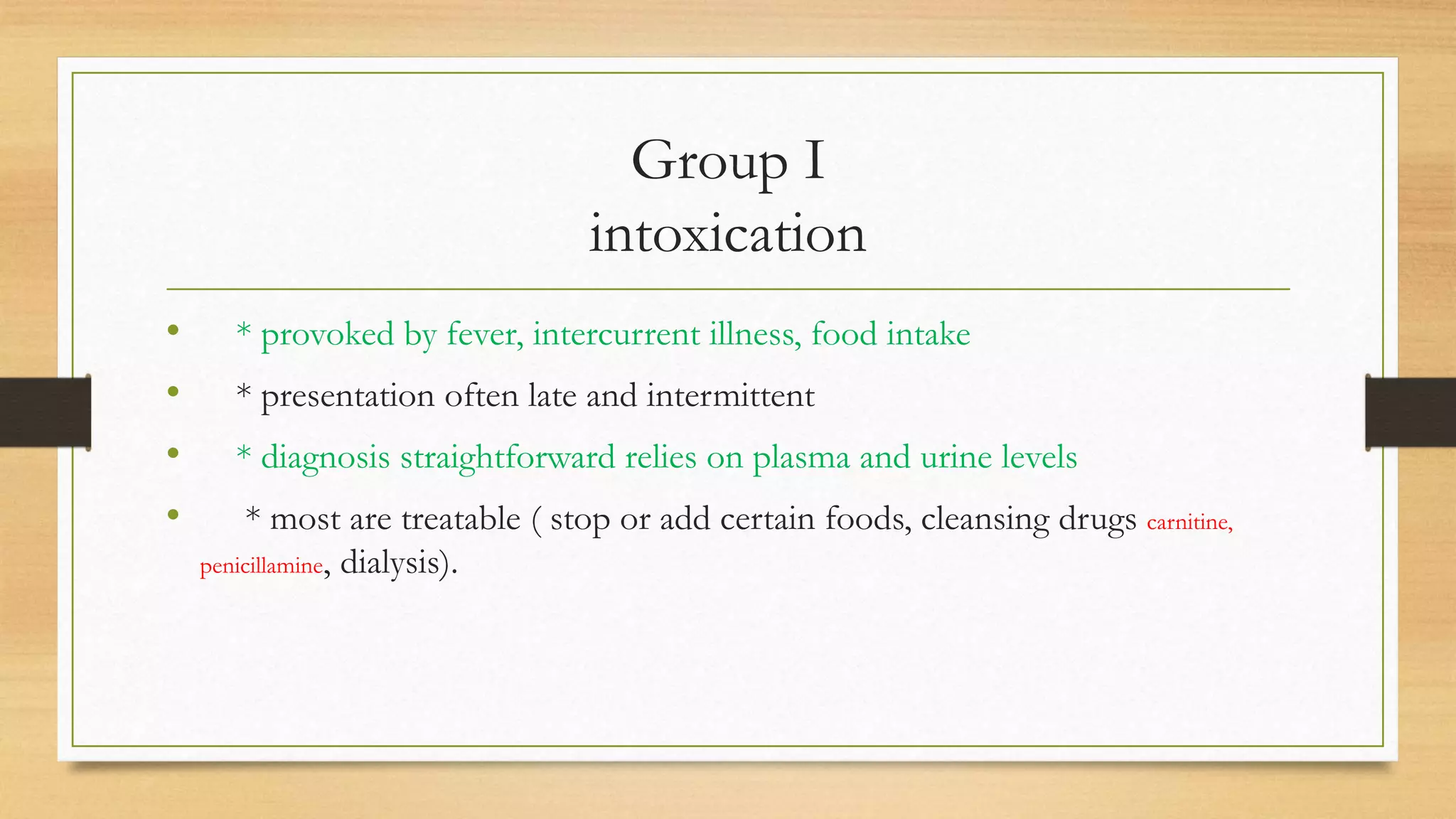
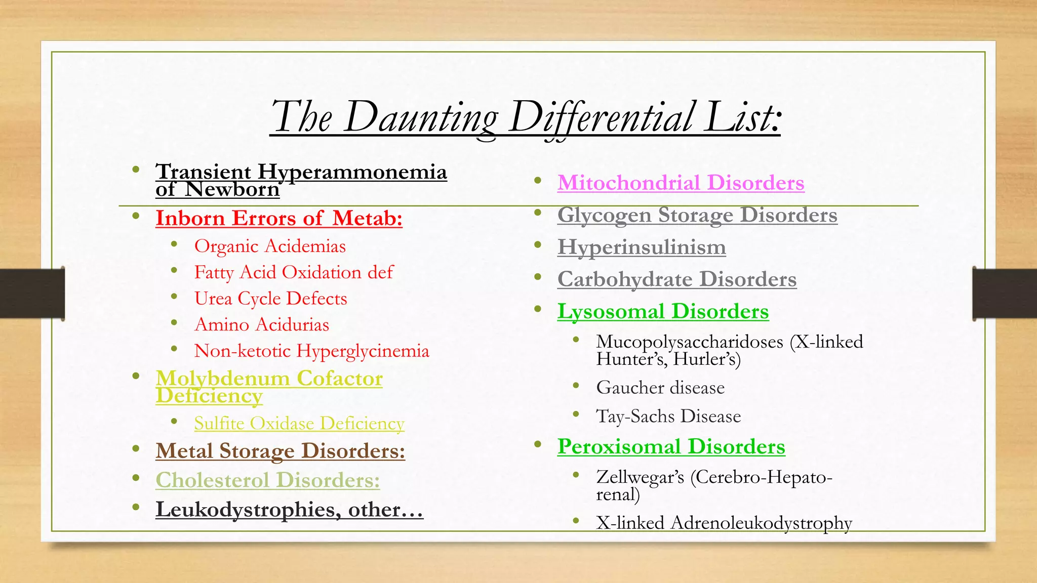
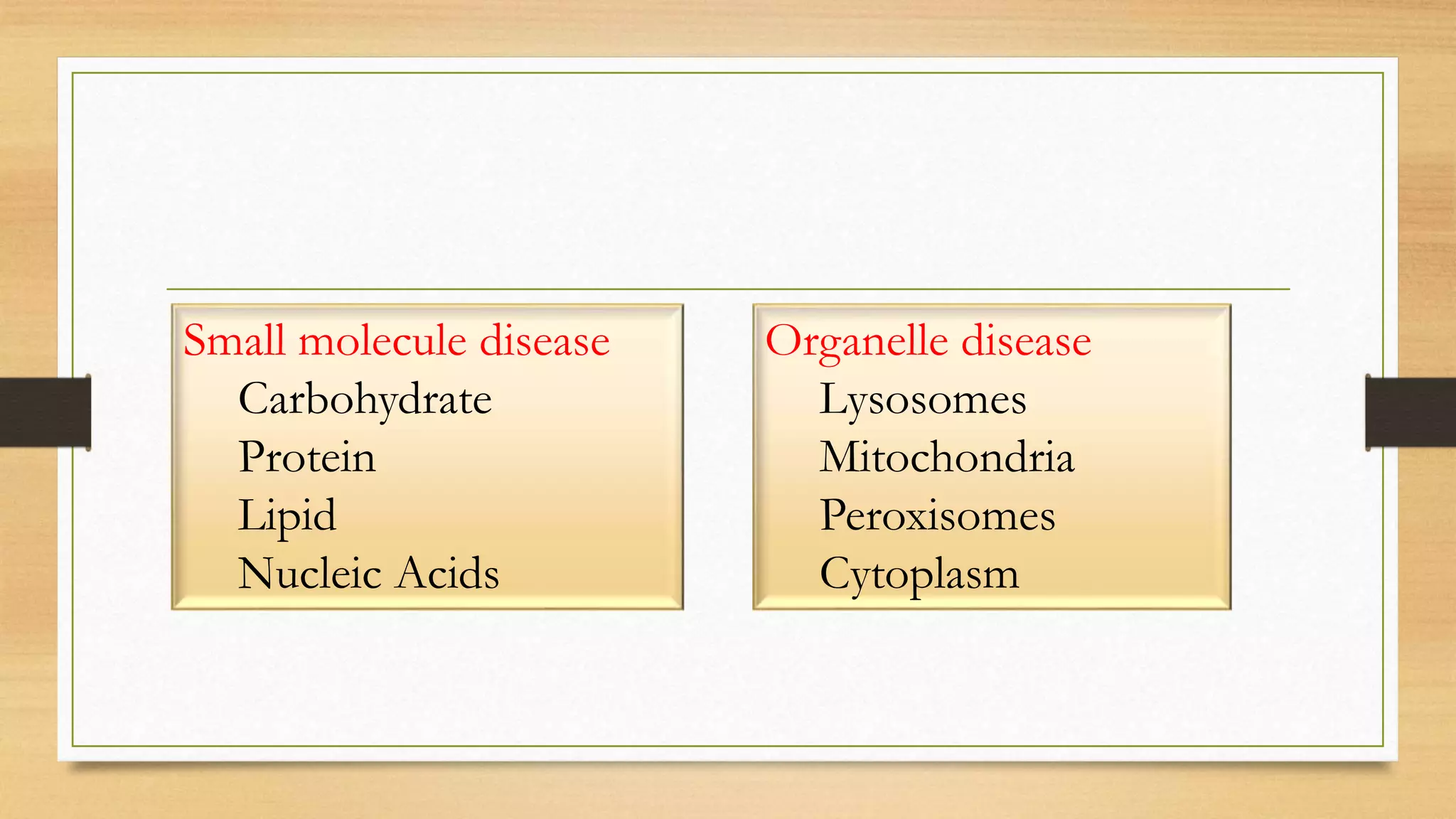
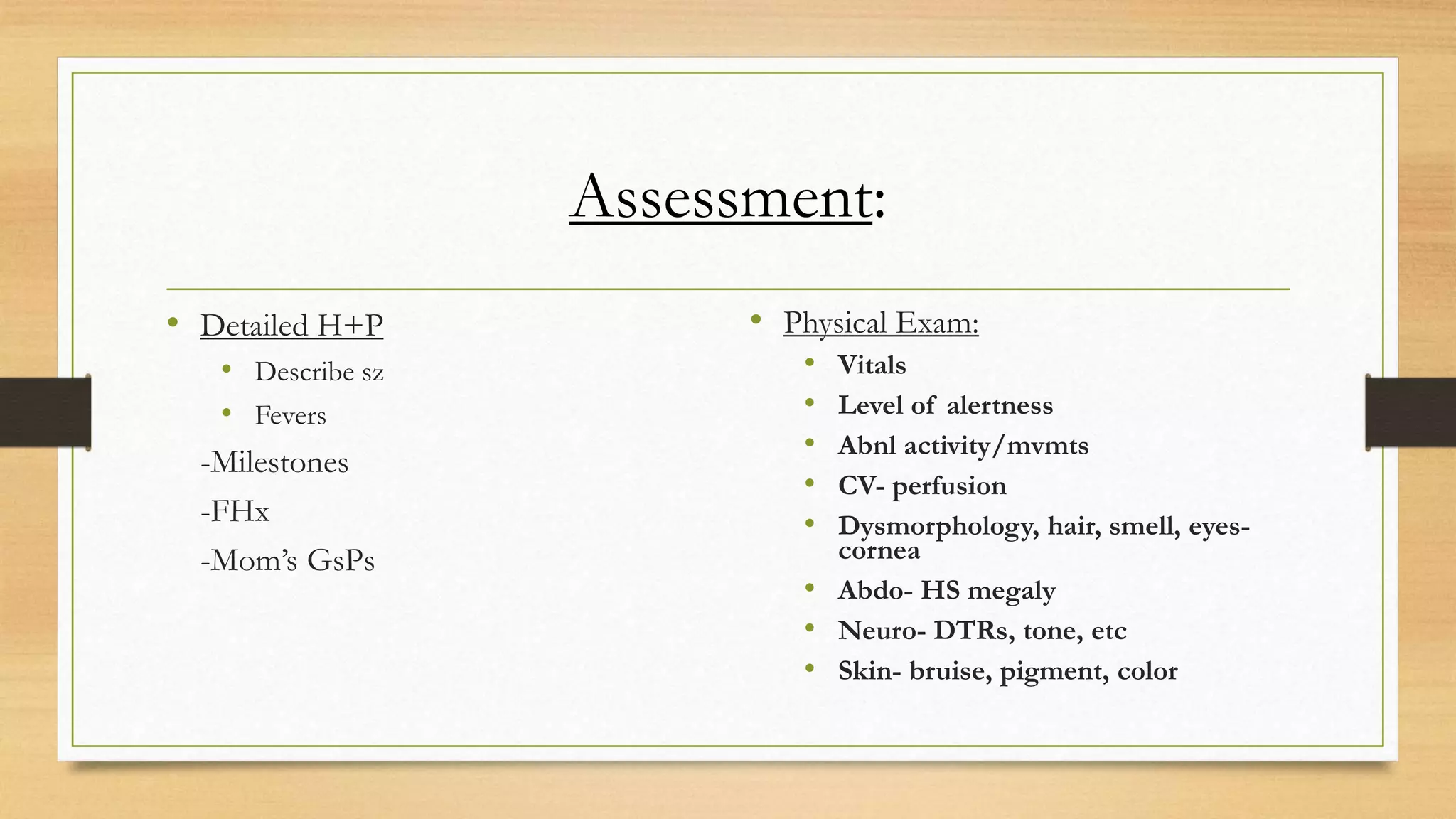
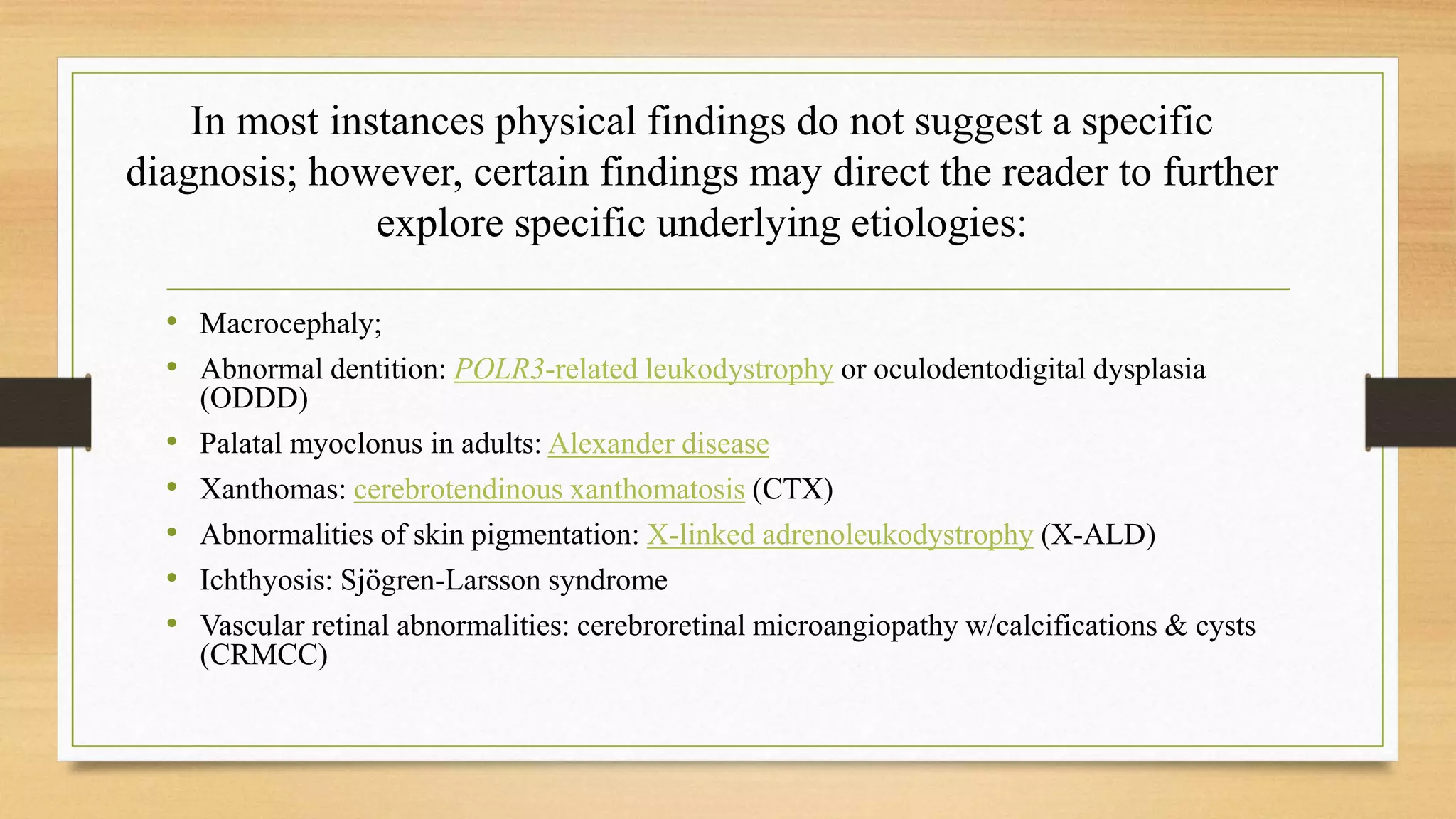
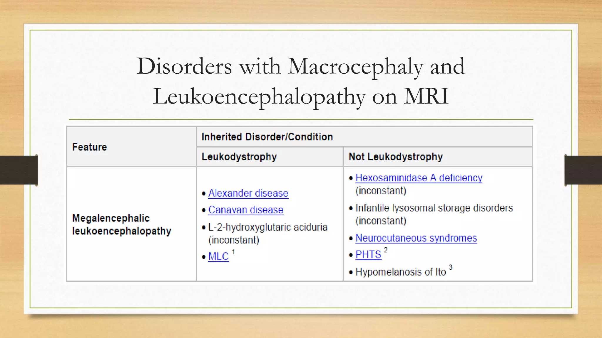
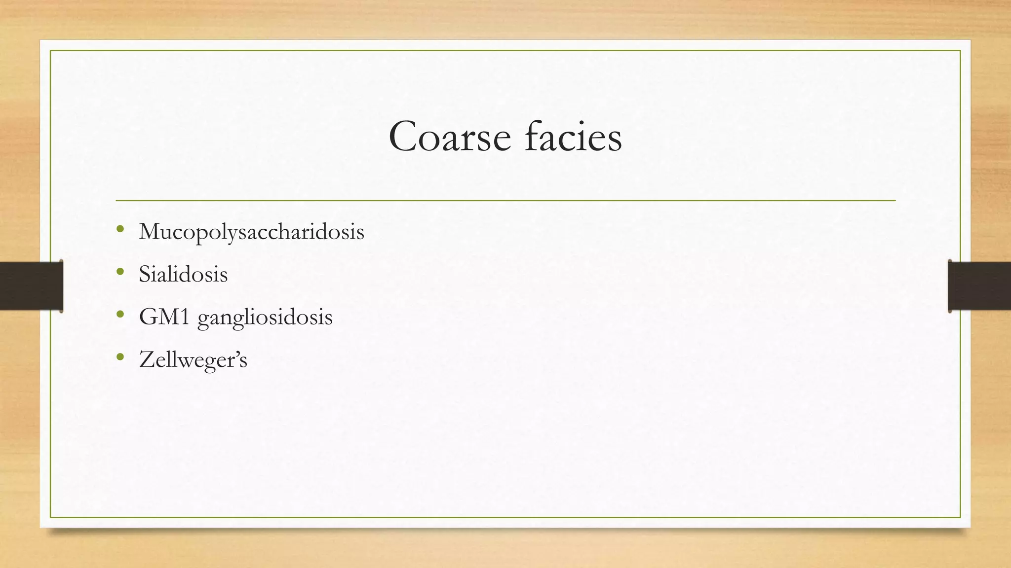
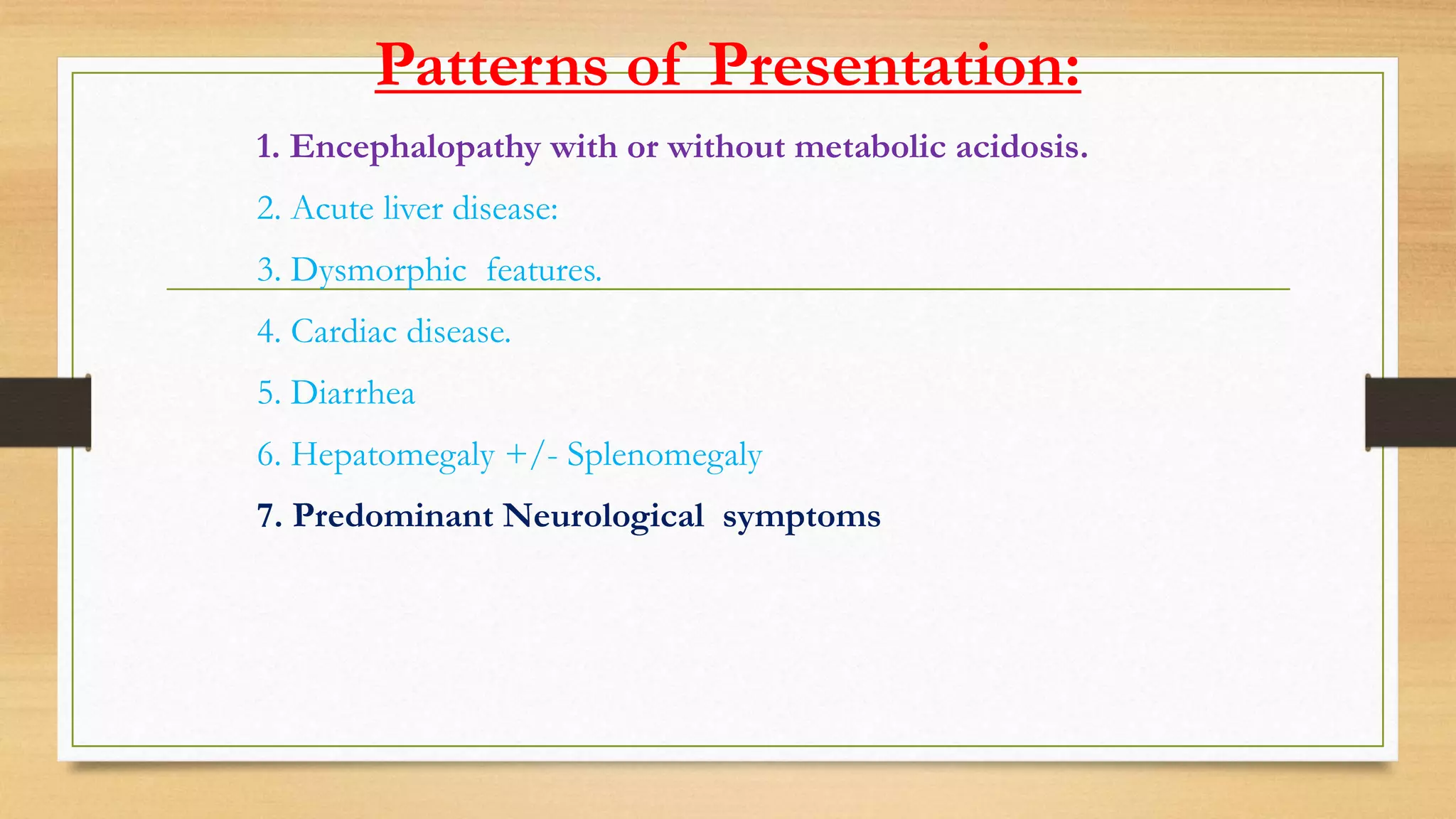
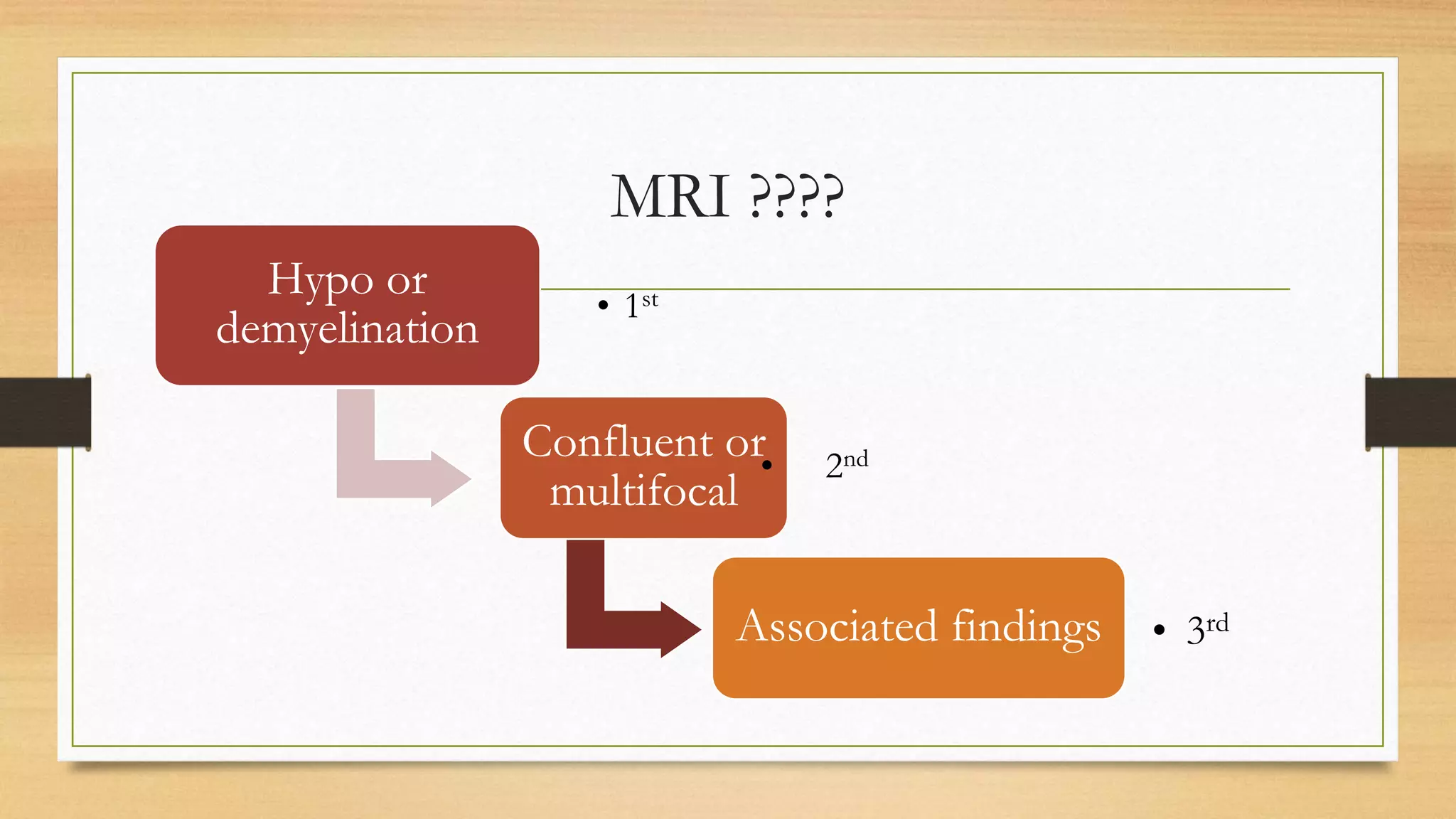
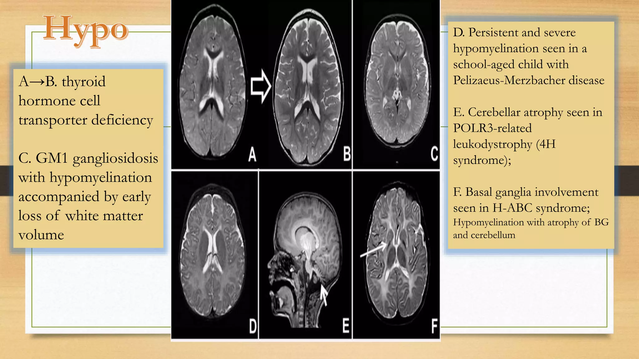
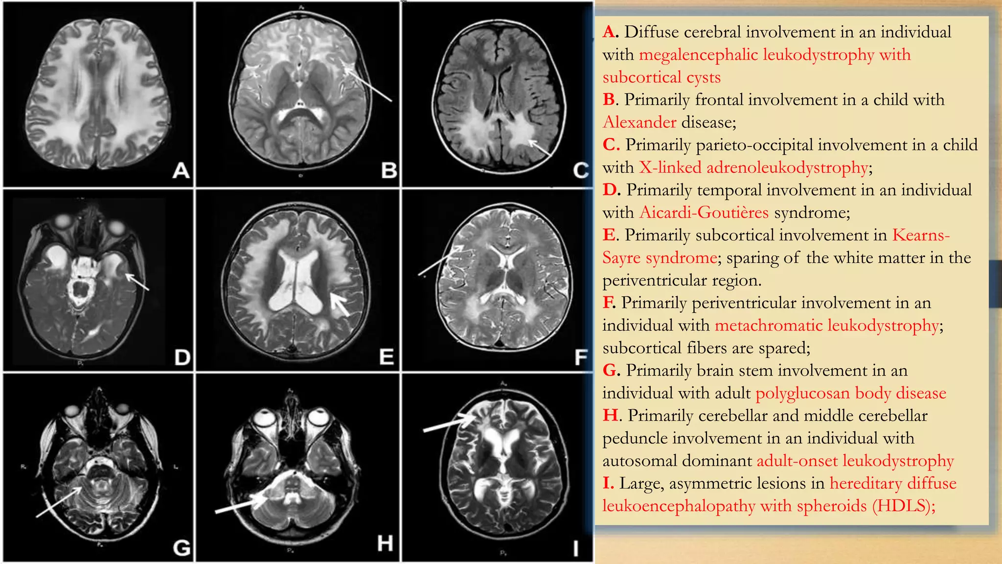
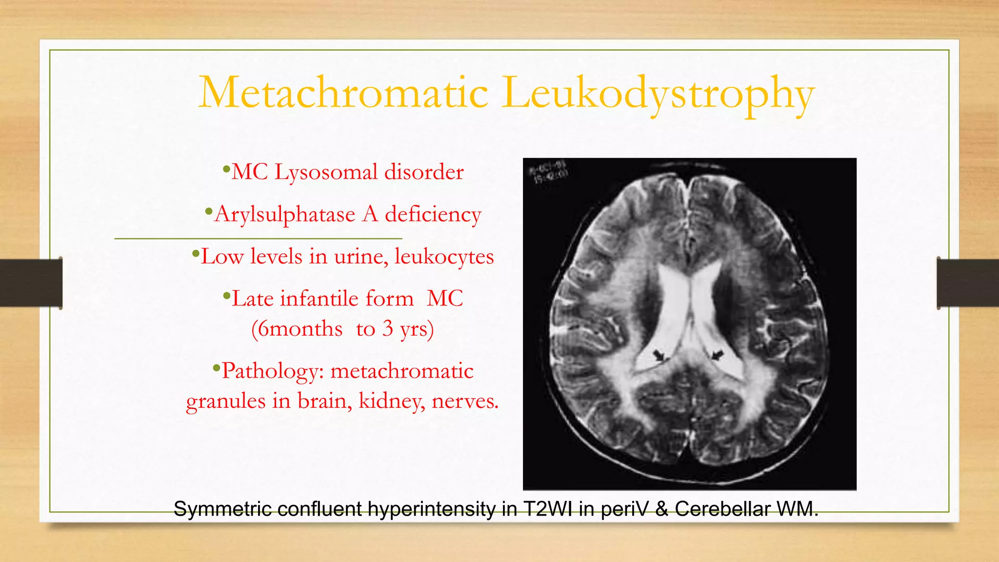
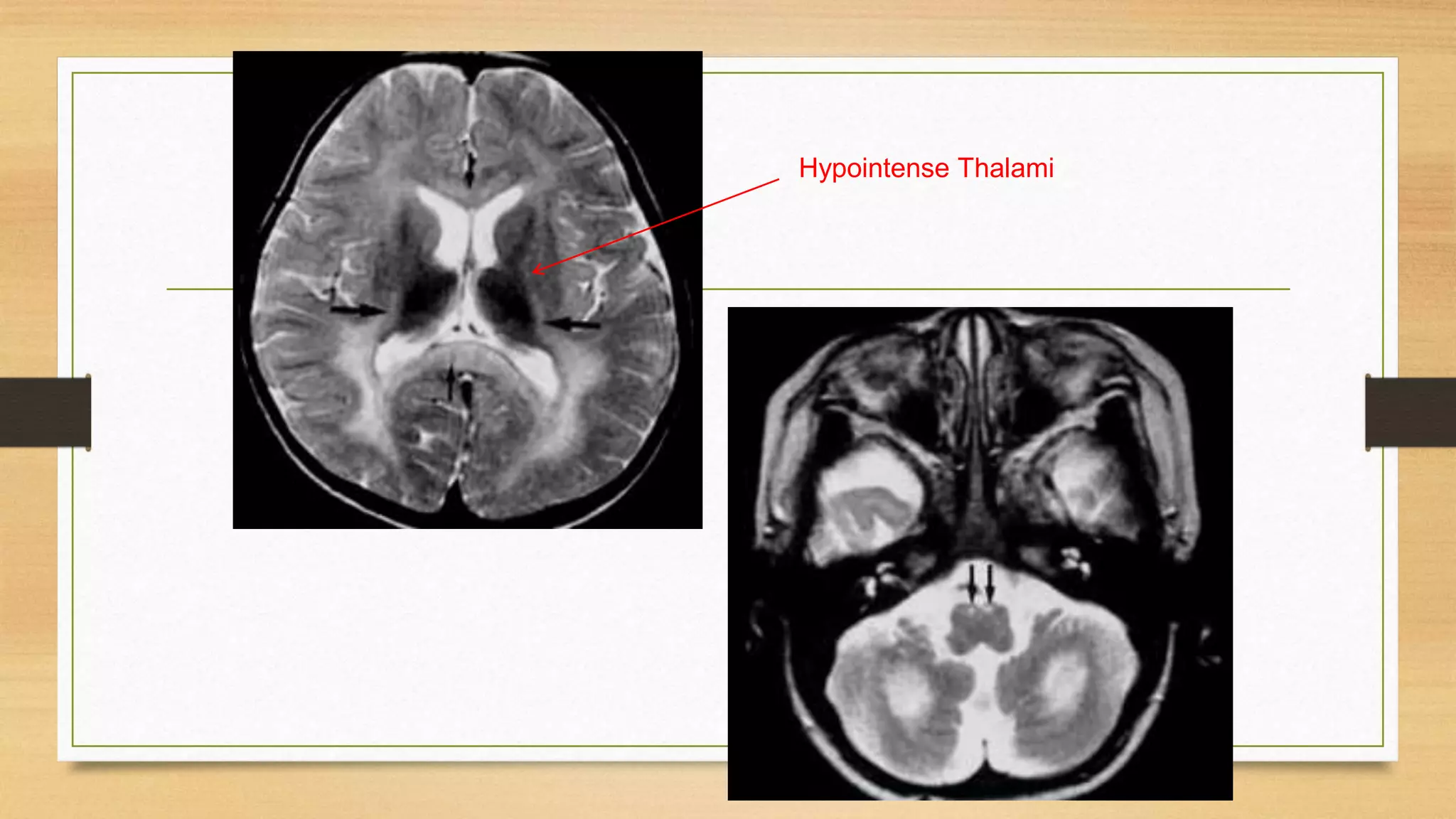
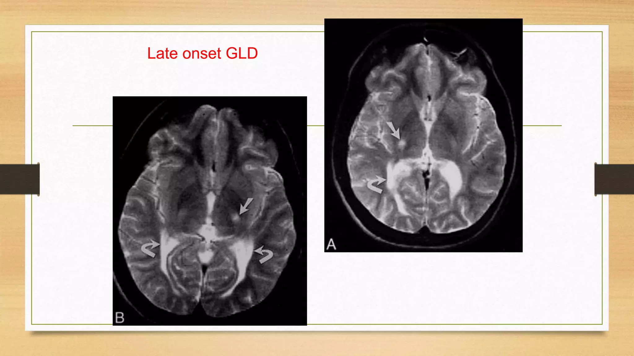
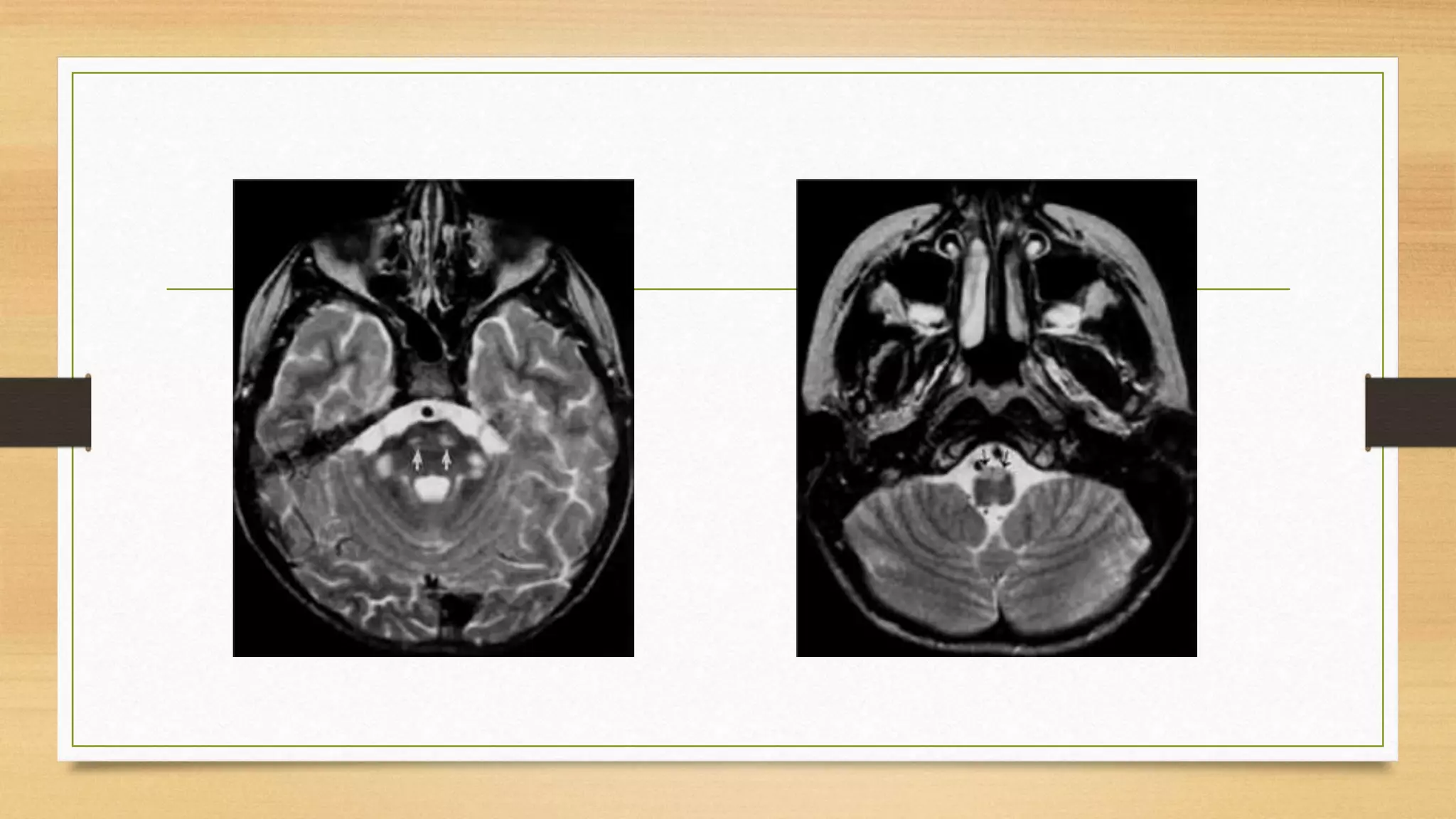
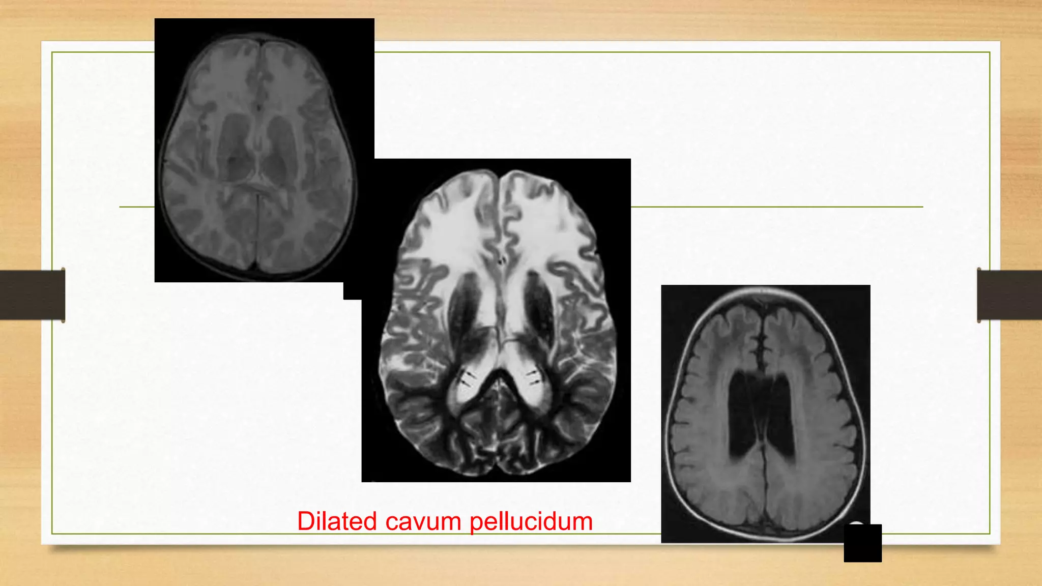
Metabolec eesoreers
ee eeuroloee
Neuro-Metabolic disorders
By
Mohammed E. Mahdy, Msc
Objectives
• Introduction
• How to suspect neurometabolic disorder?
• MRI
• Leukodystrophies
• Mitochondrial
Garrod’s hypothesis?
• enzyme
• substrate normal product
•
• enzyme
• substrate product
•
• enzyme
• substrate normal product
• toxic
Metabolic disorders can classified into:
I
Intoxication
II
Energy
metabolism
III
Complex
molecules
Group I
intoxication
• Acute or progressive intoxication from accumulation of toxic compounds proximal
to metabolic block.
• Example : phenylketonuria
• Common features:
• * fetal life not affected
• * symptom free interval
• * may be acute ( coma, liver failure, vomiting, thrombo-embolic)
• * may be chronic ( developmental delay, cardiomyopathy)
Group I
intoxication
• * provoked by fever, intercurrent illness, food intake
• * presentation often late and intermittent
• * diagnosis straightforward relies on plasma and urine levels
• * most are treatable ( stop or add certain foods, cleansing drugs carnitine,
penicillamine, dialysis).
Group II
energy metabolism
• Failure of energy production or utilization
• In ( liver, muscle, myocardium, brain, others)
• Mitochondrial or cytoplasmic
• Common features:
• * exercise intolerance and fatigue
• * hypoglycemia
• * hyperlactinemia
Group II
energy metabolism
• * hepatomegaly
• * hypotonia
• * myopathy
• * cardiomyopathy
• * S U E D I ( sudden unexpected death in infancy)
• * diagnosis more difficult needs enzymatic levels, biopsies, cell culture
• * treatment more difficult in mitochondrial than cytoplasmic
Group III
complex molecules
• Lysosomes and peroxisomes
• Common features:
• * permenent
• * progressive
• * not provoked
• * most are non treatable
Neurometabolic disorders
Different classifications
• • Clinical syndrome
• • Cellular organelle involved
• • Biochemical studies
• • Genetic mutation
The Daunting Differential List:
• Transient Hyperammonemia
of Newborn
• Inborn Errors of Metab:
• Organic Acidemias
• Fatty Acid Oxidation def
• Urea Cycle Defects
• Amino Acidurias
• Non-ketotic Hyperglycinemia
• Molybdenum Cofactor
Deficiency
• Sulfite Oxidase Deficiency
• Metal Storage Disorders:
• Cholesterol Disorders:
• Leukodystrophies, other…
• Mitochondrial Disorders
• Glycogen Storage Disorders
• Hyperinsulinism
• Carbohydrate Disorders
• Lysosomal Disorders
• Mucopolysaccharidoses (X-linked
Hunter’s, Hurler’s)
• Gaucher disease
• Tay-Sachs Disease
• Peroxisomal Disorders
• Zellwegar’s (Cerebro-Hepato-
renal)
• X-linked Adrenoleukodystrophy
Small molecule disease
Carbohydrate
Protein
Lipid
Nucleic Acids
Organelle disease
Lysosomes
Mitochondria
Peroxisomes
Cytoplasm
when do you suspect a neurometabolic disorder
??
• An infant or child presented with any of the following:
• * progressively deteriorating neurological or developemental disorder
• * diffuse neurological involvement
• * family history
• * dysmorphic features
• * progressive childhood ataxias
• * dystonias or other movement disorders
• * childhood epilepsies specially if myoclonic
• * cognitive decline
• * spinal deformity
• * other organ dysfunction ( liver, heart, GIT)
• * MRI features
Assessment:
• Detailed H+P
• Describe sz
• Fevers
-Milestones
-FHx
-Mom’s GsPs
• Physical Exam:
• Vitals
• Level of alertness
• Abnl activity/mvmts
• CV- perfusion
• Dysmorphology, hair, smell, eyes-
cornea
• Abdo- HS megaly
• Neuro- DTRs, tone, etc
• Skin- bruise, pigment, color
In most instances physical findings do not suggest a specific
diagnosis; however, certain findings may direct the reader to further
explore specific underlying etiologies:
• Macrocephaly;
• Abnormal dentition: POLR3-related leukodystrophy or oculodentodigital dysplasia
(ODDD)
• Palatal myoclonus in adults: Alexander disease
• Xanthomas: cerebrotendinous xanthomatosis (CTX)
• Abnormalities of skin pigmentation: X-linked adrenoleukodystrophy (X-ALD)
• Ichthyosis: Sjögren-Larsson syndrome
• Vascular retinal abnormalities: cerebroretinal microangiopathy w/calcifications & cysts
(CRMCC)
Disorders with Macrocephaly and
Leukoencephalopathy on MRI
with Cataract
• Galactosemia
• Homocystinuria
• Cerebrotendinous xanthomatosis
Coarse facies
• Mucopolysaccharidosis
• Sialidosis
• GM1 gangliosidosis
• Zellweger’s
Kinky hair
• Menke’s syndrome
• Multiple carboxylase deficiency
• Arginosuccinic aciduria
Menke’s ***** Wilson ????
Copper
transporting
ATPase
High
copper
ATPase
7B
ATPase
7 A
Cherry red spot
• Tay-Sachs (GM2 gangliosidosis)
• Niemann-Pick
• Gaucher’s
• Sialidosis
Abnormal odour (urine, body)
• Musty : PKU
• Caramel : Maple syrup urine disease
• Cat urine: multiple carboxylase deficiency
Patterns of Presentation:
1. Encephalopathy with or without metabolic acidosis.
2. Acute liver disease:
3. Dysmorphic features.
4. Cardiac disease.
5. Diarrhea
6. Hepatomegaly +/- Splenomegaly
7. Predominant Neurological symptoms
Encephalopathy with or without metabolic acidosis:
• Encephalopathy, seizures,and tone abnormalities are
predominant presenting features of
-organic acidemias,
-urea cycle defects, and
-congenital lactic acidosis.
• Intractable seizures are prominent in
-pyridoxine dependency,
-non-ketotic hyperglycinemia,
-molybdenum co-factor defect, and
-folinic-acid responsive seizures.
Predominant Neurological Symptoms:
a. Psychomotor delay:
-Aminoacidopathies
-Organic acidemias
-CNS storage diseases
b. Ataxia:
-Hartnup dis
-Urea cycle disorders
-Pyruvate decarboxylase deficiency
c. Extrapyramidal signs:
- Wilsons disease
d. Hypotonia :
- Zellweger synd
-Mitochondrial myopathies
-Muscle carnitine deficiency
First line investigations (metabolic screen):
• The following tests should be obtained in all babies with suspected IEM.
• 1) Complete blood count: (neutropenia and thrombocytopenia seen in propionic and
methylmalonic academia)
• 2) Arterial blood gases and electrolytes
• 3) Blood glucose
• 4) Plasma ammonia (Normal values in newborn: 90-150 mg/dl or 64-107 mmol/L)
• 5) Arterial blood lactate (Normal values: 0.5-1.6 mmol/L)
• 6) Liver function tests,
• 7) Urine ketones,
• 8) Urine reducing substances,
• 9) Serum uric acid (low in molybdenum cofactor deficiency).
Screening : urine
Screening : blood
• Glucose
• Lactate
• PH
• Ammonia
• Amino acids
Screening : refractory seizures in infancy
• Urine : sulphite, aminoadipic semialdehde
• Blood: biotinidase, pyridoxine
• CSF: pipecolic acid
MRI ????
Hypo or
demyelination
• 1st
Confluent or
multifocal
• 2nd
Associated findings • 3rd
A→B. thyroid
hormone cell
transporter deficiency
C. GM1 gangliosidosis
with hypomyelination
accompanied by early
loss of white matter
volume
D. Persistent and severe
hypomyelination seen in a
school-aged child with
Pelizaeus-Merzbacher disease
E. Cerebellar atrophy seen in
POLR3-related
leukodystrophy (4H
syndrome);
F. Basal ganglia involvement
seen in H-ABC syndrome;
Hypomyelination with atrophy of BG
and cerebellum
Confluent
Specific Associated
Multifocal
Demyelinating
A. Diffuse cerebral involvement in an individual
with megalencephalic leukodystrophy with
subcortical cysts
B. Primarily frontal involvement in a child with
Alexander disease;
C. Primarily parieto-occipital involvement in a child
with X-linked adrenoleukodystrophy;
D. Primarily temporal involvement in an individual
with Aicardi-Goutières syndrome;
E. Primarily subcortical involvement in Kearns-
Sayre syndrome; sparing of the white matter in the
periventricular region.
F. Primarily periventricular involvement in an
individual with metachromatic leukodystrophy;
subcortical fibers are spared;
G. Primarily brain stem involvement in an
individual with adult polyglucosan body disease
H. Primarily cerebellar and middle cerebellar
peduncle involvement in an individual with
autosomal dominant adult-onset leukodystrophy
I. Large, asymmetric lesions in hereditary diffuse
leukoencephalopathy with spheroids (HDLS);
A. White matter rarefaction and cysts on FLAIR
imaging in vanishing white matter disease
B. Calcium deposits and hemosiderin deposits
visible on CT in Aicardi-Goutières syndrome;
C. Contrast enhancement on T1-weighted
imaging within abnormal white matter in X-
linked adrenoleukodystrophy;
D. Cortical gray matter lesions in POLG-related
disorders;
E. Cerebellar abnormalities seen in the dentate
nucleus in L-2-hydroxyglutaric aciduria;
F. Thinning of the corpus callosum (particularly
of the genu) in hereditary spastic paraplegia 11;
arrow indicates thin corpus callosum with
anterior beaking
G. Non-calcifying basal ganglia lesions in
Alexander disease;
H. Typical brain stem involvement in AD adult-
onset leukodystrophy (ADLD);
I. Spinal cord involvement in LBSL;
Leukoencephalopathy with Brain Stem
and Spinal Cord Involvement and
Lactate Elevation
WHITE MATTER DISEASES
Leukoencephalopathies
• Demyelination Primary : MS
Secondary : ADEM, SSPE, HIV
Hypertensive
• Dysmyelination (Leukodystrophy)
Hypodense on CT ; Hypointense in T1WI and Hyper in T2WI
MRI cannot distinguish Demyelination from Dysmyelination
Classification
• Genetics
• Organelle based Lysosomal : MLD, Krabbe
Peroxisomal : ALD
Mitochondrial : MELAS, MERRF
X-linked
Adrenoleukodystrophy
Pelizaeus-Merzebacher disease
Autosomal recessive
Metachromatic Leukodystrophy
Krabbe
Canavan
Cockayne
Neonatal ALD
Unknown
Alexander disease
Some Discriminating features
• Near complete lack of
myelination
• Frontal Predominance
• Occipital Predominance
• Macrocephaly
• Enhancement
• Infarcts
• Hyperdense BG
• Dysplasia
Canavan Disease, PMD
Alexander disease
X- linked ALD
Alexander, Canavan
ALD, Alexander
MELAS, MERRF, Leigh
Krabbe, Aicardie
Zellweger
Metachromatic Leukodystrophy
•MC Lysosomal disorder
•Arylsulphatase A deficiency
•Low levels in urine, leukocytes
•Late infantile form MC
(6months to 3 yrs)
•Pathology: metachromatic
granules in brain, kidney, nerves.
Symmetric confluent hyperintensity in T2WI in periV & Cerebellar WM.
Hypointense Thalami
KRABBE DISEASE
Globoid Cell Leukodystrophy (GLD)
AR, Lysosomal Disorder
Early infantile form MC
Death by 2 yrs
Based on age of onset , MRI
findings –
≤ 2yrs (early onset)
> 2yrs (late onset)
Abnormal high signal in T2WI along the corticospinal tracts.
Late onset GLD
ADRENOLEUKODYSTROPHY
• Peroxisomal disorder
• Single enzyme defect – Classic X linked ALD, AMN
• Multiple enzyme defects – Zellweger disease, Neonatal ALD
• Symmetrical areas of WM abnormalities in pareito-occipital region, extending across splenium
• Corticospinal tract involvement
• Auditory pathway involvement (LGB)
• Contrast enhancement in leading edge of dysmyelination
ALEXANDER
DISEASE
Macrocrania
•Frontal predominance
•Enhancement
•Late diffuse cases
resemble Canavan.
Dilated cavum pellucidum
CANAVAN DISEASE
Macrocrania
•Diffuse involvement of
WM with swelling
•Subcortical U fibers
involved early
•High signal in Globus
pallidus.
Pelizaeus-Merzebacher Disease
X-linked recessive
Lack of proteolipid lipoprotein
(Lipophilin)
Diffuse involvement of the WM
with sparing of internal capsule
and subcortical U fibers
Characteristic sparing of
perivascular WM – Tigroid or
Leopard skin pattern.
Mitochondriopathies
(Multiorgan Disorders)
Skeletal muscle
Cardiac muscle
Extraocular muscle
Liver
Retina
Lens
Central nervous system
Peripheral nervous system
Mitochondrial vs Mendelian genetics?
Mitochondrial Disorders
• Involve both gray matter and white matter
Leigh disease (subacute necrotizing encephalo-myelopathy)
MERRF (Myoclonic epilepsy with ragged red fibers)
MELAS (Mitochondrial Encephalomyelopathy, Lactic Acidosis and Stroke
Like episodes)
Kearns-Sayre syndrome (KSS)
Leigh Disease
Symmetric hyperintensities in lentiform nucleus & caudate nucleus
Periventricular WM and periaquiductal GM often involved
Kearns-Sayre syndrome
MELAS and MERRF
ZELLWEGER’S SYNDROME
•Neuronal migration disorders - Heterotopias, pachygyria,
polymicrogyria
•White matter hypomyelination
•Cortical neuronal loss
Van der Knapp Syndrome
• Identified in 1995
• Vacuolating Megalencephalic Leukoencephalopathy with
Subcortical Cysts
• Near complete lack of
myelination
• Frontal Predominance
• Occipital Predominance
• Macrocephaly
• Enhancement
• Infarcts
• Hyperdense BG
• Dysplasia
Canavan Disease, PMD
Alexander disease
X- linked ALD
Alexander, Canavan
ALD, Alexander
MELAS, MERRF, Leigh
Krabbe
Zellweger
Treatment
• Dietary Restriction
• Supplement deficient product
• Stimulate alternate pathway
• Supply vitamin co-factor
• Organ transplantation
• Enzyme replacement therapy
• Gene Therapy
Metabolic disorders in neurology

Metabolic disorders in neurology

  • 1.
  • 2.
  • 3.
    Objectives • Introduction • Howto suspect neurometabolic disorder? • MRI • Leukodystrophies • Mitochondrial
  • 8.
  • 9.
    • enzyme • substratenormal product •
  • 10.
  • 11.
    • enzyme • substratenormal product • toxic
  • 13.
    Metabolic disorders canclassified into: I Intoxication II Energy metabolism III Complex molecules
  • 14.
    Group I intoxication • Acuteor progressive intoxication from accumulation of toxic compounds proximal to metabolic block. • Example : phenylketonuria • Common features: • * fetal life not affected • * symptom free interval • * may be acute ( coma, liver failure, vomiting, thrombo-embolic) • * may be chronic ( developmental delay, cardiomyopathy)
  • 15.
    Group I intoxication • *provoked by fever, intercurrent illness, food intake • * presentation often late and intermittent • * diagnosis straightforward relies on plasma and urine levels • * most are treatable ( stop or add certain foods, cleansing drugs carnitine, penicillamine, dialysis).
  • 16.
    Group II energy metabolism •Failure of energy production or utilization • In ( liver, muscle, myocardium, brain, others) • Mitochondrial or cytoplasmic • Common features: • * exercise intolerance and fatigue • * hypoglycemia • * hyperlactinemia
  • 17.
    Group II energy metabolism •* hepatomegaly • * hypotonia • * myopathy • * cardiomyopathy • * S U E D I ( sudden unexpected death in infancy) • * diagnosis more difficult needs enzymatic levels, biopsies, cell culture • * treatment more difficult in mitochondrial than cytoplasmic
  • 18.
    Group III complex molecules •Lysosomes and peroxisomes • Common features: • * permenent • * progressive • * not provoked • * most are non treatable
  • 19.
    Neurometabolic disorders Different classifications •• Clinical syndrome • • Cellular organelle involved • • Biochemical studies • • Genetic mutation
  • 20.
    The Daunting DifferentialList: • Transient Hyperammonemia of Newborn • Inborn Errors of Metab: • Organic Acidemias • Fatty Acid Oxidation def • Urea Cycle Defects • Amino Acidurias • Non-ketotic Hyperglycinemia • Molybdenum Cofactor Deficiency • Sulfite Oxidase Deficiency • Metal Storage Disorders: • Cholesterol Disorders: • Leukodystrophies, other… • Mitochondrial Disorders • Glycogen Storage Disorders • Hyperinsulinism • Carbohydrate Disorders • Lysosomal Disorders • Mucopolysaccharidoses (X-linked Hunter’s, Hurler’s) • Gaucher disease • Tay-Sachs Disease • Peroxisomal Disorders • Zellwegar’s (Cerebro-Hepato- renal) • X-linked Adrenoleukodystrophy
  • 21.
    Small molecule disease Carbohydrate Protein Lipid NucleicAcids Organelle disease Lysosomes Mitochondria Peroxisomes Cytoplasm
  • 22.
    when do yoususpect a neurometabolic disorder ?? • An infant or child presented with any of the following: • * progressively deteriorating neurological or developemental disorder • * diffuse neurological involvement • * family history • * dysmorphic features • * progressive childhood ataxias • * dystonias or other movement disorders • * childhood epilepsies specially if myoclonic
  • 23.
    • * cognitivedecline • * spinal deformity • * other organ dysfunction ( liver, heart, GIT) • * MRI features
  • 24.
    Assessment: • Detailed H+P •Describe sz • Fevers -Milestones -FHx -Mom’s GsPs • Physical Exam: • Vitals • Level of alertness • Abnl activity/mvmts • CV- perfusion • Dysmorphology, hair, smell, eyes- cornea • Abdo- HS megaly • Neuro- DTRs, tone, etc • Skin- bruise, pigment, color
  • 25.
    In most instancesphysical findings do not suggest a specific diagnosis; however, certain findings may direct the reader to further explore specific underlying etiologies: • Macrocephaly; • Abnormal dentition: POLR3-related leukodystrophy or oculodentodigital dysplasia (ODDD) • Palatal myoclonus in adults: Alexander disease • Xanthomas: cerebrotendinous xanthomatosis (CTX) • Abnormalities of skin pigmentation: X-linked adrenoleukodystrophy (X-ALD) • Ichthyosis: Sjögren-Larsson syndrome • Vascular retinal abnormalities: cerebroretinal microangiopathy w/calcifications & cysts (CRMCC)
  • 26.
    Disorders with Macrocephalyand Leukoencephalopathy on MRI
  • 27.
    with Cataract • Galactosemia •Homocystinuria • Cerebrotendinous xanthomatosis
  • 28.
    Coarse facies • Mucopolysaccharidosis •Sialidosis • GM1 gangliosidosis • Zellweger’s
  • 31.
    Kinky hair • Menke’ssyndrome • Multiple carboxylase deficiency • Arginosuccinic aciduria
  • 32.
  • 33.
    Menke’s ***** Wilson???? Copper transporting ATPase High copper ATPase 7B ATPase 7 A
  • 34.
    Cherry red spot •Tay-Sachs (GM2 gangliosidosis) • Niemann-Pick • Gaucher’s • Sialidosis
  • 35.
    Abnormal odour (urine,body) • Musty : PKU • Caramel : Maple syrup urine disease • Cat urine: multiple carboxylase deficiency
  • 36.
    Patterns of Presentation: 1.Encephalopathy with or without metabolic acidosis. 2. Acute liver disease: 3. Dysmorphic features. 4. Cardiac disease. 5. Diarrhea 6. Hepatomegaly +/- Splenomegaly 7. Predominant Neurological symptoms
  • 38.
    Encephalopathy with orwithout metabolic acidosis: • Encephalopathy, seizures,and tone abnormalities are predominant presenting features of -organic acidemias, -urea cycle defects, and -congenital lactic acidosis. • Intractable seizures are prominent in -pyridoxine dependency, -non-ketotic hyperglycinemia, -molybdenum co-factor defect, and -folinic-acid responsive seizures.
  • 39.
    Predominant Neurological Symptoms: a.Psychomotor delay: -Aminoacidopathies -Organic acidemias -CNS storage diseases b. Ataxia: -Hartnup dis -Urea cycle disorders -Pyruvate decarboxylase deficiency c. Extrapyramidal signs: - Wilsons disease d. Hypotonia : - Zellweger synd -Mitochondrial myopathies -Muscle carnitine deficiency
  • 40.
    First line investigations(metabolic screen): • The following tests should be obtained in all babies with suspected IEM. • 1) Complete blood count: (neutropenia and thrombocytopenia seen in propionic and methylmalonic academia) • 2) Arterial blood gases and electrolytes • 3) Blood glucose • 4) Plasma ammonia (Normal values in newborn: 90-150 mg/dl or 64-107 mmol/L) • 5) Arterial blood lactate (Normal values: 0.5-1.6 mmol/L) • 6) Liver function tests, • 7) Urine ketones, • 8) Urine reducing substances, • 9) Serum uric acid (low in molybdenum cofactor deficiency).
  • 41.
  • 43.
    Screening : blood •Glucose • Lactate • PH • Ammonia • Amino acids
  • 44.
    Screening : refractoryseizures in infancy • Urine : sulphite, aminoadipic semialdehde • Blood: biotinidase, pyridoxine • CSF: pipecolic acid
  • 45.
    MRI ???? Hypo or demyelination •1st Confluent or multifocal • 2nd Associated findings • 3rd
  • 46.
    A→B. thyroid hormone cell transporterdeficiency C. GM1 gangliosidosis with hypomyelination accompanied by early loss of white matter volume D. Persistent and severe hypomyelination seen in a school-aged child with Pelizaeus-Merzbacher disease E. Cerebellar atrophy seen in POLR3-related leukodystrophy (4H syndrome); F. Basal ganglia involvement seen in H-ABC syndrome; Hypomyelination with atrophy of BG and cerebellum
  • 47.
  • 49.
    A. Diffuse cerebralinvolvement in an individual with megalencephalic leukodystrophy with subcortical cysts B. Primarily frontal involvement in a child with Alexander disease; C. Primarily parieto-occipital involvement in a child with X-linked adrenoleukodystrophy; D. Primarily temporal involvement in an individual with Aicardi-Goutières syndrome; E. Primarily subcortical involvement in Kearns- Sayre syndrome; sparing of the white matter in the periventricular region. F. Primarily periventricular involvement in an individual with metachromatic leukodystrophy; subcortical fibers are spared; G. Primarily brain stem involvement in an individual with adult polyglucosan body disease H. Primarily cerebellar and middle cerebellar peduncle involvement in an individual with autosomal dominant adult-onset leukodystrophy I. Large, asymmetric lesions in hereditary diffuse leukoencephalopathy with spheroids (HDLS);
  • 50.
    A. White matterrarefaction and cysts on FLAIR imaging in vanishing white matter disease B. Calcium deposits and hemosiderin deposits visible on CT in Aicardi-Goutières syndrome; C. Contrast enhancement on T1-weighted imaging within abnormal white matter in X- linked adrenoleukodystrophy; D. Cortical gray matter lesions in POLG-related disorders; E. Cerebellar abnormalities seen in the dentate nucleus in L-2-hydroxyglutaric aciduria; F. Thinning of the corpus callosum (particularly of the genu) in hereditary spastic paraplegia 11; arrow indicates thin corpus callosum with anterior beaking G. Non-calcifying basal ganglia lesions in Alexander disease; H. Typical brain stem involvement in AD adult- onset leukodystrophy (ADLD); I. Spinal cord involvement in LBSL; Leukoencephalopathy with Brain Stem and Spinal Cord Involvement and Lactate Elevation
  • 51.
    WHITE MATTER DISEASES Leukoencephalopathies •Demyelination Primary : MS Secondary : ADEM, SSPE, HIV Hypertensive • Dysmyelination (Leukodystrophy) Hypodense on CT ; Hypointense in T1WI and Hyper in T2WI MRI cannot distinguish Demyelination from Dysmyelination
  • 52.
    Classification • Genetics • Organellebased Lysosomal : MLD, Krabbe Peroxisomal : ALD Mitochondrial : MELAS, MERRF X-linked Adrenoleukodystrophy Pelizaeus-Merzebacher disease Autosomal recessive Metachromatic Leukodystrophy Krabbe Canavan Cockayne Neonatal ALD Unknown Alexander disease
  • 55.
    Some Discriminating features •Near complete lack of myelination • Frontal Predominance • Occipital Predominance • Macrocephaly • Enhancement • Infarcts • Hyperdense BG • Dysplasia Canavan Disease, PMD Alexander disease X- linked ALD Alexander, Canavan ALD, Alexander MELAS, MERRF, Leigh Krabbe, Aicardie Zellweger
  • 56.
    Metachromatic Leukodystrophy •MC Lysosomaldisorder •Arylsulphatase A deficiency •Low levels in urine, leukocytes •Late infantile form MC (6months to 3 yrs) •Pathology: metachromatic granules in brain, kidney, nerves. Symmetric confluent hyperintensity in T2WI in periV & Cerebellar WM.
  • 57.
  • 58.
    KRABBE DISEASE Globoid CellLeukodystrophy (GLD) AR, Lysosomal Disorder Early infantile form MC Death by 2 yrs Based on age of onset , MRI findings – ≤ 2yrs (early onset) > 2yrs (late onset) Abnormal high signal in T2WI along the corticospinal tracts.
  • 59.
  • 60.
    ADRENOLEUKODYSTROPHY • Peroxisomal disorder •Single enzyme defect – Classic X linked ALD, AMN • Multiple enzyme defects – Zellweger disease, Neonatal ALD • Symmetrical areas of WM abnormalities in pareito-occipital region, extending across splenium • Corticospinal tract involvement • Auditory pathway involvement (LGB) • Contrast enhancement in leading edge of dysmyelination
  • 63.
  • 64.
  • 65.
    CANAVAN DISEASE Macrocrania •Diffuse involvementof WM with swelling •Subcortical U fibers involved early •High signal in Globus pallidus.
  • 68.
    Pelizaeus-Merzebacher Disease X-linked recessive Lackof proteolipid lipoprotein (Lipophilin) Diffuse involvement of the WM with sparing of internal capsule and subcortical U fibers Characteristic sparing of perivascular WM – Tigroid or Leopard skin pattern.
  • 70.
    Mitochondriopathies (Multiorgan Disorders) Skeletal muscle Cardiacmuscle Extraocular muscle Liver Retina Lens Central nervous system Peripheral nervous system
  • 71.
  • 72.
    Mitochondrial Disorders • Involveboth gray matter and white matter Leigh disease (subacute necrotizing encephalo-myelopathy) MERRF (Myoclonic epilepsy with ragged red fibers) MELAS (Mitochondrial Encephalomyelopathy, Lactic Acidosis and Stroke Like episodes) Kearns-Sayre syndrome (KSS)
  • 73.
    Leigh Disease Symmetric hyperintensitiesin lentiform nucleus & caudate nucleus Periventricular WM and periaquiductal GM often involved
  • 74.
  • 75.
  • 76.
    ZELLWEGER’S SYNDROME •Neuronal migrationdisorders - Heterotopias, pachygyria, polymicrogyria •White matter hypomyelination •Cortical neuronal loss
  • 77.
    Van der KnappSyndrome • Identified in 1995 • Vacuolating Megalencephalic Leukoencephalopathy with Subcortical Cysts
  • 78.
    • Near completelack of myelination • Frontal Predominance • Occipital Predominance • Macrocephaly • Enhancement • Infarcts • Hyperdense BG • Dysplasia Canavan Disease, PMD Alexander disease X- linked ALD Alexander, Canavan ALD, Alexander MELAS, MERRF, Leigh Krabbe Zellweger
  • 80.
    Treatment • Dietary Restriction •Supplement deficient product • Stimulate alternate pathway • Supply vitamin co-factor • Organ transplantation • Enzyme replacement therapy • Gene Therapy