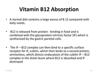
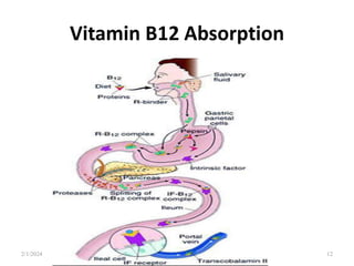
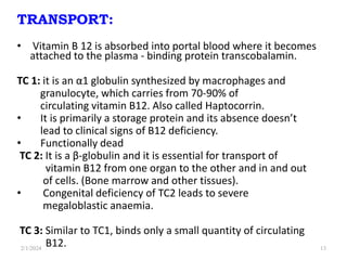
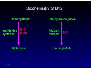
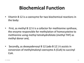
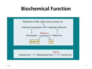
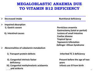
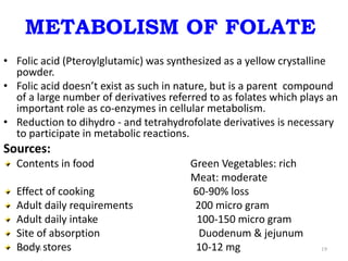
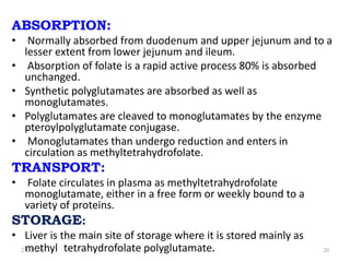
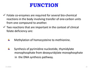
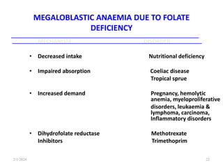
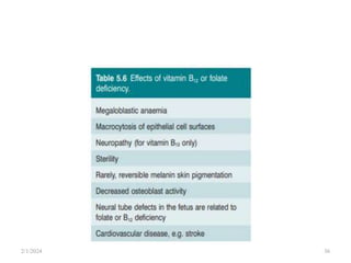
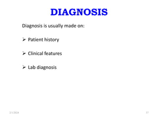
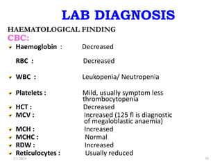
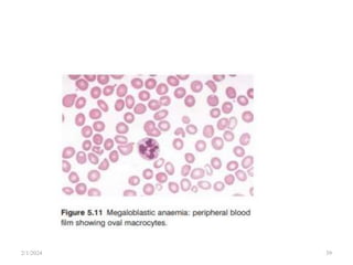
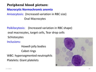
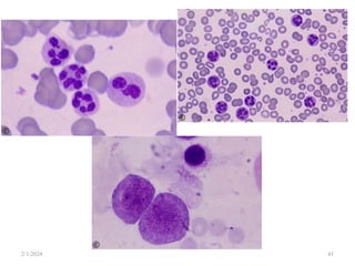
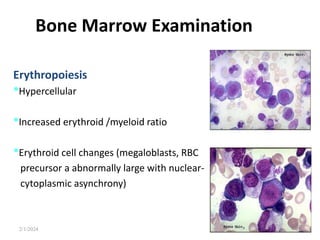
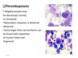
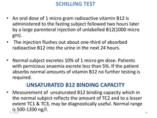
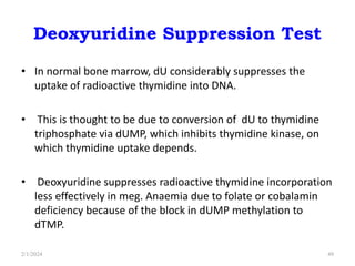
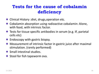
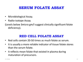
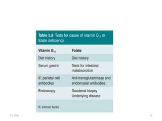
3. Macrocytic Anemias
• In macrocytic anaemia the red cells are abnormally large
(mean corpuscular volume, MCV > 98 fL)
• There are several causes but they can be broadly subdivided
into megaloblastic and non-megaloblastic, based on the
appearance of developing erythroblasts in the bone marrow.
Megaloblastic :
• Vitamin B12 or folate deficiency
Non Megaloblastic:
• Alcohol, liver disease, myelodysplasia, aplastic anaemia, etc.
2/1/2024 3
4. MEGALOBLASTIC ANAEMIA
The megaloblastic anaemias are a group of disorders characterized
by
o Presence Megaloblast in Bone marrow,
o Oval macrocytic cells and Hyper segmented Neutrophils, giant
platelets
o Pancytopenia.
The cause is usually deficiency of either cobalamin (vitamin B 12 ) or
folate, but megaloblastic anaemia may arise because of inherited or
acquired abnormalities affecting the metabolism of these vitamins
or because of defects in DNA synthesis not related to
cobalamin or folate.
2/1/2024 4
5. Megaloblast
• Megaloblast describes a bone marrow cell that is part of the
factory making red cells that has become abnormally large
and has an abnormal appearance.
• Usually large oval shaped cells with large nucleus, open
chromatin, and due to nuclear cytoplasmic asynchronisim .
2/1/2024 5
6. CAUSES
• Cobalamin deficiency or abnormalities of cobalamin
metabolism.
• Folate deficiency or abnormalities of folate metabolism.
• Therapy with antifolate drugs. (e.g. methotrexate)
• Independent of either cobalamin or folate deficiency and
refractory to cobalamin and folate therapy.
• Therapy with drugs interfering with synthesis of DNA. (e.g.
cytosine arabinoside)
2/1/2024 6
8. Vitamin B12 Metabolism
Sources:
• This vitamin is synthesized in nature by microorganisms;
animals acquire it by eating other animal foods, by internal
production from intestinal bacteria (not in humans) or by
eating bacterially contaminated foods.
•
The vitamin consists of a small group of compounds, the
cobalamins, which have the same basic structure, with a
cobalt atom at the centre of a corrin ring which is attached
to a nucleotide portion
• The vitamin is found in foods of animal origin such as liver,
meat, fish and dairy products but does not occur in fruit,
cereals or vegetables.
2/1/2024 8
10. Vitamin B12 Absorption
• A normal diet contains a large excess of B 12 compared with
daily needs.
• B12 is released from protein - binding in food and is
combined with the glycoprotein intrinsic factor (IF) which is
synthesized by the gastric parietal cells.
• The IF – B12 complex can then bind to a specific surface
receptor for IF, cubilin, which then binds to a second protein,
amnionless, which directs endocytosis of the cubilin IF – B12
complex in the distal ileum where B12 is absorbed and IF
destroyed
2/1/2024 10
13. TRANSPORT:
• Vitamin B 12 is absorbed into portal blood where it becomes
attached to the plasma - binding protein transcobalamin.
TC 1: it is an α1 globulin synthesized by macrophages and
granulocyte, which carries from 70-90% of
circulating vitamin B12. Also called Haptocorrin.
• It is primarily a storage protein and its absence doesn’t
lead to clinical signs of B12 deficiency.
• Functionally dead
TC 2: It is a β-globulin and it is essential for transport of
vitamin B12 from one organ to the other and in and out
of cells. (Bone marrow and other tissues).
• Congenital deficiency of TC2 leads to severe
megaloblastic anaemia.
TC 3: Similar to TC1, binds only a small quantity of circulating
B12.
2/1/2024 13
14. Storage sites of Vitamin B12
• The total amount of vitamin B12 in the body is 2 to 5 mg
(adequate for 2 to 4 years).
• The major site of storage is the liver.
• Vitamin B12 is excreted through the bile and shedding of
intestinal epithelial cells.
• Most of the excreted vitamin B12 is again absorbed in the
intestine (enterohepatic circulation).
2/1/2024 14
15. FUNCTIONS
Vitamin B12 acting as co-enzyme for two important
biochemical reactions in humans:
The conversion of methylmalonyl-CoA to succinyl-CoA.
Synthesis of methionine from homocystine.
Vitamin B12 is required for maintenance of the
integrity of nervous system.
2/1/2024 15
16. Biochemical Function
• Vitamin B 12 is a coenzyme for two biochemical reactions in
the body:
• First, as methyl B 12 it is a cofactor for methionine synthase,
the enzyme responsible for methylation of homocysteine to
methionine using methyl tetrahydrofolate (methyl THF) as
methyl donor and,
• Secondly, as deoxyadenosyl B 12 (ado B 12 ) it assists in
conversion of methylmalonyl coenzyme A (CoA) to succinyl
CoA.
2/1/2024 16
18. MEGALOBLASTIC ANAEMIA DUE
TO VITAMIN B12 DEFICIENCY
MECHANISM DISORDER
Decreased intake Nutritional deficiency
Impaired absorption
i). Gastric causes Pernicious anaemia
Gastrectomy (total or partial)
ii). Intestinal causes Lesions of small intestine
Coeliac disease
Tropical Sprue
Tapeworm Infestation
Zollinger -Ellison Syndrome
Abnormalities of cobalamin metabolism
i). Transport protein defects Inherited TC 2 deficiency
ii). Congenital intrinsic factor Present before the age of two
deficiency years
iii). Congenital methylmalonic acidaemia Infants are ill from birth
and aciduria
2/1/2024 18
19. METABOLISM OF FOLATE
• Folic acid (Pteroylglutamic) was synthesized as a yellow crystalline
powder.
• Folic acid doesn’t exist as such in nature, but is a parent compound
of a large number of derivatives referred to as folates which plays an
important role as co-enzymes in cellular metabolism.
• Reduction to dihydro - and tetrahydrofolate derivatives is necessary
to participate in metabolic reactions.
Sources:
Contents in food Green Vegetables: rich
Meat: moderate
Effect of cooking 60-90% loss
Adult daily requirements 200 micro gram
Adult daily intake 100-150 micro gram
Site of absorption Duodenum & jejunum
Body stores 10-12 mg
2/1/2024 19
20. ABSORPTION:
• Normally absorbed from duodenum and upper jejunum and to a
lesser extent from lower jejunum and ileum.
• Absorption of folate is a rapid active process 80% is absorbed
unchanged.
• Synthetic polyglutamates are absorbed as well as
monoglutamates.
• Polyglutamates are cleaved to monoglutamates by the enzyme
pteroylpolyglutamate conjugase.
• Monoglutamates than undergo reduction and enters in
circulation as methyltetrahydrofolate.
TRANSPORT:
• Folate circulates in plasma as methyltetrahydrofolate
monoglutamate, either in a free form or weekly bound to a
variety of proteins.
STORAGE:
• Liver is the main site of storage where it is stored mainly as
methyl tetrahydrofolate polyglutamate.
2/1/2024 20
21. FUNCTION
Folate co-enzymes are required for several bio-chemical
reactions in the body involving transfer of one-carbon units
from one compound to another.
Two reactions that are important in the context of clinical
folate deficiency are:
Methylation of homocysteine to methionine.
Synthesis of pyrimidine nucleotide, thymidylate
monophosphate from deoxyuridylate monophosphate
in the DNA synthesis pathway.
2/1/2024 21
24. Pathophysiology
• In megaloblastic anaemia, the anaemia results from failure
of the megaloblastic bone marrow to compensate for a
moderate reduction in red cell life span.
• Red cell survival studies have shown the presence of mild
haemolysis.
• Lack of vitamin B12 or folate causing slowing of DNA
synthesis in developing erythroblasts with an accumulation
of cells in premitotic phase of cell cycle.
• Some of these cells die within the marrow.
• The neutropenia and thrombocytopenia also appear to
result from ineffective production by abnormal precursor
cells in the marrow.
2/1/2024 24
26. PATHOGENESIS
• Both vitamin B12 and folic acid are required for ordered
DNA synthesis.
• But due to deficiency of vitmain B12 & folic acid in
megaloblastic anaemia DNA synthesis is impaired or
blocked in rapidly dividing cells.
• As a result the cells proliferates abnormally and increases
in size and become megaloblasts which are fully
heamoglobinized cells and abnormal in appearance and
function.
• Because of their increased size they occupy much of the
space in marrow and they disturb other cell lines too.
2/1/2024 26
27. MASKED MEGALOBLASTIC
ANAEMIA
• In certain megaloblastic anaemias , iron deficiency is
sometimes present at the same time as folate or B12
deficiency.
• Associated iron deficiency may partly mask the typical
haematological features of megaloblastic anaemia.
• PF showes double population (dimorphic blood picture) some
red cells being oval and well haemoglobinized, and others
small and poorly haemoglobinized.
2/1/2024 27
28. MASKED MEGALOBLASTIC
ANAEMIA
• In other cases masking takes the form of a lesser degree of
macrocytosis, so that most cells are of normal size and MCV is
normal or even mildly reduced.
• However, careful scrutiny of blood film shows a small no. of
oval macrocytes and hypersegmented neutrophils.
Other disorders:
• Thalassaemia, Infection, chronic renal disease, rheumatoid
arthritis.
2/1/2024 28
29. Pernicious Anemia
• This is caused by autoimmune attack on the gastric mucosa
leading to atrophy of the stomach. The wall of the stomach
becomes thin.
• There is achlorhydria and secretion of IF is absent or almost
absent. Serum gastrin levels are raised.
• Helicobater pylori infection may initiate an autoimmune
gastritis which presents in younger subjects as iron deficiency
and in the elderly as pernicious anaemia.
• More females than males are aff ected (1.6 : 1), with a peak
occurrence at 60 years, and there may be associated
autoimmune disease including the autoimmune
polyendocrine syndrome
2/1/2024 29
30. CLINICAL MANIFESTATIONS
Features of anaemia: Pallor, anorexia, weight loss,
diarrhoea.
i). Macrocytic megaloblastic anaemia.
ii). Glossitis.
iii). Subacute combined degeneration
(demyelination) of spinal cord due to vit B12
deficiency
iv). Perephral neuropathy and Neural tube defects
due to Vit B12 or Folate deficiency
NOTE: Deficiency of folate doesn’t produce sub-acute
combined degeneration of spinal cord, but peripheral
neuropathy is occasionally seen.
2/1/2024 30
43. Thrombopoiesis
• Megakaryocytes may
be decreased, normal,
or increased.
• Maturation, however, is distinctly
abnormal.
• Some larger than normal forms can
be found with separation
of nuclear lobes and
fregments
2/1/2024 43
44. Grannulopoiesis
• Is abnormal large as
typical granulocytes giant
metamyelocytes(30µm)
and bands with loose,
open chromatin in the
nuclei are diagnostic.
• Myelocytes show poor
granulation more mature
stages
2/1/2024 44
47. SPECIAL TESTS
Serum vitamin B12 assay:
It is performed by two ways:
• Microbiological assay.
• Radio-isotope assay.
Radioactive vitamin B12 absorption test:
The ability of body to
absorb vitamin B12 can be assessed by measuring the
absorption of a small dose of Co- labelled vitamin B12.
The test is called Schilling test.
2/1/2024 47
48. SCHILLING TEST
• An oral dose of 1 micro gram radioactive vitamin B12 is
administered to the fasting subject followed two hours later
by a large parenteral injection of unlabelled B12(1000 micro
gm).
• The injection flushes out about one-third of absorbed
radioactive B12 into the urine in the next 24 hours.
• Normal subject excretes 10% of 1 micro gm dose. Patients
with pernicious anaemia excrete less than 5%. If the patient
absorbs normal amounts of vitamin B12 no further testing is
required.
UNSATURATED B12 BINDING CAPACITY
• Measurement of unsaturated B12 binding capacity which in
the normal subject reflects the amount of TC2 and to a lesser
extent TC1 & TC3, may be diagnostically useful. Normal range
is 500-1200 ng/l.
2/1/2024 48
49. Deoxyuridine Suppression Test
• In normal bone marrow, dU considerably suppresses the
uptake of radioactive thymidine into DNA.
• This is thought to be due to conversion of dU to thymidine
triphosphate via dUMP, which inhibits thymidine kinase, on
which thymidine uptake depends.
• Deoxyuridine suppresses radioactive thymidine incorporation
less effectively in meg. Anaemia due to folate or cobalamin
deficiency because of the block in dUMP methylation to
dTMP.
2/1/2024 49
50. Tests for the cause of cobalamin
deficiency
Clinical History :diet, drugs,operation etc.
Cobalamin absorption using radioactive cobalamin: Alone,
with food, with intrinsic factor.
Tests for tissue specific antibodies in serum (e.g. IF, parietal
cells etc)
Endoscopy with gastric biopsy.
Measurement of intrinsic factor in gastric juice after maximal
stimulation. (rarely performed)
Small intestinal studies.
Stool for fish tapeworm ova.
2/1/2024 50
51. SERUM FOLATE ASSAY
• Microbiological Assay.
• Radio-isotope Assay.
(Levels below 3micro gm/l suggest clinically significant folate
deficiency).
RED CELL FOLATE ASSAY
• Red cells contain 20-50 times as much folate as serum.
• It is usually a more reliable indicator of tissue folate stores
than the serum folate.
• It reflects mean folate that existed in plasma during
maturation of precursors.
2/1/2024 51
52. Parietal cell antibodies:
• Serum antibodies to surface membrane
and cytoplasmic antigens of gastric parietal cells are found in at
least 85% of patients with pernicious anaemia.
Intrinsic factor antibodies:
Two types of antibodies are found:
Blocking antibodies.
Binding antibodies.
2/1/2024 52