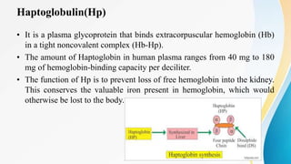
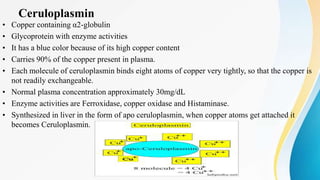
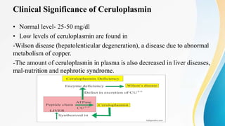
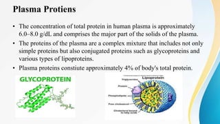
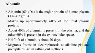
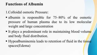
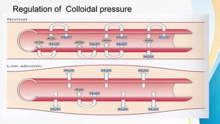
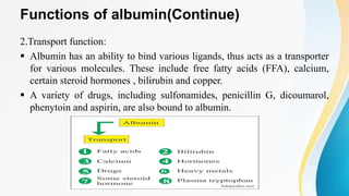
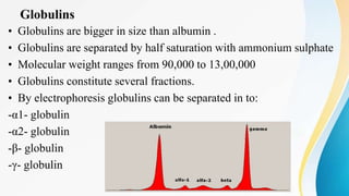
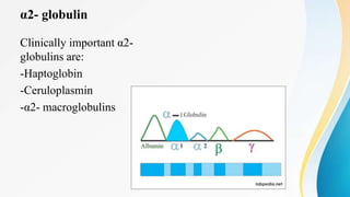
Plasma is the liquid portion of blood that remains after red and white blood cells are removed. It contains water, salts, enzymes, antibodies, and proteins. The major plasma proteins are albumin, globulins, and fibrinogen. Albumin is the most abundant plasma protein and plays important roles in maintaining colloidal osmotic pressure and transporting molecules like fatty acids, hormones, and bilirubin. Low albumin levels can cause edema. Globulins are divided into alpha, beta, and gamma classes, and include proteins like ceruloplasmin, transferrin, C-reactive protein, and haptoglobin that have clinical significance as markers of disease states.
3. Plasma
Introduction:
Plasma is a straw colored liquid portion of blood
It is obtained as supenatant when anti-cogulated blood is centrifuged leaving the cells
components as precipitate
55-60% of blood is made of plasma
Plasma consists of water, electrolytes, metabolites , nutrients , hormones ,
immunoglobulins and other protiens.
5. Plasma Protiens
• The concentration of total protein in human plasma is approximately
6.0–8.0 g/dL and comprises the major part of the solids of the plasma.
• The proteins of the plasma are a complex mixture that includes not only
simple proteins but also conjugated proteins such as glycoproteins and
various types of lipoproteins.
• Plasma proteins constiute approximately 4% of body's total protein.
6. Plasma Proteins (Continue)
• Almost all plasma proteins except immunoglobulins are synthesized in
liver.
• Plasma proteins are generally synthesized on membrane -bound
polyribosomes.
• Most plasma proteins are glycoproteins.
• Plasma proteins forms 7% of the solids in plasma
• Their normal values(7.4 gm%) ranges from (6.4 – 8.3 gm%)
7. Classification of Plasma Proteins
• The plasma proteins consist of :
1)albumin (3.5-5 g/dl)
2)globulins (2.5-3.5 g/dl)
3)fibrinogen (200-400 mg/dl)
8.
9. Albumin
• Albumin (69 kDa) is the major protein of human plasma
(3.4–4.7 g/dL)
• Makes up approximately 60% of the total plasma
protein.
• About 40% of albumin is present in the plasma, and the
other 60% is present in the extracellular space.
• Half life of albumin is about 20 days.
• Migrates fastest in electrophoresis at alkaline pH and
precipitates last in salting out methods
10.
11. Structure of Albumin
• Mature human albumin consists of one polypeptide
chain of 585 amino acids and contains 17 disulfide
bonds
• It has an ellipsoidal shape, which means that it does not
increase the viscosity of the plasma as much as an
elongated molecule such as fibrinogen does.
• Has a relatively low molecular mass about 69 kDa
• Has an iso-electric pH of 4.7
12.
13. Synthesis of albumin
• The liver produces about 12 g of albumin per day.
• Albumin is initially synthesized as a preproprotein
• Albumin is synthesized in the liver as preproalbumin which
has an N-terminal peptide that is removed before the
nascent protein is released from the rough endoplasmic
reticulum. The product, proalbumin, is in turn cleaved in the
Golgi vesicles to produce the secreted albumin.
14.
15. Functions of Albumin
1.Colloidal osmotic Pressure:
albumin is responsible for 75–80% of the osmotic
pressure of human plasma due to its low molecular
weight and large concentration
It plays a predominant role in maintaining blood volume
and body fluid distribution.
Hypoalbuminemia leads to retention of fluid in the tissue
spaces(Edema)
17. Functions of albumin(Continue)
2.Transport function:
Albumin has an ability to bind various ligands, thus acts as a transporter
for various molecules. These include free fatty acids (FFA), calcium,
certain steroid hormones , bilirubin and copper.
A variety of drugs, including sulfonamides, penicillin G, dicoumarol,
phenytoin and aspirin, are also bound to albumin.
18. Functions of Albumin(Continue)
3.Nutritive Function
Albumin serves as a source of amino acids for tissue protein
synthesis to a limited extent, particularly in nutritional
deprivation of amino acids.
4.Buffering Function
Among the plasma proteins, albumin has the maximum
buffering capacity due to its high concentration and the
presence of large number of histidine residues, which
contribute maximally towards maintenance of acid base
balance.
19. Clinical Significance of Albumin
1)BLOOD-BRAIN BARRIER:
Albumin-fatty acid complex cannot cross blood-brain
barrier and hence fatty acids cannot be taken up by
brain.
Loosely bound bilirubin to albumin can be easily
replaced by drugs like aspirin
In new born if such drugs are given, the released
bilirubin gets deposited in brain causing Kernicterus.
20. Clinical Significance of Albumin(Continue)
2)PROTEIN-BOUND CALCIUM:
Calcium level in blood is lowered in hypo-albuminemia
Thus , even though total calcium level in blood is
lowered, ionised calcium level may be normal, so tetany
may not occur.
Calcium is lowered by 0.8 mg/dl for a fall of 1g/dl of
albumin
21. Clinical Significance of Albumin(Continue)
3)THERAPEUTIC USE:
Human albumin is therapeutically useful to treat
burns,hemorrhage and shock.
4)EDEMA:
Hypo-albuminemia will result in tissue edema
Eg: a)malnutrition
b)nephrotic syndrome
c)cirrhosis of liver
d)chronic congestive cardiac failure.
22. Hypo-albuminemia
• Hypoalbuminemia (or hypoalbuminaemia) is a medical
sign in which the level of albumin in the blood is low.
This can be due to decreased production in the liver,
increased loss in the gastrointestinal tract or kidneys,
increased use in the body, or abnormal distribution
between body compartments.
• Diagnostic Level:
Level below 3.5 grams per deciliter
23. Causes of Hypo-albuminemia
1.CIRRHOSIS OF LIVER:
Cirrhosis is a late stage of scarring (fibrosis) of the liver caused by many forms of
liver diseases and conditions, such as hepatitis and chronic alcoholism.In this
disease synthesis of albumin is decreased.
2.MALNUTRITION:
Availability of amino acids is reduce and so albumin synthesis is affected.
3.NEPHROTIC SYNDROME:
Permeability of kidney glomerular membrane is defective , so that albumin is
excreted in large quantities.
24.
25. Albuminuria
• Presence of albumin in urine is called albuminuria.
• It is always pathological.
• Seen in:
a)Nephrotic syndrome(large quantities)
b)Acute nephritis
c)Inflammatory conditions of urinary tract.
• Detection of albumin in urine is done by heat and acetic acid
test.
26. MICRO-ALBUMINURIA:
• In micro-albuminuria small quantity of albumin (30-300 mg/dl)
is seen in urine
• It is estimated by RIA(Radioimmunoassay)
• Increased levels of microalbuminuria is an indication of early
involvement of renal tissue in diabetic patients
27. Hypoproteinemia
• Hypoproteinemia is a condition in which a person has very low
levels of protein in the blood. Proteins are essential to every
part of the human body. Bones, muscles, skin, and nearly every
vital organ or tissue contain them. The body needs protein to
function and survive and must get it through food.
• Since albumin is the major protein present in the blood, any
condition causing lowering of albumin will lead to reduce total
proteins in blood
28. HYPERALBUMINEMIA :
• Hyperalbuminemia is an increased concentration of
albumin in the blood. Typically, this condition is due to
dehydration. Hyperalbuminemia has also been associated
with high protein diets.
• Increased levels of plasma albumin are present only in
acute dehydration and have no clinical significance
ANALBUMINAEMIA:
• Analbuminemia is a rare hereditary abnormality in which
plasma albumin concentration is usually less than1.0gm/L
29. Globulins
• Globulins are bigger in size than albumin .
• Globulins are separated by half saturation with ammonium sulphate
• Molecular weight ranges from 90,000 to 13,00,000
• Globulins constitute several fractions.
• By electrophoresis globulins can be separated in to:
-α1- globulin
-α2- globulin
-β- globulin
-γ- globulin
30. Synthesis of globulins
• α and β globulins are synthesized in the liver.
• Y globulins are synthesized in plasma cells and B-cells of
lymphoid tissues (Reticulo-endothelial system)
• Synthesis of Y globulins is increased in chronic infections,
chronic liver diseases, auto immune diseases, leukemias,
lymphomas and various other malignancies.
31. α Globulins
• They are glycoproteins
• Based on electrophoretic mobility , they are sub classified in to
α1 and α2 globulins
• α1 globulins
• Examples:
-α1antitrypsin
-Orosomucoid (α1 acid glycoprotein)
-α1-fetoprotein (AFP)
32. α1 Globulins
α1-antitrypsin:
Also called α1-antiprotease
It is a single-chain protein of 394 amino acids, contains three oligosaccharide chains
It is the major component (> 90%) of the α 1fraction of human plasma.
It is synthesized by hepatocytes and macrophages and is the principal serine protease
inhibitor of human plasma.
It inhibits trypsin, elastase, and certain other proteases by forming complexes with them.
33. Polymorphic forms of α1-antitrypsin
• The most common alleles are the M variants, which are subdivided into six M
subtypes, homozygous or heterozygous for the M1, M2, M3 and M4 alleles.
When M variants are inherited in homozygous or heterozygous form, AAT
serum levels are supposed to be normal.The common variants that lead to AAT
plasma deficiency (AATD) are Z and S, which could result in early-onset
chronic obstructive pulmonary diseases, these including emphysema and
chronic bronchitis, as well as liver disease, expressed as neonatal cholestasis,
that may give rise to juvenile cirrhosis or a slowly progressive liver disease in
adults. Thus, individuals who are homozygous for AATD alleles can develop
liver or lung diseases. Nevertheless, AATD heterozygotes, which have inherited
one normal allele, have sufficient amounts of AAT and are therefore less prone
to diseases.
34.
35. Clinical consscequences of α1-antitrypsin Deficiency
Emphysema:
• Normally antitrypsin protects the lung tissue from proteases (active
elastase) released from macrophages
• Forms a complex with protease and inactivates it.
• In its deficiency, the active elastase destroys the lung tissue by
proteolysis.
36. α1-fetoprotein(AFP)
• Present in high concentration in fetal blood during mid
pregnancy
• Normal concentration in healthy adult-< 1µg/100ml
• Level increases during pregnancy
• Clinically considered a tumor marker for the diagnosis of
hepatocellular carcinoma or teratoblastomas
38. Haptoglobulin(Hp)
• It is a plasma glycoprotein that binds extracorpuscular hemoglobin (Hb)
in a tight noncovalent complex (Hb-Hp).
• The amount of Haptoglobin in human plasma ranges from 40 mg to 180
mg of hemoglobin-binding capacity per deciliter.
• The function of Hp is to prevent loss of free hemoglobin into the kidney.
This conserves the valuable iron present in hemoglobin, which would
otherwise be lost to the body.
39. Clinical significance of Haptoglobulin
• Concentration rises in inflammatory conditions
• Half-life of haptoglobin is approximately 5 days, the half-life of
the Hb-Hp complex is about 90 minutes, the complex being
rapidly removed from plasma by hepatocytes.
• Thus, when haptoglobin is bound to hemoglobin, it is cleared from
the plasma about 80 times faster than normally.
• The level of haptoglobin falls rapidly in hemolytic anemias.
• Free Hp level or Hp binding capacity depicts the degree of
intravascular hemolysis.
40. Ceruloplasmin
• Copper containing α2-globulin
• Glycoprotein with enzyme activities
• It has a blue color because of its high copper content
• Carries 90% of the copper present in plasma.
• Each molecule of ceruloplasmin binds eight atoms of copper very tightly, so that the copper is
not readily exchangeable.
• Normal plasma concentration approximately 30mg/dL
• Enzyme activities are Ferroxidase, copper oxidase and Histaminase.
• Synthesized in liver in the form of apo ceruloplasmin, when copper atoms get attached it
becomes Ceruloplasmin.
41. Clinical Significance of Ceruloplasmin
• Normal level- 25-50 mg/dl
• Low levels of ceruloplasmin are found in
-Wilson disease (hepatolenticular degeneration), a disease due to abnormal
metabolism of copper.
-The amount of ceruloplasmin in plasma is also decreased in liver diseases,
mal-nutrition and nephrotic syndrome.
42. α2- macroglobulins
• Major component of α2 proteins
• Comprises 8–10% of the total plasma protein in humans.
• Tetrameric protein with molecular weight of 725,000.
• Synthesized by hepatocytes and macrophages
• Inactivates all the proteases and thus is an important in vivo anticoagulant.
• Carrier of many growth factors
• Normal serum level:130-300 mg/dl
• Concentration is markedly increased in nephrotic syndrome, since other proteins are lost
through urine in this condition.
43. β- globulin
• Beta globulins are a group of globular proteins in plasma that are
more mobile in alkaline or electrically charged solutions than
gamma globulins, but less mobile than alpha globulins
• β Globulins of clinical importance are:
-Transferrin
-C-reactive protein
-Haemopexin
-Complement C1q
-β Lipoprotein(LDL)
44. Transferrin
• Transferrin (Tf) is a β 1-globulin with a molecular mass of approximately 76 kDa.
• It is a glycoprotein and is synthesized in the liver.
• About 20 polymorphic forms of transferrin have been found.
• It plays a central role in the body's metabolism of iron because it transports iron (2 mol
of Fe3+ per mole of Tf) in the circulation to sites where iron is required, eg, from the gut
to the bone marrow and other organs.
• Approximately 200 billion red blood cells (about 20 mL) are catabolized per day,
releasing about 25 mg of iron into the body—most of which is transported by
transferrin.
45. Clinical significance of Transferrin
• The concentration of transferrin in plasma is approximately 300 mg/dL.
• This amount of transferrin can bind 300 g of iron per deciliter, so that this
represents the total iron-binding capacity of plasma.
• However, the protein is normally only one-third saturated with iron.
• In iron deficiency anemia, the protein is even less saturated with iron, whereas in
conditions of storage of excess iron in the body (eg, hemochromatosis) the
saturation with iron is much greater than one-third.
46. C-reactive protein
• So named because it reacts with C-polysaccharide of capsule of pneumococci
• Molecular weight of 115-140 kD
• Synthesized in liver
• Can stimulate complement activity and macrophages
• Acute phase protein- Concentration rises in inflammatory conditions
• Clinically important marker to predict the risk of coronary heart disease
47. Haemopexin
• Molecular weight 57,000-80,000
• Normal level in adults-0.5 to 1.0 gm/L
• Low level at birth, reaches adult value within first year of life
• Synthesized in liver
• Function is to bind haem formed from breakdown of Hb and other
haemoproteins
• Low levels may be found in certain pathological conditions other
than haemolytic disease, namely renal and liver diseases. The
concentration is increased in diabetes mellitus, infections and
carcinoma.
48. Complement C1q
• First complement factor to bind antibody
• Binding takes place at the Fc region of IgG or IgM
• Binding triggers the classical complement pathway
• Thermo labile, destroyed by heating
• Normal level – 0.15 gm/L
• Molecular weight-400,000
• Can bind heparin and bivalent ions
• Decreased level is used as an indicator of circulating Ag –Ab complex.
• High levels are found in chronic infections
49. Gamma Globulins
• They are immunoglobulins with antibody activity
• They occupy the gamma region on electrophoresis
• Immunoglobulins play a key role in the defense mechanisms of the
body
• There are five types of immunoglobulins:
IgG, IgA, IgM, IgD, and IgE.
50. Functions of Immunoglobulins
Immunoglobulins Functions
IgG Main antibody in the secondary response.
Opsonizes bacteria, Fixes complement, neutralizes
bacterial toxins and viruses and crosses the
placenta.
IgA Secretory IgA prevents attachment of bacteria and
viruses to mucous membranes. Does not fix
complement.
IgM Produced in the primary response to an antigen.
Fixes complement. Does not cross the placenta.
Antigen receptor on the surface of B cells.
IgD Uncertain. Found on the surface of many B cells as
well as in serum.
IgE Mediates immediate hypersensitivity.Defends
against worm infections. Does not fix complement.
51. Fibrinogen
• Also called clotting factor1
• Constitutes 4-6% of total protein
• Precipitated with 1/5 th saturation with ammonium sulphate
• Large asymmetric molecule,highly elongated with axial ratio of 20:1
• Imparts maximum viscosity to blood,synthesized in liver
• Made up of 6 polypeptide chains,chains are linked together by S-S linkages
• Amino terminal end is highly negative due to the presence of glutamic acid
• Negative charge contributes to its solubility in plasma and prevents aggregation
due to electrostatic repulsions between the fibrinogen molecules.
52. Multiple Myeloma
• A malignant proliferation of plasma cells
• Results in an abnormally high concentration of serum immunoglobulins, usually
IgG or IgA
Bence Jones Proteins:
• In multiple myeloma, more light chains are produced than heavy chains and
enter the bloodstream
• Because they are of relatively low m.wt, they pass through glomerular
membrane and appear in the urine, these protein chains of low m.wt are known
as Bence Jones Proteins
• Bence Jones proteins have the remarkable characteristic of precipitating on
heating urine from 450 – 600 C and redissolve when the heating is continued
above 800 C
53. Methods of sepration of Plasma Proteins
• Precipitation by salting out.
• Cohn’s fractional precipitation method.
• Electrophoresis separation of protein fractions.
• Immunoelectrophoresis technique.
• Ultra-centrifugation technique.
54. Sepration of plasma proteins using salt
• In clinical laboratory, separation is usually done by salts.
• Thus , fibrinogen is precipitated by 10% and globulins by 22%
concentration of sodium sulphate
Ammonium sulphate will precipitate :
• albumin by full-saturation
• globulin by half-saturation
55. Electrophoresis
• The most common method of analyzing plasma proteins is by
electrophoresis.
• The term electrophoresis refers to the movement of charged particles
through an electrolyte when subjected to an electric field
• In clinical laboratory , cellulose acetate is widely used as a supporting
medium.
• Its use permits resolution , after staining , of plasma proteins into five bands
, designated albumin , α1 , α2 , β and γ fractions, respectively
• The stained strip of cellulose acetate is called electrophoretogram.
• The amounts of these five bands can be conveniently quantified by use of
Densitometric scanning machines.
• Characteristic changes in the amounts of one or more of these five bands are
found in many diseases.
56.
57. Serum Uric Acid
• A uric acid blood test, also known as a serum uric acid
measurement, determines how much uric acid is present in your
blood. The test can help determine how well your body produces
and removes uric acid
• Uric acid is a chemical produced when your body breaks down
foods that contain organic compounds called purines.
• Uric acid is the final product of purine catabolism in human
beings.
• Approximately two thirds of total body urate is produced
endogenously, while the remaining one third is accounted for by
dietary purines.
58. Serum uric acid(Continue)
• Approximately 70% of the urea produced daily is excreted by
the kidneys, while the rest is eliminated by the intestines.
• The blood levels of uric acid are a function of the balance
between the breakdown of purines and the rate of uric acid
excretion.
• Alterations in this balance may account for hyperuricemia.
59. Uric acid production and excretion
1.Uric acid, an end product of purine metabolism, is produced from both dietary and endogenous
sources.
2. Its formation results from the conversion of adenine and guanine of nucleoproteins
and nucleotides.
3. Xanthine oxidase catalyzes the reaction that occurs as the final step in the degradation of
purines to uric acid.
4. The body ultimately excretes uric acid via the kidneys (300–600 mg/day; two-thirds of total
uric acid) and via the gastrointestinal (GI) tract (100–300 mg/day; one-third of the total uric
acid).
5. Uric acid has no known biological function.
6. The body has a total uric acid content of 1.0 to 1.2 g; the daily turnover rate is 600 to 800 mg.
7. At a pH of 4.0 to 5.0 (i.e., in urine), uric acid exists as a poorly soluble free acid; at
physiological pH, it exists primarily as monosodium urate salt.
60.
61. Hyperuricemia
• Hyperuricemia is an elevated uric acid level in the blood. The normal upper limit
is 6.8mg/dL, and anything over 7 mg/dL is considered saturated, and symptoms
can occur. This elevated level is the result of increased production, decreased
excretion of uric acid, or a combination of both processes.
Hyperuricemia symptoms
• severe pain in your joints.
• joint stiffness.
• difficulty moving affected joints.
• redness and swelling.
• misshapen joints.
62. Mechanism of Hyperuricemia
• There are two main mechanisms of hyperuricemia:
1. Under excretion e.g chronic kidney disease
2. Overproduction e.g rapid cell death such as in
cancer chemotherapy or a combination of these two
mechanisms!!
63. Gout
• Gout is a disorder of purine metabolism, characterized by elevated serum uric acid
level.
• Higher level of serum uric acid may be due to overproduction or impaired
excretion of uric acid.
• It is characterized by interment attacks of acute arthritis produced by deposition of
sodium urate crystals in joints. ( Uric acid has low water solubility and gets
precipitated in the joints, kidney and subcutaneous tissues.)
• Secondary hyperuricemia may result due to excessive production (breakdown of
proteins and nucleic acids during cancer chemotherapy) or decreased excretion
(due to the use of thiazides, loop diuretics, ethambutol, clofibrate etc.) of uric acid
64.
65. Acute Gout
• This clinical presentation of gout is characterized by painful arthritic attacks of sudden
onset.
Pathogenesis
• Monosodium urate crystals form in articular tissues; this process sets off an inflammatory
reaction.
• Trauma, exposure to cold, dietary overindulgence, or another triggering event may be
involved in the development of the acute attack.
Signs and symptoms
• The initial attack is abrupt, usually occurring at night or in the early morning as synovial
fluid is reabsorbed.
• This severe arthritic pain progressively worsens and generally involves only one or a few
joints.
• The affected joints typically become hot, swollen, and extremely tender.
66. Diagnostic Criteria:
1. Definitive diagnosis of gouty arthritis can be made by demonstration of monosodium urate
crystals in the synovial fluid of affected joints. These needle-shaped crystals are termed
negatively birefringent when viewed through a polarized light microscope.
2. Serum analysis usually reveals an above-normal uric acid level: however, this finding is not
specific for acute gout. Other common serum findings include leukocytosis and a moderately
elevated erythrocyte sedimentation rate.
3. A dramatic therapeutic response to colchicine may be helpful in establishing the diagnosis, but
this is not absolute because other causes of acute arthritis may respond as well.
4. When fluid cannot be aspirated from the affected joint, a diagnosis of gout is supported by:
• A prior history of acute monoarticular arthritis (especially of the big toe) followed by a
symptom-free period
• The presence of hyperuricemia
• Rapid resolution of symptoms after colchicine therapy
5. Other conditions that may mimic gout include pseudogout (calcium pyrophosphate dehydrate
crystal disease) or septic arthritis.
67. Treatment Goals
1. To Relieve Pain and Inflammation
2. To Terminate the acute Attack
3. To Restore Normal Function to the Affected joints
Therapy:
1. General Therapeutic Principles
a. The affected joint (or joints) should be immobilized.
b. Anti-inflammatory drug therapy should begin immediately. For maximal
therapeutic effectiveness, these drugs should be kept on hand so that the patient
may begin therapy as soon as a subsequent attack begins.
2. Specific drugs
• Nonsteroidal anti-inflammatory drugs (NSAIDs) • Colchicine & Corticosteroids
Editor's Notes
جگر روزانہ تقریبا g 12 جی البمومین تیار کرتا ہے ، جو کل ہیپاٹک پروٹین ترکیب کا تقریبا 25٪ اور اس کے آدھے خستہ پروٹین کی نمائندگی کرتا ہے۔
Phenotype:the set of observable characteristics of an individual resulting from the interaction of its genotype with the environment.
Emphysema is a lung condition that causes shortness of breath.
NE=Neutrophil elastase burden
Nephrotic syndrome is a kidney disorder that causes your body to pass too much protein in your urine. Nephrotic syndrome is usually caused by damage to the clusters of small blood vessels in your kidneys that filter waste and excess water from your blood.