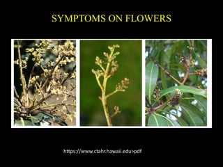
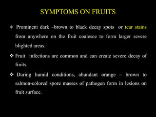
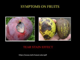
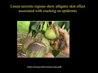
A Critique of the Proposed National Education Policy Reform
MANGO ANTHRACNOSE
1. MANGO ANTHRACNOSE
COLLEGE OF AGRICULTURAL TECHNOLOGY
(Affiliated to Tamil Nadu Agricultural University, Coimbatore-3)
Kullapuram (Po),ViaVaigai Dam, Theni-625 562
STUDENT
Miss. ABIRAMI.C
ID. No. 2015021003
COURSE TEACHER
Dr. PARTHASARATHY S
Asst. Professor (Plant Pathology)
2. SIGNIFICANCE
Post harvest loses of mango in several tropical countries
(Brazil, Pakistan) ranges from 15% in dry season to 70% in
rainy season.
China and India postharvest losses of mango due to
anthracnose is 15% - 25%.
The incidence of this disease reach 100% in fruit under wet
conditions.
30 – 60% yield loss on mango occurs different countries of
world (Aken,2006:Chowdhury and Rahim,2009)
4. CAUSAL ORGANISM
• Anamorph stage (asexual stage)
Colletotrichum gloeosporioides (Penz and Sacc)
• Teleomorph ( sexual stage )
Glomerella cingulata (Fitzell, 1979 and Prakash, 1990)
5. SYSTEMATIC POSITION
KINGDOM : Fungi
DIVISION : Ascomycota
CLASS : Sordariomycetes
ORDER : Glomerellales
FAMILY : Glomerellaceae
GENUS : Colletotrichum
SPECIES : C. gloeosporioides
6. SYMPTOMS ON LEAVES
Small blister like spots develop on the leaves and
twigs.
Young leaves wither and dry Tender twigs wither and
die back symptom appears.
8. SYMPTOMS ON FLOWERS
Infections on the panicles (form clusters) start as
small black or dark- brown spots.
These can enlarge, coalesce and kill the flowers.
The panicles become prone to disintegration.
10. SYMPTOMS ON FRUITS
Prominent dark –brown to black decay spots or tear stains
from anywhere on the fruit coalesce to form larger severe
blighted areas.
Fruit infections are common and can create severe decay of
fruits.
During humid conditions, abundant orange – brown to
salmon-colored spore masses of pathogen form in lesions on
fruit surface.
12. Linear necrotic regions show alligator skin effect
associated with cracking on epidermis
https://www.ctahr.hawaii.edu>pdf
13. SYMPTOMS ON STEM AND BRANCHES
• Twig dieback occurs when severe elongated,
blackened lesions form on stems and twig die
back apically.
• Abundant sporulation of the pathogen covers
the decomposed points of infection.
15. PATHOGEN CHARACTERS
Conidiophore is simple, branched hyphae on which
conidia are produced.
Conidia is an asexual non-motile fungi spore that
develop externally from the cell that formed it.
17. EPIDEMIOLOGY
High humidity
Temperature of 24°C – 32°C.
Frequent rains.
Moist conditions favours the development of
disease (October to November)
19. Dissemination
Conidia are dispersed passively by splashing of rain or
irrigation water.
Inoculation
Spores land on infectious sites (panicles, leaves,
branch terminals)
Symptoms and disease development
Black, sunken, rapidly expanding lesions develop in
fruiting bodies (Acervuli).
20. Pathogen reproduction
Sticky masses of conidia are produced in fruiting
body (Acervuli).
Pathogen survival
The pathogen survives between seasons on infected
and defoliated branch terminals and mature leaves.
21. MODE OF INFECTION
Primary spread
Air borne conidia and infected plant debris are
primary source of infection.
Secondary spread
Rain splashes and irrigation water are secondary
source of infection.
22. INTEGARATED DISEASE MANAGEMENT
Spray P. fluorescens (FP7) at 3 weeks interval
commencing from October at 5 g/like on flower branches,
5 – 7 sprays one to be given on flowers and bunches.
Spray twice with Carbendazim (0.1%) at 15 days interval
during flowering to control blossom infection.
23. Spraying of Mancozeb 2 g/lit Thiophanate methyl 1 g/lit or
chlorothalonil 2 g/lit 3 times at 15 days interval.
Spray copper fungicides (0.3%) for the control of foliar
infection.
Before storage, treat with hot water (50 – 55ºC) for 15
minutes or dip in Benomyl solution (500 ppm) or
Thiobendazole (1000 ppm) for 5 minutes.