Basal cell carcinoma (BCC) is the most common form of skin cancer, and its occurrence on the face is particularly significant due to cosmetic and functional considerations. Management typically involves a multi-pronged approach, considering factors such as tumor size, location, histological subtype, patient comorbidities. Etiology and Risk Factors
* Ultraviolet (UV) Radiation (main culprit)
* Genetic Predisposition
* Immunosuppression
* Fair Skin Phenotype
* Age
* Previous BCC/Skin Cancer History
* Environmental Exposures (e.g., arsenic)
3. Clinical Presentation and Diagnosis
* Common Clinical Subtypes (Nodular, Superficial, Morpheaphorm/Infiltrative, Pigmented, Basosquamous)
* Dermoscopy in Diagnosis
* Biopsy (Excisional, Incisional, Shave, Punch) and Histopathological Confirmation
4. Staging and Risk Stratification
* Clinical vs. Pathological Staging
* High-Risk Features (size, location, histological subtype, perineural invasion, recurrence, immunosuppression). Treatment Modalities for Facial BCC
**5.1. Surgical Excision (Gold Standard)**
* **Standard Surgical Excision:**
* Indications, technique, and margins
* Advantages and disadvantages
* Histopathological margin control
* **Mohs Micrographic Surgery (MMS):**
* Principle and detailed process (sequential excision and microscopic examination of margins)
* Indications for facial BCC (high-risk, cosmetically sensitive areas, recurrent lesions)
* Advantages (tissue sparing, highest cure rates)
* Limitations and patient selection
**5.2. Non-Surgical Treatment Options**
* **Topical Therapies:**
* **Imiquimod (Aldara, Zyclara):** Mechanism of action, indications (superficial BCC), application, side effects, efficacy.
* **5-Fluorouracil (5-FU) (Carac, Efudex):** Mechanism, indications (superficial BCC), application, side effects, efficacy.
* Other topical agents (e.g., ingenol mebutate, tazarotene)
* **Photodynamic Therapy (PDT):**
* Principle (photosensitizer + light), indications (superficial BCC, large field cancerization), advantages (cosmetic outcome), limitations (pain, photosensitivity, recurrence rates).
* **Cryosurgery:**
* Mechanism (freezing), indications (small, low-risk, superficial BCC), advantages (minimally invasive), disadvantages (scarring, pigment changes, lack of margin control).
* **Curettage and Electrodessication (C&D):**
* Technique (scraping + cautery), indications (small, low-risk BCC on trunk/extremities, less favored on face due to scarring and lack of margin control), efficacy, limitations.
* **Radiation Therapy:**
* Mechanism, indications (elderly/infirm, large/inoperable tumors, adjuvant therapy, recurrent lesions), types (external beam, brachytherapy), advantages, side effects (local skin reactions, long-term scarring, potential for secondary malignancies).
* **Systemic Therapies (for Advanced/Metastatic BCC):**


![➔ BCC is nowadays thought to arise from stem cells of the hair follicle
➔ BCC is one of the most highly mutated human tumours (i.e. tumour
mutational burden [TMB] is 65 mutations/megabases, compared with
14 mutations/ megabases for melanoma)](https://image.slidesharecdn.com/nrwin3aeteqm06i5pps2-management-of-basal-cell-carcinoma-250616151929-b753e3d4/75/Management-of-Basal_cell_carcinoma-of-Head-and-neck-pptx-6-2048.jpg)


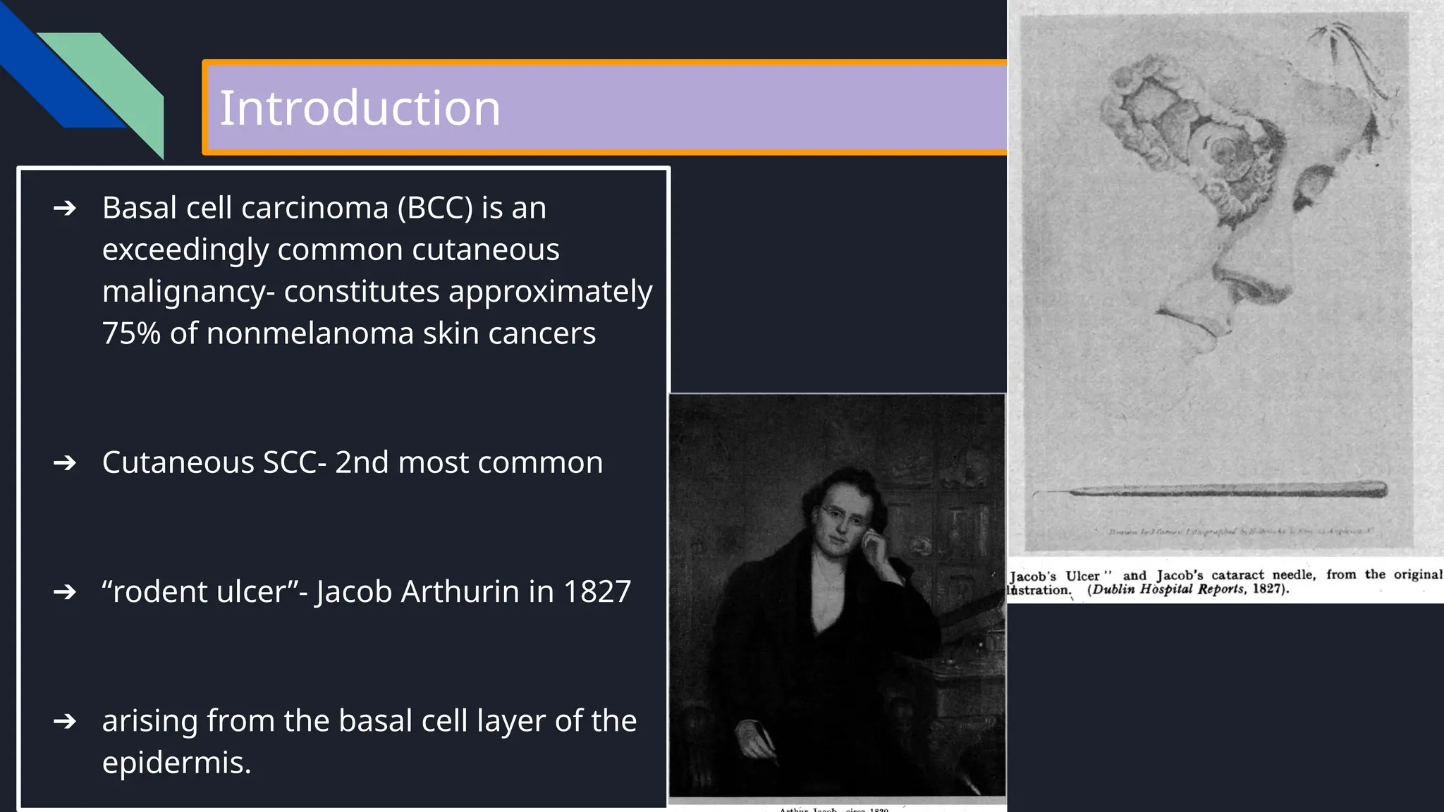
![Imaging
not routinely indicated for early-stage disease in the clinically node-negative
patient
useful to perform to delineate the overall extent of the tumour when perineural
invasion (magnetic resonance imaging [MRI]) or bony invasion (computed
tomography [CT]) is suspected
When to plan imaging?
A. clinically node-positive patient
B. recurrent tumours
C. For advanced tumours
D. Patients with clinically perineural invasion or aggressive histological type](https://image.slidesharecdn.com/nrwin3aeteqm06i5pps2-management-of-basal-cell-carcinoma-250616151929-b753e3d4/75/Management-of-Basal_cell_carcinoma-of-Head-and-neck-pptx-23-2048.jpg)