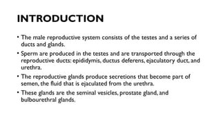
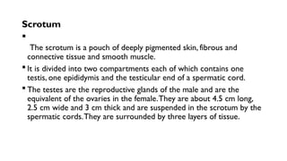
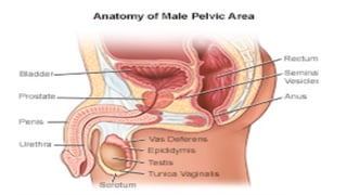
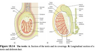
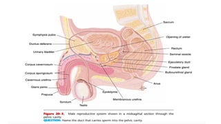
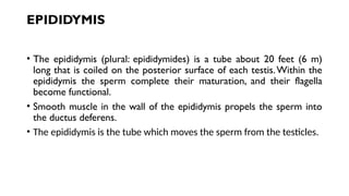
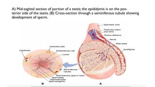
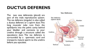
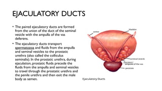
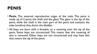
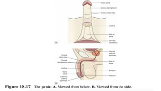
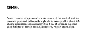
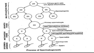
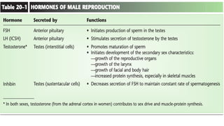
Male Reproductive Anatomy
and Physiology
SESSION
EACC, YEAR I
2022
COURSE OUTLINE
• INTRODUCTION
• DEFINE DIFFERENTS PARTS FOR MALE REPRODUCTIVE SYSTEM
INTRODUCTION
• The male reproductive system consists of the testes and a series of
ducts and glands.
• Sperm are produced in the testes and are transported through the
reproductive ducts: epididymis, ductus deferens, ejaculatory duct, and
urethra.
• The reproductive glands produce secretions that become part of
semen, the fluid that is ejaculated from the urethra.
• These glands are the seminal vesicles, prostate gland, and
bulbourethral glands.
Scrotum

The scrotum is a pouch of deeply pigmented skin, fibrous and
connective tissue and smooth muscle.
 It is divided into two compartments each of which contains one
testis, one epididymis and the testicular end of a spermatic cord.
 The testes are the reproductive glands of the male and are the
equivalent of the ovaries in the female.They are about 4.5 cm long,
2.5 cm wide and 3 cm thick and are suspended in the scrotum by the
spermatic cords.They are surrounded by three layers of tissue.
Scrotum
• Scrotum. The bag of skin that holds and helps to protect the testicles.
The testicles make sperm and, to do this, the temperature of the
testicles needs to be cooler than the inside of the body. This is why
the scrotum is located outside of the body.
Scrotum
• Tunica vaginalis
This is a double membrane, forming the outer covering of the testes, and is a
downgrowth of the abdominal and pelvic peritoneum. During early fetal life, the
testes develop in the lumbar region of the abdominal cavity just below the
kidneys.
• Tunica albuginea
This is a fibrous covering beneath the tunica vaginalis that surrounds the testes.
Ingrowths form septa,
dividing the glandular structure of the testes into lobules.
• Tunica vasculosa
This consists of a network of capillaries supported by delicate connective
tissue.
TESTES
• The testes are located in the scrotum, a sac of skin between the
upper thighs.
• The temperature within the scrotum is about35.5 c, slightly lower
than body temperature, which is necessary for the production of
viable sperm.
• In the male fetus, the testes develop near the kidneys, then descend
into the scrotum just before birth.
• Cryptorchidism is the condition in which the testes fail to descend,
and the result is sterility unless the testes are surgically placed in the
scrotum.
TESTES
• Each testis is about 1.5 inches long by 1 inch wide (4 cm by 2.5 cm) and is divided
internally into lobes.
• Each lobe contains several seminiferous tubules, in which spermatogenesis takes place.
• Among the spermatogonia of the seminiferous tubules are sustentacular (Sertoli) cells,
which produce the hormone inhibin when stimulated by testosterone. Between the
loops of the seminiferous tubules are interstitial cells, which produce testosterone
when stimulated by luteinizing hormone (LH) from the anterior pituitary gland.
• Besides its role in the maturation of sperm, testosterone is also responsible for the
male secondary sex characteristics, which begin to develop at puberty (Table 20–1).
A sperm cell consists of several parts, which are shown in Fig. 20–1.
• The head contains the 23 chromosomes.
TESTES
• On the tip of the head is the acrosome,which is similar to a
lysosome and contains enzymes to digest the membrane of an egg
cell.Within the middle piece are mitochondria that produce ATP.
• The flagellum provides motility, the capability of the sperm cell to
move. It is the beating of the flagellum that requires energy from ATP.
Sperm from the seminiferous tubules enter a tubu-lar network called
the rete testis, then enter the epi-didymis, the first of the
reproductive ducts.
EPIDIDYMIS
• The epididymis (plural: epididymides) is a tube about 20 feet (6 m)
long that is coiled on the posterior surface of each testis.Within the
epididymis the sperm complete their maturation, and their flagella
become functional.
• Smooth muscle in the wall of the epididymis propels the sperm into
the ductus deferens.
• The epididymis is the tube which moves the sperm from the testicles.
A) Mid-sagittal section of portion of a testis; the epididymis is on the pos-
terior side of the testis. (B) Cross-section through a seminiferous tubule showing
development of sperm.
DUCTUS DEFERENS
The two vasa deferentia (plural) are
part of the male reproductive system.
The vas deferens (singular) is also called
a ductus deferens or a sperm duct.This
long muscular tube runs from the
epididymis into the pelvic cavity behind
your bladder and connects to your
urethra through a structure called the
ejaculatory duct. The vas deferens is
surrounded by a spermatic cord and
transports mature sperm to the urethra
before you ejaculate.
EJACULATORY DUCTS
• The paired ejaculatory ducts are formed
from the union of the duct of the seminal
vesicle with the ampulla of the vas
deferens.
• The ejaculatory ducts transport
spermatozoa and fluids from the ampulla
and seminal vesicles to the prostatic
urethra (also called the colliculus
seminalis). In the prostatic urethra, during
ejaculation, prostatic fluids precede the
fluids from the ampulla and seminal vesicles
to travel through the prostatic urethra and
the penile urethra and then exit the male
body as semen.
EJACULATORY DUCTS
• Each of the two ejaculatory ducts receives sperm from the ductus
deferens and the secretion of the seminal vesicle on its own side.
Both ejaculatory ducts empty into the single urethra.
• The ejaculatory ducts are two tubes about 2 cm long, each formed by
the union of the duct from a seminal vesicle and a deferent duct.
• They pass through the prostate gland and join the prostatic urethra,
carrying seminal fluid and spermatozoa to the urethra.
• The ejaculatory ducts are composed of the same layers of tissue as
the seminal vesicles
SEMINAL VESICLES
The paired seminal vesicles are posterior to the urinary bladder.
Their secretion contains fructose to provide an energy source for
sperm and is alkaline to enhance sperm motility.
The duct of each seminal vesicle joins the ductus deferens on that side
to form the ejaculatory duct.
SEMINAL VESICLES
The paired seminal vesicles are
posterior to the urinary bladder.
Their secretion contains fructose
to provide an energy source for
sperm and is alkaline to enhance
sperm motility.
The duct of each seminal vesicle
joins the ductus deferens on that
side to form the ejaculatory duct.
PROSTATE GLAND
A muscular gland just below the urinary bladder, the prostate gland is
about 1.2 inches high by 1.6 inches wide by 0.8 inch deep (3 cm by 4
cm by 2 cm, about the size of a walnut).
• It surrounds the first inch of the ure- thra as it emerges from the
bladder.
• The glandular tissue of the prostate secretes an alkaline fluid that
helps maintain sperm motility.The smooth muscle of the prostate
gland contracts during ejacula-tion to contribute to the expulsion of
semen from the urethra.
PROSTATE GLAND(FUNCTIONS)
• The prostate gland secretes a thin, milky fluid that makes up about
30% of semen, and gives it its milky appearance.
• It contains a clotting enzyme, which thickens the semen in the vagina,
increasing the likelihood of semen being retained close to the cervix.
BULBOURETHRAL GLANDS
•
Also called Cowper’s glands, the bulbourethral glands are about the size of peas and
are located below the prostate gland; they empty into the urethra.
• Their alkaline secretion coats the interior of the urethra just before ejaculation,
which neutralizes any acidic urine
that might be present.
• You have probably noticed that the secretions of the male reproductive glands are
alkaline.
• This is important because the cavity of the female vagina has an acidic pH created by
the normal flora, the natural bacterial population of the vagina.
• The alkalinity of seminal fluid helps neutralize the acidic vaginal pH and permits sperm
motility in what might otherwise be an unfavorable environment.
URETHRA—
 The male urethra provides a common pathway for the flow of urine
and semen, the combined secretions of the male reproductive organs.
It is about 19 to 20 cm long and consists of three parts.
 The urethra is the last of the ducts through which semen travels, and
its longest portion is enclosed within the penis.
 The penis is an external genital organ; its distal end is called the glans
penis and is covered with a fold of skin called the prepuce or foreskin.
•
URETHRA
• This is a tube that allows urine to flow outside the body. It is also the
channel for semen to pass during ejaculation. The brain signals the
bladder muscles to tighten.This squeezes urine out of the bladder.At
the same time, the brain signals the sphincter muscles to relax to let
urine exit the bladder through the urethra. When all the signals
happen in the correct order, normal urination happens.
PENIS
• Penis. The external reproductive organ of the male. The penis is
made up of 2 parts, the shaft and the glans.The glans is the tip of the
penis, while the shaft is the main part of the penis and contains the
tube (urethra) that drains the bladder.
• All boys are born with a foreskin, or a covering over the tip of the
penis. Some boys are circumcised. This means that this covering of
skin is removed. Other boys are not circumcised and may have skin
that covers the tip of the penis.
SEMEN
Semen consists of sperm and the secretions of the seminal vesicles,
prostate gland, and bulbourethral glands; its average pH is about 7.4.
During ejaculation, approximately 2 to 4 mL of semen is expelled.
Each milliliter of semen contains about 100 million sperm cells.
Spermatogenesis
• Spermatogenesis is the process of sperm formation,
Spermatogenesis occurs in the testes.
• The process of meiosis is used during spermatogenesis to produce
mature sperm cells.
• Meiosis is a type of cell division where the genetic material is halved.
This is important for sexual reproduction, as during fertilization a
sperm and an egg fuse and combine their genetic material.Thus, a new
organism is created with the original amount of genetic material.
Spermatogenesis
• The process of formation of sperms is called spermatogenesis. It
occurs in the male gonads testis.Testes are made up of many
seminiferous tubules lined by germinal epithelium. Cells of this layer
divide to form spermatozoa in the following steps:
Spermatogenesis
• (1) Multiplication Phase: At maturity, the primordial germ cells divide
by mitosis to produce a large number of spermatogonia. Type A
spermatogonia is the stem cells which divide to form spermatogonia.
Type B spermatogonia are the precursors of sperms.
• (2) Growth Phase: Type B spermatogonium actively grows to a
primary spermatocyte. It obtains nourishment from the nursing cells.
Spermatogenesis
• (3) Maturation Phase: Each primary spermatocyte undergoes two
maturation divisions.The first maturation division is reductional and
forms two haploid daughter cells called secondary spermatocytes.
Both secondary spermatocytes then undergo second maturation
division to form four haploid spermatids.
• (4) Spermiogenesis: It is the process of transformation of spermatic to
a spermatozoan.The spermatozoa are then known as sperms.The
four spermatid becomes the head of the sperm, the Golgi apparatus,
containing proteolytic enzymes, becomes the acrosome cap.
• THANKYOU

Male system .pptxxxxxxxxxxxxxxxxxxxxxxxx

  • 1.
    Male Reproductive Anatomy andPhysiology SESSION EACC, YEAR I 2022
  • 2.
    COURSE OUTLINE • INTRODUCTION •DEFINE DIFFERENTS PARTS FOR MALE REPRODUCTIVE SYSTEM
  • 3.
    INTRODUCTION • The malereproductive system consists of the testes and a series of ducts and glands. • Sperm are produced in the testes and are transported through the reproductive ducts: epididymis, ductus deferens, ejaculatory duct, and urethra. • The reproductive glands produce secretions that become part of semen, the fluid that is ejaculated from the urethra. • These glands are the seminal vesicles, prostate gland, and bulbourethral glands.
  • 4.
    Scrotum  The scrotum isa pouch of deeply pigmented skin, fibrous and connective tissue and smooth muscle.  It is divided into two compartments each of which contains one testis, one epididymis and the testicular end of a spermatic cord.  The testes are the reproductive glands of the male and are the equivalent of the ovaries in the female.They are about 4.5 cm long, 2.5 cm wide and 3 cm thick and are suspended in the scrotum by the spermatic cords.They are surrounded by three layers of tissue.
  • 5.
    Scrotum • Scrotum. Thebag of skin that holds and helps to protect the testicles. The testicles make sperm and, to do this, the temperature of the testicles needs to be cooler than the inside of the body. This is why the scrotum is located outside of the body.
  • 8.
    Scrotum • Tunica vaginalis Thisis a double membrane, forming the outer covering of the testes, and is a downgrowth of the abdominal and pelvic peritoneum. During early fetal life, the testes develop in the lumbar region of the abdominal cavity just below the kidneys. • Tunica albuginea This is a fibrous covering beneath the tunica vaginalis that surrounds the testes. Ingrowths form septa, dividing the glandular structure of the testes into lobules. • Tunica vasculosa This consists of a network of capillaries supported by delicate connective tissue.
  • 9.
    TESTES • The testesare located in the scrotum, a sac of skin between the upper thighs. • The temperature within the scrotum is about35.5 c, slightly lower than body temperature, which is necessary for the production of viable sperm. • In the male fetus, the testes develop near the kidneys, then descend into the scrotum just before birth. • Cryptorchidism is the condition in which the testes fail to descend, and the result is sterility unless the testes are surgically placed in the scrotum.
  • 11.
    TESTES • Each testisis about 1.5 inches long by 1 inch wide (4 cm by 2.5 cm) and is divided internally into lobes. • Each lobe contains several seminiferous tubules, in which spermatogenesis takes place. • Among the spermatogonia of the seminiferous tubules are sustentacular (Sertoli) cells, which produce the hormone inhibin when stimulated by testosterone. Between the loops of the seminiferous tubules are interstitial cells, which produce testosterone when stimulated by luteinizing hormone (LH) from the anterior pituitary gland. • Besides its role in the maturation of sperm, testosterone is also responsible for the male secondary sex characteristics, which begin to develop at puberty (Table 20–1). A sperm cell consists of several parts, which are shown in Fig. 20–1. • The head contains the 23 chromosomes.
  • 12.
    TESTES • On thetip of the head is the acrosome,which is similar to a lysosome and contains enzymes to digest the membrane of an egg cell.Within the middle piece are mitochondria that produce ATP. • The flagellum provides motility, the capability of the sperm cell to move. It is the beating of the flagellum that requires energy from ATP. Sperm from the seminiferous tubules enter a tubu-lar network called the rete testis, then enter the epi-didymis, the first of the reproductive ducts.
  • 13.
    EPIDIDYMIS • The epididymis(plural: epididymides) is a tube about 20 feet (6 m) long that is coiled on the posterior surface of each testis.Within the epididymis the sperm complete their maturation, and their flagella become functional. • Smooth muscle in the wall of the epididymis propels the sperm into the ductus deferens. • The epididymis is the tube which moves the sperm from the testicles.
  • 14.
    A) Mid-sagittal sectionof portion of a testis; the epididymis is on the pos- terior side of the testis. (B) Cross-section through a seminiferous tubule showing development of sperm.
  • 15.
    DUCTUS DEFERENS The twovasa deferentia (plural) are part of the male reproductive system. The vas deferens (singular) is also called a ductus deferens or a sperm duct.This long muscular tube runs from the epididymis into the pelvic cavity behind your bladder and connects to your urethra through a structure called the ejaculatory duct. The vas deferens is surrounded by a spermatic cord and transports mature sperm to the urethra before you ejaculate.
  • 16.
    EJACULATORY DUCTS • Thepaired ejaculatory ducts are formed from the union of the duct of the seminal vesicle with the ampulla of the vas deferens. • The ejaculatory ducts transport spermatozoa and fluids from the ampulla and seminal vesicles to the prostatic urethra (also called the colliculus seminalis). In the prostatic urethra, during ejaculation, prostatic fluids precede the fluids from the ampulla and seminal vesicles to travel through the prostatic urethra and the penile urethra and then exit the male body as semen.
  • 17.
    EJACULATORY DUCTS • Eachof the two ejaculatory ducts receives sperm from the ductus deferens and the secretion of the seminal vesicle on its own side. Both ejaculatory ducts empty into the single urethra. • The ejaculatory ducts are two tubes about 2 cm long, each formed by the union of the duct from a seminal vesicle and a deferent duct. • They pass through the prostate gland and join the prostatic urethra, carrying seminal fluid and spermatozoa to the urethra. • The ejaculatory ducts are composed of the same layers of tissue as the seminal vesicles
  • 18.
    SEMINAL VESICLES The pairedseminal vesicles are posterior to the urinary bladder. Their secretion contains fructose to provide an energy source for sperm and is alkaline to enhance sperm motility. The duct of each seminal vesicle joins the ductus deferens on that side to form the ejaculatory duct.
  • 19.
    SEMINAL VESICLES The pairedseminal vesicles are posterior to the urinary bladder. Their secretion contains fructose to provide an energy source for sperm and is alkaline to enhance sperm motility. The duct of each seminal vesicle joins the ductus deferens on that side to form the ejaculatory duct.
  • 20.
    PROSTATE GLAND A musculargland just below the urinary bladder, the prostate gland is about 1.2 inches high by 1.6 inches wide by 0.8 inch deep (3 cm by 4 cm by 2 cm, about the size of a walnut). • It surrounds the first inch of the ure- thra as it emerges from the bladder. • The glandular tissue of the prostate secretes an alkaline fluid that helps maintain sperm motility.The smooth muscle of the prostate gland contracts during ejacula-tion to contribute to the expulsion of semen from the urethra.
  • 21.
    PROSTATE GLAND(FUNCTIONS) • Theprostate gland secretes a thin, milky fluid that makes up about 30% of semen, and gives it its milky appearance. • It contains a clotting enzyme, which thickens the semen in the vagina, increasing the likelihood of semen being retained close to the cervix.
  • 22.
    BULBOURETHRAL GLANDS • Also calledCowper’s glands, the bulbourethral glands are about the size of peas and are located below the prostate gland; they empty into the urethra. • Their alkaline secretion coats the interior of the urethra just before ejaculation, which neutralizes any acidic urine that might be present. • You have probably noticed that the secretions of the male reproductive glands are alkaline. • This is important because the cavity of the female vagina has an acidic pH created by the normal flora, the natural bacterial population of the vagina. • The alkalinity of seminal fluid helps neutralize the acidic vaginal pH and permits sperm motility in what might otherwise be an unfavorable environment.
  • 23.
    URETHRA—  The maleurethra provides a common pathway for the flow of urine and semen, the combined secretions of the male reproductive organs. It is about 19 to 20 cm long and consists of three parts.  The urethra is the last of the ducts through which semen travels, and its longest portion is enclosed within the penis.  The penis is an external genital organ; its distal end is called the glans penis and is covered with a fold of skin called the prepuce or foreskin. •
  • 24.
    URETHRA • This isa tube that allows urine to flow outside the body. It is also the channel for semen to pass during ejaculation. The brain signals the bladder muscles to tighten.This squeezes urine out of the bladder.At the same time, the brain signals the sphincter muscles to relax to let urine exit the bladder through the urethra. When all the signals happen in the correct order, normal urination happens.
  • 25.
    PENIS • Penis. Theexternal reproductive organ of the male. The penis is made up of 2 parts, the shaft and the glans.The glans is the tip of the penis, while the shaft is the main part of the penis and contains the tube (urethra) that drains the bladder. • All boys are born with a foreskin, or a covering over the tip of the penis. Some boys are circumcised. This means that this covering of skin is removed. Other boys are not circumcised and may have skin that covers the tip of the penis.
  • 27.
    SEMEN Semen consists ofsperm and the secretions of the seminal vesicles, prostate gland, and bulbourethral glands; its average pH is about 7.4. During ejaculation, approximately 2 to 4 mL of semen is expelled. Each milliliter of semen contains about 100 million sperm cells.
  • 28.
    Spermatogenesis • Spermatogenesis isthe process of sperm formation, Spermatogenesis occurs in the testes. • The process of meiosis is used during spermatogenesis to produce mature sperm cells. • Meiosis is a type of cell division where the genetic material is halved. This is important for sexual reproduction, as during fertilization a sperm and an egg fuse and combine their genetic material.Thus, a new organism is created with the original amount of genetic material.
  • 30.
    Spermatogenesis • The processof formation of sperms is called spermatogenesis. It occurs in the male gonads testis.Testes are made up of many seminiferous tubules lined by germinal epithelium. Cells of this layer divide to form spermatozoa in the following steps:
  • 32.
    Spermatogenesis • (1) MultiplicationPhase: At maturity, the primordial germ cells divide by mitosis to produce a large number of spermatogonia. Type A spermatogonia is the stem cells which divide to form spermatogonia. Type B spermatogonia are the precursors of sperms. • (2) Growth Phase: Type B spermatogonium actively grows to a primary spermatocyte. It obtains nourishment from the nursing cells.
  • 34.
    Spermatogenesis • (3) MaturationPhase: Each primary spermatocyte undergoes two maturation divisions.The first maturation division is reductional and forms two haploid daughter cells called secondary spermatocytes. Both secondary spermatocytes then undergo second maturation division to form four haploid spermatids. • (4) Spermiogenesis: It is the process of transformation of spermatic to a spermatozoan.The spermatozoa are then known as sperms.The four spermatid becomes the head of the sperm, the Golgi apparatus, containing proteolytic enzymes, becomes the acrosome cap.
  • 37.