Download to read offline

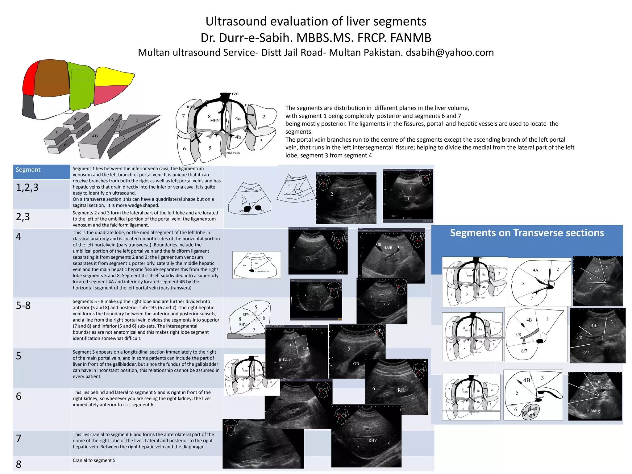

1) Segment 1 lies between the inferior vena cava, ligamentum venosum, and left branch of the portal vein. It is unique in receiving branches from both the right and left portal veins and draining directly into the inferior vena cava. 2) Segments 2 and 3 form the lateral part of the left lobe and are located to the left of structures including the umbilical portion of the portal vein and falciform ligament. 3) Segment 4, also known as the quadrate lobe or medial segment of the left lobe, is bounded by structures including the umbilical portion of the left portal vein and falciform ligament. It is subdivided into segments


![20 colon-por-enema643[1]](https://cdn.slidesharecdn.com/ss_thumbnails/20-colon-por-enema6431-120404135003-phpapp01-thumbnail.jpg?width=640&height=640&fit=bounds)


