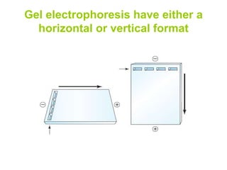
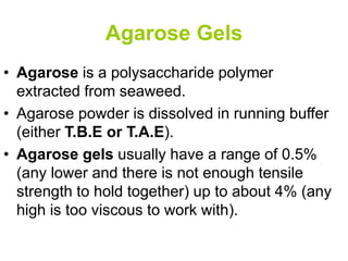
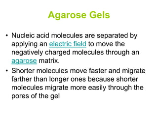
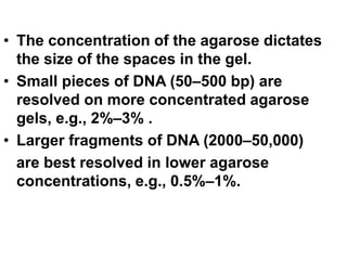
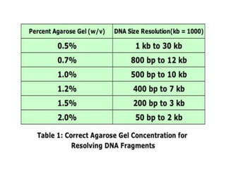
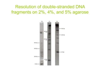
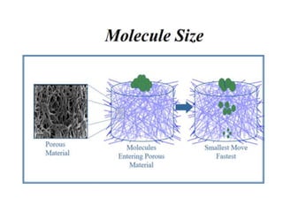
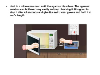
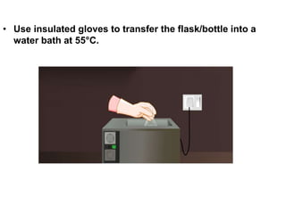
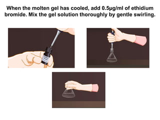
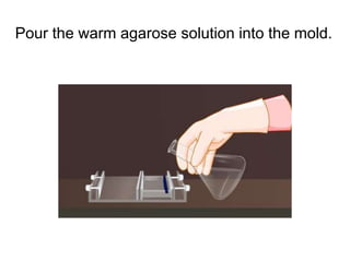
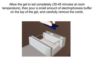
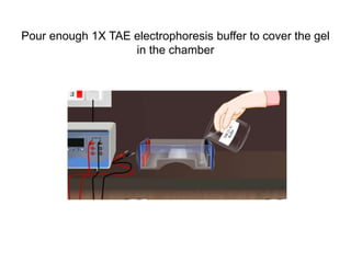
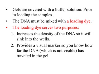
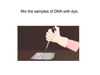
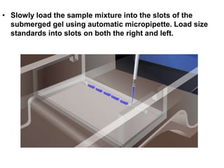
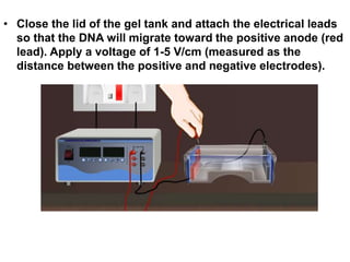
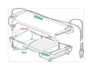
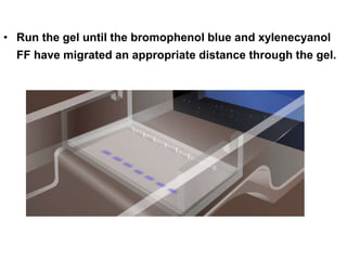
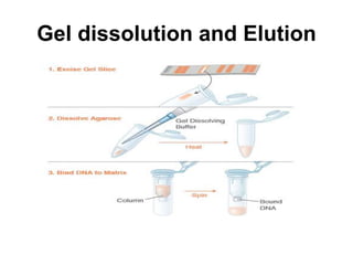
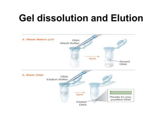
Electrophoresis
Electrophoresis is the movement of molecules by
an electric current .This is practically done in a matrix
to limit migration and contain the migrating material.
Electrophoresis is routinely applied to the
analysis of proteins and nucleic acids
Gel Systems
• Agarose Gels
• Polyacrylamide Gels
• Capillary Electrophoresis
Gel electrophoresis have either a
horizontal or vertical format
Agarose Gels
• Agarose is a polysaccharide polymer
extracted from seaweed.
• Agarose powder is dissolved in running buffer
(either T.B.E or T.A.E).
• Agarose gels usually have a range of 0.5%
(any lower and there is not enough tensile
strength to hold together) up to about 4% (any
high is too viscous to work with).
Agarose Gels
• Nucleic acid molecules are separated by
applying an electric field to move the
negatively charged molecules through an
agarose matrix.
• Shorter molecules move faster and migrate
farther than longer ones because shorter
molecules migrate more easily through the
pores of the gel
• The concentration of the agarose dictates
the size of the spaces in the gel.
• Small pieces of DNA (50–500 bp) are
resolved on more concentrated agarose
gels, e.g., 2%–3% .
• Larger fragments of DNA (2000–50,000)
are best resolved in lower agarose
concentrations, e.g., 0.5%–1%.
Resolution of double-stranded DNA
fragments on 2%, 4%, and 5% agarose
3. Buffer: a fluid mixture of water and ions.
*A buffer is a chemical system that maintains a relatively constant
pH even when strong acids or bases are added. Buffer solutions
contain either a weak acid or weak base and one of their salts.
Because a change in pH can alter the charge on a particle, it is
important to use a buffer solution when separating during
electrophoresis.
Electrophoresis Buffer
• TAE (Tris -acetate-EDTA) and TBE (Tris-borate-EDTA) –
pH buffer. PH 8.0.
1. Tris a pH buffer.
2. Acetic acid provide ions to support conductivity and
maintain pH.
3. EDTA, prevent breake down of molecules. “all dissolved
in water”.
Notes:
Use of water will produce no migration.
Agarose dissolved in electrophoresis buffer.
Procedure
• Prepare sufficient electrophoresis buffer (usually 1x TAE )
to fill the electrophoresis tank and to cast the gel
• Prepare a solution of agarose in electrophoresis buffer at
an appropriate concentration.
For this usually 2 grams of agarose is added to 100ml of
electrophoresis buffer.
• .
• Heat in a microwave oven until the agarose dissolves. The agarose
solution can boil over very easily so keep checking it. It is good to
stop it after 45 seconds and give it a swirl. wear gloves and hold it at
arm's length
• Use insulated gloves to transfer the flask/bottle into a
water bath at 55°C.
When the molten gel has cooled, add 0.5µg/ml of ethidium
bromide. Mix the gel solution thoroughly by gentle swirling.
Pour the warm agarose solution into the mold.
Allow the gel to set completely (30-45 minutes at room
temperature), then pour a small amount of electrophoresis buffer
on the top of the gel, and carefully remove the comb.
Pour enough 1X TAE electrophoresis buffer to cover the gel
in the chamber
• Gels are covered with a buffer solution. Prior
to loading the samples.
• The DNA must be mixed with a loading dye.
• The loading dye serves two purposes:
1. Increases the density of the DNA so it will
sink into the wells.
2. Provides a visual marker so you know how
far the DNA (which is not visible) has
traveled in the gel.
Mix the samples of DNA with dye.
• Slowly load the sample mixture into the slots of the
submerged gel using automatic micropipette. Load size
standards into slots on both the right and left.
• Close the lid of the gel tank and attach the electrical leads
so that the DNA will migrate toward the positive anode (red
lead). Apply a voltage of 1-5 V/cm (measured as the
distance between the positive and negative electrodes).
• Run the gel until the bromophenol blue and xylenecyanol
FF have migrated an appropriate distance through the gel.
Visualization of DNA
• Monitoring the progress of the electrophoresis:
• tracking dyes visible to naked eye during run
₋ Xylene cyanol (migrates with ̴ 5.0kb fragments)
₋ Bromophenol blue (migrates with fragments of a few hundred
base pairs)
₋ Orange G (migrates with fragments of ̴ 50bp)
• DNA may be visualized using ethidium
bromide which, when intercalated into
DNA, fluoresce under ultraviolet light.
Recovery of DNA
The basic methods for recovery of DNA
from gel slices are:
1. Electroelution
2. Elution by diffusion
3. Gel dissolution
4. Freeze squeeze
Electoelution
• The gel piece containing the fragment is
excised and placed in a dialysis bag with
buffer.
• Application of electric current in the form of
electrophoresis causes the DNA to migrate
out of the gel into the dialysis bag with buffer.
Gel dissolution and Elution
Freeze Squeeze DNA
Recovery
Freeze 'N Squeeze DNA gel extraction spin columns provide a
quick and effective means to purify double-stranded DNA
fragments from TAE- or TBE-buffered agarose gels. Unlike other
purification methods which are often time-consuming or utilize
toxic chaotropic materials, the Freeze 'N Squeeze spin column
method purifies via filtration, using short centrifugation to draw
50–23,000 bp of DNA out of agarose gel slices that have been
quickly frozen and thawed.

GEL ELECTROPHORESIS.ppt

  • 2.
    Electrophoresis Electrophoresis is themovement of molecules by an electric current .This is practically done in a matrix to limit migration and contain the migrating material. Electrophoresis is routinely applied to the analysis of proteins and nucleic acids
  • 3.
    Gel Systems • AgaroseGels • Polyacrylamide Gels • Capillary Electrophoresis
  • 4.
    Gel electrophoresis haveeither a horizontal or vertical format
  • 5.
    Agarose Gels • Agaroseis a polysaccharide polymer extracted from seaweed. • Agarose powder is dissolved in running buffer (either T.B.E or T.A.E). • Agarose gels usually have a range of 0.5% (any lower and there is not enough tensile strength to hold together) up to about 4% (any high is too viscous to work with).
  • 6.
    Agarose Gels • Nucleicacid molecules are separated by applying an electric field to move the negatively charged molecules through an agarose matrix. • Shorter molecules move faster and migrate farther than longer ones because shorter molecules migrate more easily through the pores of the gel
  • 7.
    • The concentrationof the agarose dictates the size of the spaces in the gel. • Small pieces of DNA (50–500 bp) are resolved on more concentrated agarose gels, e.g., 2%–3% . • Larger fragments of DNA (2000–50,000) are best resolved in lower agarose concentrations, e.g., 0.5%–1%.
  • 9.
    Resolution of double-strandedDNA fragments on 2%, 4%, and 5% agarose
  • 11.
    3. Buffer: afluid mixture of water and ions. *A buffer is a chemical system that maintains a relatively constant pH even when strong acids or bases are added. Buffer solutions contain either a weak acid or weak base and one of their salts. Because a change in pH can alter the charge on a particle, it is important to use a buffer solution when separating during electrophoresis.
  • 12.
    Electrophoresis Buffer • TAE(Tris -acetate-EDTA) and TBE (Tris-borate-EDTA) – pH buffer. PH 8.0. 1. Tris a pH buffer. 2. Acetic acid provide ions to support conductivity and maintain pH. 3. EDTA, prevent breake down of molecules. “all dissolved in water”. Notes: Use of water will produce no migration. Agarose dissolved in electrophoresis buffer.
  • 14.
    Procedure • Prepare sufficientelectrophoresis buffer (usually 1x TAE ) to fill the electrophoresis tank and to cast the gel
  • 15.
    • Prepare asolution of agarose in electrophoresis buffer at an appropriate concentration. For this usually 2 grams of agarose is added to 100ml of electrophoresis buffer.
  • 17.
  • 18.
    • Heat ina microwave oven until the agarose dissolves. The agarose solution can boil over very easily so keep checking it. It is good to stop it after 45 seconds and give it a swirl. wear gloves and hold it at arm's length
  • 19.
    • Use insulatedgloves to transfer the flask/bottle into a water bath at 55°C.
  • 20.
    When the moltengel has cooled, add 0.5µg/ml of ethidium bromide. Mix the gel solution thoroughly by gentle swirling.
  • 21.
    Pour the warmagarose solution into the mold.
  • 22.
    Allow the gelto set completely (30-45 minutes at room temperature), then pour a small amount of electrophoresis buffer on the top of the gel, and carefully remove the comb.
  • 23.
    Pour enough 1XTAE electrophoresis buffer to cover the gel in the chamber
  • 24.
    • Gels arecovered with a buffer solution. Prior to loading the samples. • The DNA must be mixed with a loading dye. • The loading dye serves two purposes: 1. Increases the density of the DNA so it will sink into the wells. 2. Provides a visual marker so you know how far the DNA (which is not visible) has traveled in the gel.
  • 25.
    Mix the samplesof DNA with dye.
  • 26.
    • Slowly loadthe sample mixture into the slots of the submerged gel using automatic micropipette. Load size standards into slots on both the right and left.
  • 27.
    • Close thelid of the gel tank and attach the electrical leads so that the DNA will migrate toward the positive anode (red lead). Apply a voltage of 1-5 V/cm (measured as the distance between the positive and negative electrodes).
  • 29.
    • Run thegel until the bromophenol blue and xylenecyanol FF have migrated an appropriate distance through the gel.
  • 30.
    Visualization of DNA •Monitoring the progress of the electrophoresis: • tracking dyes visible to naked eye during run ₋ Xylene cyanol (migrates with ̴ 5.0kb fragments) ₋ Bromophenol blue (migrates with fragments of a few hundred base pairs) ₋ Orange G (migrates with fragments of ̴ 50bp) • DNA may be visualized using ethidium bromide which, when intercalated into DNA, fluoresce under ultraviolet light.
  • 32.
    Recovery of DNA Thebasic methods for recovery of DNA from gel slices are: 1. Electroelution 2. Elution by diffusion 3. Gel dissolution 4. Freeze squeeze
  • 33.
    Electoelution • The gelpiece containing the fragment is excised and placed in a dialysis bag with buffer. • Application of electric current in the form of electrophoresis causes the DNA to migrate out of the gel into the dialysis bag with buffer.
  • 35.
  • 37.
    Freeze Squeeze DNA Recovery Freeze'N Squeeze DNA gel extraction spin columns provide a quick and effective means to purify double-stranded DNA fragments from TAE- or TBE-buffered agarose gels. Unlike other purification methods which are often time-consuming or utilize toxic chaotropic materials, the Freeze 'N Squeeze spin column method purifies via filtration, using short centrifugation to draw 50–23,000 bp of DNA out of agarose gel slices that have been quickly frozen and thawed.