
Recommended
More Related Content
Similar to Kampala international university Glucagon, insulin & oral hypoglycemic drugs.ppt
Similar to Kampala international university Glucagon, insulin & oral hypoglycemic drugs.ppt (20)
More from YIKIISAAC
More from YIKIISAAC (20)
Recently uploaded
Recently uploaded (20)
Kampala international university Glucagon, insulin & oral hypoglycemic drugs.ppt
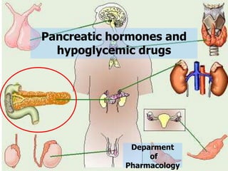
- 1. Pancreatic hormones and hypoglycemic drugs Deparment of Pharmacology
- 2. BIBLIOGRAPHY Goodman and Gilman’s. The pharmacological basis of therapeutics. Basic and Clinical Pharmacology. Bertram G. Katzung. Pharmacology. Rang and Dale’s. 6th edition. Pharmacology. 3rd edition. Lippincott´s.
- 3. SUMMARY 1. Glucose 2. Glucagon 3. Insulin 4. Oral hypoglycemic drugs.
- 4. The single most important source of energy for almost all tissues being utilised for both glycolysis and the tricarboxylic acid (TCA) cycle. Most tissues can also utilise fatty acids via the β-oxidation pathway. The brain is unusual in that, it can only utilise glucose and ketone bodies but the maintenance of an adequate plasma glucose concentration is especially important for the functioning of the CNS. Under normal circumstances the blood glucose level is maintained within a narrow range. However, under some circumstances it may fall outside that range and remain consistently low or high. When this occurs, the situation becomes potentially dangerous. Glucose
- 5. Mechanism of insulin release When glucose levels increase there is an entry of glucose to the pancreatic β cells, glucose is oxidise to G6P with a release of intracellular ATP. The K+-ATP dependant channels located at the cell membrane of the pancreas get close when the ATP attach them and the exflux of K+ through these channels stop. This provoke a change in the resting membrane potential (relative depolarization) and the specific voltage value needed for opening the Ca2+ voltage gated channels at the pancretatic membranes is reached. When these channels open, the Ca2+ flow into the cell and these is the stimulus for the insulin release from the vesicles (exocitosis). Insulin increases the cell permeability to glucose making its blood levels lower preventing hyperglicemia.
- 6. Following its absorption from the gut into the bloodstream any glucose that is not immediately required for energy production is transformed and stored either as glycogen or triglyceride via insulin-mediated processes. In fasting state, plasma glucose level is maintained by glycogenolysis and gluconeogenesis. Main regulators of these processes and, of the plasma glucose level are: Insulin, Glucagon, Adrenalin, Cortisol & GH Glucose Regulation
- 7. EFFECT OF HORMONES ON BLOOD GLUCOSE
- 8. Pancreatic hormones products Endocrine: (islets of Langerhans) Glucagon (A or α-Cells) Insulin (B or β-Cells) Somatostatin (D-Cells): Inhibits glucagon and insulin secretion. It is the major inhibitor of GH synthesis and release. Exocrine: Digestive enzymes
- 9. It is a hormone secreted by the alpha cells of the islets of Langerhans, when the blood glucose concentration falls. Has several functions that are diametrically opposed to those of insulin. Most important of these functions is to increase the blood glucose concentration, an effect that is exactly the opposite that of insulin. It is a large polypeptide, of a chain of 29 amino acids. Has a molecular weight of 3485 Da. Also called the hyperglycemic hormone. Glucagon
- 10. Its major effects on glucose metabolism: 1. Breakdown of liver glycogen (glycogenolysis). 2. Increased gluconeogenesis in the liver. - Both of these effects greatly enhance the availability of glucose to the other organs of the body. - The most dramatic effect of glucagon is it's ability to cause glycogenolysis in the liver, which in turn increase the blood glucose concentration within minutes. Glucagon
- 11. In very high concentration it also: 1. Enhances the strength of the heart. 2. Increases blood flow in some tissues especially in the kidneys. 3. Enhances bile secretion. 4. Inhibits gastric acid secretion. All these effects are probably of minimal importance in the normal function of the body. Glucagon
- 12. It can be given IM or SC as well as IV. Treatment of hypoglycemia in unconscious patients (who cannot drink); unlike IV glucose, it can be administered by non-medical personnel (e.g. spouses or ambulance crew). It is useful if obtaining IV access is difficult. Treatment of acute cardiac failure precipitated by β-adrenoceptor antagonists (to treat the bradycardia and hypoglycemia). Glucagon Clinical Uses Responses of activation of β2-Adrenoceptors: promote glycogenolysis (breakdown of glycogen to glucose), therefore β blockers leads to hypoglycemia in diabetic patiens.
- 13. Insulin It is a small protein with molecular weight of 5808 Da. It is a polypeptide hormone with 2 amino acid chains linked by disulfide (-S-S-) bridges. To initiate it’s effects on target cells, insulin first binds with and activates a membrane receptors protein. It is the activates receptor, not the insulin, that causes the subsequent effects of insulin.
- 14. Insulin cont. Within seconds after insulin binds with it’s membrane receptors, the membrane of about 80% of the body’s cells markedly increase their uptake of glucose and amino acids and regulate glycogen metabolism and triglycerides in the cells. This is especially true of muscle cells and adipose cells but not true of most neurons in the brain. The cell membrane becomes more permeable to many amino acids, potassium ions, and phosphate ions, causing increased transport of these substances into cells.
- 15. Insulin cont. When insulin is secreted into the blood, it’s circulated almost entirely in an unbound form; it has plasma half life averages only about 6 min, so that it is mainly cleared from the circulation within 10-15 min. Except for the portion of the insulin that combines with receptors in the target cells, the remained is degraded by the enzyme insulinase mainly in the liver, to a lesser extent in the kidneys and muscles.
- 16. Actions of Insulin (anabolic hormone) Tyr kinase Insulin reduces blood glucose Insulin Insulin α β P P P P Glucose Recruits additional glucose transporter Increased formation of glycogen, protein and fat Increased glucose uptake Increased glucose utilisation Decreased formation of glucose from glycogen, fat and protein Promote cell division and growth Cell wall
- 17. In summary insulin: • Increases glucose uptake into the muscle and fat via Glut-4 enhancing glucose metabolism. • Increase glycogen, protein and fat synthesis. • Decreases gluconeogenesis (synthesis of glucose from non-carbohydrate sources). • Decreases glycogenolysis (glycogen breakdown). • Increase glycolysis (glucose utilisation). • Increases glycogenesis (glycogen synthesis). • Increases lipogenesis. • Decreases lipolysis. • Decreases protein breakdown.
- 18. Comparison between Insulin & Glucagon Insulin Is released by β-cells in the Islets of Langerhans in the pancreas. Responds to high levels of blood sugar; is released when someone has a meal and needs to store extra energy. Lack of insulin or response to insulin leads to diabetes. Glucagon Is released by α-cells in the Islets of Langerhans in the pancreas. Responds to low levels of blood sugar; is released when someone hasn’t eaten or requires extra energy. Basically, glucagon is the opposite of insulin.
- 19. Diabetes mellitus Elevation of blood glucose. Relative or absolute insulin deficiency. It is aggravated by an excess of glucagon. Polydipsia, polyphagia, polyuria and weight loss. Can be classified in: a) DM Type 1 (Insulin dependant). b) DM Type 2 (Non-insulin dependant). c) DM Type 3 (It is due to mutations of genes). d) DM Type 4 (Gestational diabetes).
- 21. Absolute or relative lack of insulin (Diabetes Mellitus) Retinophaty Nephropathy Neurophaty Cardiovascular Complications Hyperglycemia - Hypertension is 2 to 3 times more common in diabetes. - Both hypertension and diabetes are risk factors for CVS and other complications.
- 22. Dehydration
- 24. Gangrene
- 27. Insulin preparations As drug, insulin is destroyed enzymatically in the GIT and must be given parenterally. Usually is given SC but the absorption into the circulation is variable and depends on local blood flow. Consequently it is used IV or IM in emergencies. Once it has entered the blood flow it is bound by specialized receptors that are found on the membranes of most tissues. The dose of insulin is adjusted on an individual basis. Insulin therapy in diabetic patients should be instituted temporarily during intercurrent illness such as myocardial infarction, infection, trauma and coma. Hypoglycaemic effect of insulin is enhanced by β-blockers. (β2-Adrenoceptors Promote glycogenolysis)
- 28. Administration of Insulin in Diabetes The normal daily insulin requirement depends on body mass, sugar intake and energy expenditure. In most individuals it varies between 60 and 75 units per day. Insulin is used in Type 1 diabetes (a deficiency of insulin), and in a minority of patients with Type 2 diabetes (insensitivity to insulin).
- 29. Administration of Insulin in Diabetes cont. Injections are usually given on the anterior abdominal wall or thigh, but other sites may also be used. Patients should not use the same injection site continuously. The injection through scar tissue is less painful but the problem is erratic absorption & side effect of insulin lipodystrophy. Children and elderly patients requiring insulin should be taught to inject themselves.
- 30. Insulin Lipodystrophy (Loss of local fat deposits in patients as a complication of repeated insulin injections into the same subcutaneous tissue)
- 31. Insulin Preparations Insulin for clinical use was once either porcine or bovine but is now almost entirely human (made by recombinant DNA technology). Short/Rapid acting insulins Examples include: Soluble insulin Insulin lispro – a synthetic insulin, in which a lysine and a proline are switched. It is even shorter acting than soluble insulin. Short acting insulins are injected at the start of a meal and mediate their effect for 30-60 min. They permit a patient to match closely insulin requirement (blood glucose) with the dose of insulin.
- 32. Insulin Preparations cont. Medium or Intermediate acting insulins Isophane insulin: Insulin may be complexed with protamine. This renders the insulin molecule less soluble, and it is distributed from the injection site more slowly than soluble insulin. Its effect lasts for several hours. Isophane insulin may be given in a combined preparation with soluble insulin (Mixtard). Mixtard insulin is convenient and easy to manage. The control of blood glucose is often sub-optimal however.
- 33. Medium or Intermediate acting insulins Insulin zinc suspension: Insulin may be prepared in a suspension with zinc salts. Again, the consequence is reduced solubility of insulin, and delayed distribution of insulin from the injection site. The duration of action is similar to, or a little longer than, isophane insulin. Insulin-zinc suspensions cannot be given in a combined injection with soluble insulin. The zinc salts are in excess in insulin-zinc suspension, and in combination with soluble insulin, all the insulin becomes bound to zinc. Insulin Preparations cont.
- 34. Insulin Preparations cont. Long or Slow acting Insulins Protamine zinc insulin: In this preparation, insulin is complexed to both protamine and zinc. Its effect is long lasting. Glarg-insulin: This is another chemically modified recombinant insulin, containing the amino acid glargine. The structure forms a micro-precipitate at the neutral pH of an injection site, and is very slowly absorbed. Glarg-insulin is a new and relatively expensive preparation, and it is used to provide the background low level insulin requirement, which can be supplemented at meal times by soluble insulin or insulin lispro. The latter regime is now considered to provide the best cover for patients with Type 1 diabetes.
- 36. Insulin ADR Risk of hypoglycemia (is common and, if very severe, can cause brain damage). The treatment is to take a sweet drink or snack or, if the patient is unconscious, to give IV glucose or IM glucagon. Insulin Lypodystrophy. Rebound hyperglycaemia ('Somogyi effect') can follow insulin-induced hypoglycaemia, because of the release of counter-regulatory hormones (glucagon, epinephrine, cortisol, GH). Allergy to human insulin is unusual but can occur. Insulin resistance as a consequence of antibody formation is rare.
- 38. Insulin secretagogues Sulfonylureas Meglitinide analogs Promote insulin release
- 39. Sulfonylureas Mechanism of action: Stimulate β-islet cells to release insulin, by blocking KATP channels, which leads to depolarisation of the cell membrane, Ca2+ influx and insulin secretion. PANCREATIC ISLET Insulin – β-Cell Glibenclamide blocks KATP channel Ca2+
- 40. Sulfonylureas They can lower blood sugar in non-diabetic patients. First-generation: Chlorpropamide, tolbutamide. Second-generation: They are more potent than first generation- Glyburide, glipizide, glimepiride, glibenclamide, gliclazide, gliquidone. They are effective only when some residual pancreatic β-cell activity is present. Tolbutamide: short acting, t½ = 4 h, very weak action. Glipizide, gliclazide: t½ = 7 h Glibenclamide: t½ = 10 h Chlorpropamide: t½ > 15 h (should no longer be used).
- 41. ADR: Weight gain, Hyperinsulinemia, Hypoglicemia which is most frequently seen with longer acting agents (chlorpropamide and glibenclamide), Allergic skin rashes, Bone marrow suppression (rare). Sulfonylureas (or their active metabolites) are excreted in the urine. The risk of hypoglycaemia is therefore greater in patients with renal failure and in the elderly. Contraindications: Hepatic or renal failure. Pregnant women with DM2 should be treated with insulin. (Sulfonylureas traverse the placenta, can deplete insulin from the fetal pancreas). Sulfonylureas
- 42. Meglitinide analogs They include: Repaglinide, Nateglinide Similar action to sulfonylureas, inhibitors of KATP channels, but have a short duration of action, and cause less hypoglycaemia. Their action is dependent on functioning pancreatic β-cells. They bind to a distinct site on the sulfonylurea receptor of KATP channels, initiating a series of reactions culminating in the release of insulin. They are given shortly before meals, and there is evidence that they stimulate appetite less than sulfonylureas. They are categorized as postprandial glucose regulators. Combined therapy with metformin or glitazones is better than the monotherapy. Contraindications: Pregnant women with DM2.
- 43. Insulin sensitizers Biguanides Thiazolidinediones or glitazones Impruve insulin action. Lower blood sugar by improving target-cell response to insulin without increasing pancreatic insulin secretion.
- 44. Biguanides Only drug of this class: Metformin. Lowers blood glucose by accelerating the metabolism of glucose 6-phosphate to pyruvate, thus increases glucose uptake and conversion to G-6-P by hexokinase. It reduces hepatic gluconeogenesis. It requires insulin for its action but does not promote insulin secretion. It also reduces low-density and very low- density lipoproteins.
- 45. ADR: While preventing hyperglycaemia, does not cause hypoglycaemia. Commonly nausea – dose related. Also appetite is suppressed. Biguanides may (rarely) cause lactic acidosis. Clinical uses: Are widely used in type 2 diabetes (especially among obese patients) and are recommended in combination with sulfonylureas, glitazones or insulin in type 2 diabetes – there is a reduced incidence of diabetic retinopathy and other complications. Excretion: Unchanged in the urine. Biguanides
- 46. They include: Ciglitazone, troglitazone, rosiglitazone, pioglitazone. The drugs increase sensitivity to insulin, but their effects are not seen maximally for several weeks (4-6) after the drugs is started. They are used as adjunctive therapy (sulfonylurea or biguanide in type 2 diabetic patients). They reduce hepatic gluconeogenesis, and increase uptake of glucose into muscle. Their effect is mediated by activation of a nuclear receptor called peroxisome proliferator-activated receptor- γ (PPARγ). ADR: The principle adverse effect is hepatotoxicity, but also GI disturbances, headache, weight gain and fatigue and occasionally anaemia. Contraindications: Pregnant and nursing mothers. Glitazones
- 47. Acarbose, miglitol and voglibosa inhibit GI α-glucosidase. It delays carbohydrate absorption, and delivers more undigested carbohydrate to the colon. This leads to flatulence, diarrhoea, abdominal discomfort and bloating. The drug is most use in obese type 2 diabetics. Contraindications: Patients with chronic or inflammatory bowel disease. Pregnancy Alpha-glucosidase inhibitors
- 49. THANK YOU