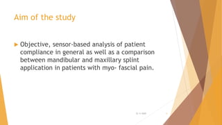
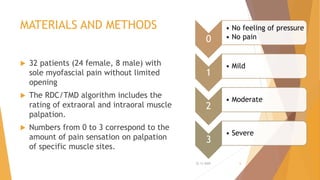
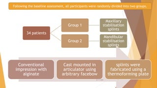
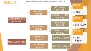
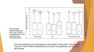
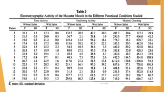
This document summarizes a study that used intraoral sensors to objectively monitor patient compliance with stabilization splint therapy for myofascial pain. Some key findings:
- 32 patients were randomly assigned maxillary or mandibular splints equipped with sensors recording wear time.
- Overall compliance was 44.4% for maxillary and 44.2% for mandibular splints, with no significant difference between the groups.
- Patients with greater pain wore their splints significantly more. Wear time decreased over the 3 observation periods as pain reduced with treatment success.
- The study demonstrated intraoral sensors are an effective way to objectively monitor splint wear compared to subjective reports.
journal cub presentation on Bps denture/biofunctional prosthetic systemNAMITHA ANAND
watch video links below for better understanding
https://www.youtube.com/watch?v=_sR2Ip5p9RE
its a series of videos 1-7 beautiful videos explaining the construction of BPS DENTURES - step by step
Duplication of complete denture prosthesis / endodontic coursesIndian dental academy
Indian Dental Academy: will be one of the most relevant and exciting
training center with best faculty and flexible training programs
for dental professionals who wish to advance in their dental
practice,Offers certified courses in Dental
implants,Orthodontics,Endodontics,Cosmetic Dentistry, Prosthetic
Dentistry, Periodontics and General Dentistry.
Diagnosis and treatment planning in implants/ cosmetic dentistry trainingIndian dental academy
Indian Dental Academy: will be one of the most relevant and exciting
training center with best faculty and flexible training programs
for dental professionals who wish to advance in their dental
practice,Offers certified courses in Dental
implants,Orthodontics,Endodontics,Cosmetic Dentistry, Prosthetic
Dentistry, Periodontics and General Dentistry.
Attachments in implant retained overdentures/ cosmetic dentistry trainingIndian dental academy
Indian Dental Academy: will be one of the most relevant and exciting
training center with best faculty and flexible training programs
for dental professionals who wish to advance in their dental
practice,Offers certified courses in Dental
implants,Orthodontics,Endodontics,Cosmetic Dentistry, Prosthetic
Dentistry, Periodontics and General Dentistry.
Recent advances in prosthodontics / crown & bridge courses by indian dental a...Indian dental academy
The Indian Dental Academy is the Leader in continuing dental education , training dentists in all aspects of dentistry and offering a wide range of dental certified courses in different formats.
Indian dental academy provides dental crown & Bridge,rotary endodontics,fixed orthodontics,
Dental implants courses.for details pls visit www.indiandentalacademy.com ,or call
0091-9248678078
Impression procedures for compromised ridges/cosmetic dentistry coursesIndian dental academy
The Indian Dental Academy is the Leader in continuing dental education , training dentists in all aspects of dentistry and
offering a wide range of dental certified courses in different formats.for more details please visit
www.indiandentalacademy.com
Indian Dental Academy: will be one of the most relevant and exciting training center with best faculty and flexible training programs for dental professionals who wish to advance in their dental practice,Offers certified courses in Dental implants,Orthodontics,Endodontics,Cosmetic Dentistry, Prosthetic Dentistry, Periodontics and General Dentistry.
Orientation jaw relation /certified fixed orthodontic courses by Indian denta...Indian dental academy
The Indian Dental Academy is the Leader in continuing dental education , training dentists in all aspects of dentistry and offering a wide range of dental certified courses in different formats.
Indian dental academy provides dental crown & Bridge,rotary endodontics,fixed orthodontics,
Dental implants courses.for details pls visit www.indiandentalacademy.com ,or call
0091-9248678078
Postoperative recovery after mandibular third molar surgery. By Dr. Akhila Damodar { dr.akhila.n@gmail.com }
This study sought to evaluate postoperative recovery after mandibular third molar surgery, with and without the use of sutures.
Objective: Tennis elbow is an inflammatory condition of the common extensor origin over the lateral epicondyle. This condition does not affect tennis players only. It often follows an injury or sudden contraction of the common extensor origin.There is many treatments and approaches towards Tennis elbow but physiotherapy is the best modern conservative treatment. The aim of this study is to evaluate the effectiveness of movement with mobilization in reducing pain and increasing strength in patients with chronic lateral epicondylitis. Design and setting: A randomized controlled study design was used to examine the differences between conventional physical therapy and physical therapy with manual mobilization approach for study duration of 15 days. Subjects: Twelve subjects of both male and female gender were divided into 2 groups. Experimental group treated with ultrasound therapy, mobilization and progressive resisted exercises. Control group treated with ultrasound therapy and progressive resisted exercises only the results were analyzed. The procedure was done in Physiotherapy Department at Masterskill college of Nursing and health. Outcome Measurement: Two outcome measures were used. NPRS for the measurement of severity of pain and various weighted sand bags (0.25 kg to 2kg) were used to measure the strength. Results: The data shows a significant difference in the post test values of pain and strength between experimental group and control group. Experimental group shows much decrease in pain and increase in strength than the control group. Conclusion: The study concludes that the manual mobilization with movement along with ultrasound therapy and progressive resisted exercises is effective in reducing pain and increasing strength than that of progressive resisted exercise along with ultra sound therapy in adults with chronic lateral epicondylitis.
Total knee replacement (TKR) is one of the most commonly done surgical procedures, with over 150,000 total knee replacements and THR performed annually in England and Wales in the National Health Service (NHS). In India although clear-cut data is not available but the incidence is increasing. In the US, 431,000 TKRs are performed yearly and the utilization of TKR has increased over the last two decades, especially among younger patients .TKR may be associated with severe post-operative pain. The International Association for the Study of Pain (IASP) has defined pain as “an unpleasant sensory or emotional experience associated with actual or potential tissue damage, or described in terms of such damage”. The Joint Commission on Accreditation of Healthcare Organizations (JCAHO) has made adequate pain management a priority and has deemed monitoring pain as the “fifth” vital sign.
Clinical Experience in Maxillary and Mandibular division block for Trigeminal...iosrphr_editor
The IOSR Journal of Pharmacy (IOSRPHR) is an open access online & offline peer reviewed international journal, which publishes innovative research papers, reviews, mini-reviews, short communications and notes dealing with Pharmaceutical Sciences( Pharmaceutical Technology, Pharmaceutics, Biopharmaceutics, Pharmacokinetics, Pharmaceutical/Medicinal Chemistry, Computational Chemistry and Molecular Drug Design, Pharmacognosy & Phytochemistry, Pharmacology, Pharmaceutical Analysis, Pharmacy Practice, Clinical and Hospital Pharmacy, Cell Biology, Genomics and Proteomics, Pharmacogenomics, Bioinformatics and Biotechnology of Pharmaceutical Interest........more details on Aim & Scope).
Temporomandibular joint, a facial joint commonly undergoes internal derangement due to the abnormal position of the articular disc in relation to the condyle. Internal derangement of the TMJ is explained in detail in this presentation.
Management of tmd symptoms with photobiomodulation therapyNishu Priya
Conservative approaches, such as soft diets, anti-inflammatory drugs and photobiomodulation therapy (PBMT) or low-level laser therapy (LLLT), have been used to manage TMD.
Lasers have proven to be successful in clinical settings and treatments of soft tissues, musculoskeletal pain, bone regeneration, dentinal hypersensitivity, and provide reduction in symptoms and improved function.
The mechanism of action in PBMT is via absorption of light, with deeply penetrating wavelengths ranging from 630 nm to 1300 nm, to stimulate tissues with direct irradiation to achieve analgesic and anti-inflammatory effects.
The output energy in PBMT does not affect skin temperature and is classified as a soft laser, which increases lymphatic flow, reduces edema and prostaglandin E2 (PGE2) and cyclooxygenase (COX) levels.
A systematic review for pain management reported placebo vs LLLT for practical and clinically relevant parameters using 700nm to 1200nm.
HOT NEW PRODUCT! BIG SALES FAST SHIPPING NOW FROM CHINA!! EU KU DB BK substit...GL Anaacs
Contact us if you are interested:
Email / Skype : kefaya1771@gmail.com
Threema: PXHY5PDH
New BATCH Ku !!! MUCH IN DEMAND FAST SALE EVERY BATCH HAPPY GOOD EFFECT BIG BATCH !
Contact me on Threema or skype to start big business!!
Hot-sale products:
NEW HOT EUTYLONE WHITE CRYSTAL!!
5cl-adba precursor (semi finished )
5cl-adba raw materials
ADBB precursor (semi finished )
ADBB raw materials
APVP powder
5fadb/4f-adb
Jwh018 / Jwh210
Eutylone crystal
Protonitazene (hydrochloride) CAS: 119276-01-6
Flubrotizolam CAS: 57801-95-3
Metonitazene CAS: 14680-51-4
Payment terms: Western Union,MoneyGram,Bitcoin or USDT.
Deliver Time: Usually 7-15days
Shipping method: FedEx, TNT, DHL,UPS etc.Our deliveries are 100% safe, fast, reliable and discreet.
Samples will be sent for your evaluation!If you are interested in, please contact me, let's talk details.
We specializes in exporting high quality Research chemical, medical intermediate, Pharmaceutical chemicals and so on. Products are exported to USA, Canada, France, Korea, Japan,Russia, Southeast Asia and other countries.
Ozempic: Preoperative Management of Patients on GLP-1 Receptor Agonists Saeid Safari
Preoperative Management of Patients on GLP-1 Receptor Agonists like Ozempic and Semiglutide
ASA GUIDELINE
NYSORA Guideline
2 Case Reports of Gastric Ultrasound
New Directions in Targeted Therapeutic Approaches for Older Adults With Mantl...i3 Health
i3 Health is pleased to make the speaker slides from this activity available for use as a non-accredited self-study or teaching resource.
This slide deck presented by Dr. Kami Maddocks, Professor-Clinical in the Division of Hematology and
Associate Division Director for Ambulatory Operations
The Ohio State University Comprehensive Cancer Center, will provide insight into new directions in targeted therapeutic approaches for older adults with mantle cell lymphoma.
STATEMENT OF NEED
Mantle cell lymphoma (MCL) is a rare, aggressive B-cell non-Hodgkin lymphoma (NHL) accounting for 5% to 7% of all lymphomas. Its prognosis ranges from indolent disease that does not require treatment for years to very aggressive disease, which is associated with poor survival (Silkenstedt et al, 2021). Typically, MCL is diagnosed at advanced stage and in older patients who cannot tolerate intensive therapy (NCCN, 2022). Although recent advances have slightly increased remission rates, recurrence and relapse remain very common, leading to a median overall survival between 3 and 6 years (LLS, 2021). Though there are several effective options, progress is still needed towards establishing an accepted frontline approach for MCL (Castellino et al, 2022). Treatment selection and management of MCL are complicated by the heterogeneity of prognosis, advanced age and comorbidities of patients, and lack of an established standard approach for treatment, making it vital that clinicians be familiar with the latest research and advances in this area. In this activity chaired by Michael Wang, MD, Professor in the Department of Lymphoma & Myeloma at MD Anderson Cancer Center, expert faculty will discuss prognostic factors informing treatment, the promising results of recent trials in new therapeutic approaches, and the implications of treatment resistance in therapeutic selection for MCL.
Target Audience
Hematology/oncology fellows, attending faculty, and other health care professionals involved in the treatment of patients with mantle cell lymphoma (MCL).
Learning Objectives
1.) Identify clinical and biological prognostic factors that can guide treatment decision making for older adults with MCL
2.) Evaluate emerging data on targeted therapeutic approaches for treatment-naive and relapsed/refractory MCL and their applicability to older adults
3.) Assess mechanisms of resistance to targeted therapies for MCL and their implications for treatment selection
ARTIFICIAL INTELLIGENCE IN HEALTHCARE.pdfAnujkumaranit
Artificial intelligence (AI) refers to the simulation of human intelligence processes by machines, especially computer systems. It encompasses tasks such as learning, reasoning, problem-solving, perception, and language understanding. AI technologies are revolutionizing various fields, from healthcare to finance, by enabling machines to perform tasks that typically require human intelligence.
micro teaching on communication m.sc nursing.pdfAnurag Sharma
Microteaching is a unique model of practice teaching. It is a viable instrument for the. desired change in the teaching behavior or the behavior potential which, in specified types of real. classroom situations, tends to facilitate the achievement of specified types of objectives.
Tom Selleck Health: A Comprehensive Look at the Iconic Actor’s Wellness Journeygreendigital
Tom Selleck, an enduring figure in Hollywood. has captivated audiences for decades with his rugged charm, iconic moustache. and memorable roles in television and film. From his breakout role as Thomas Magnum in Magnum P.I. to his current portrayal of Frank Reagan in Blue Bloods. Selleck's career has spanned over 50 years. But beyond his professional achievements. fans have often been curious about Tom Selleck Health. especially as he has aged in the public eye.
Follow us on: Pinterest
Introduction
Many have been interested in Tom Selleck health. not only because of his enduring presence on screen but also because of the challenges. and lifestyle choices he has faced and made over the years. This article delves into the various aspects of Tom Selleck health. exploring his fitness regimen, diet, mental health. and the challenges he has encountered as he ages. We'll look at how he maintains his well-being. the health issues he has faced, and his approach to ageing .
Early Life and Career
Childhood and Athletic Beginnings
Tom Selleck was born on January 29, 1945, in Detroit, Michigan, and grew up in Sherman Oaks, California. From an early age, he was involved in sports, particularly basketball. which played a significant role in his physical development. His athletic pursuits continued into college. where he attended the University of Southern California (USC) on a basketball scholarship. This early involvement in sports laid a strong foundation for his physical health and disciplined lifestyle.
Transition to Acting
Selleck's transition from an athlete to an actor came with its physical demands. His first significant role in "Magnum P.I." required him to perform various stunts and maintain a fit appearance. This role, which he played from 1980 to 1988. necessitated a rigorous fitness routine to meet the show's demands. setting the stage for his long-term commitment to health and wellness.
Fitness Regimen
Workout Routine
Tom Selleck health and fitness regimen has evolved. adapting to his changing roles and age. During his "Magnum, P.I." days. Selleck's workouts were intense and focused on building and maintaining muscle mass. His routine included weightlifting, cardiovascular exercises. and specific training for the stunts he performed on the show.
Selleck adjusted his fitness routine as he aged to suit his body's needs. Today, his workouts focus on maintaining flexibility, strength, and cardiovascular health. He incorporates low-impact exercises such as swimming, walking, and light weightlifting. This balanced approach helps him stay fit without putting undue strain on his joints and muscles.
Importance of Flexibility and Mobility
In recent years, Selleck has emphasized the importance of flexibility and mobility in his fitness regimen. Understanding the natural decline in muscle mass and joint flexibility with age. he includes stretching and yoga in his routine. These practices help prevent injuries, improve posture, and maintain mobilit
Lung Cancer: Artificial Intelligence, Synergetics, Complex System Analysis, S...Oleg Kshivets
RESULTS: Overall life span (LS) was 2252.1±1742.5 days and cumulative 5-year survival (5YS) reached 73.2%, 10 years – 64.8%, 20 years – 42.5%. 513 LCP lived more than 5 years (LS=3124.6±1525.6 days), 148 LCP – more than 10 years (LS=5054.4±1504.1 days).199 LCP died because of LC (LS=562.7±374.5 days). 5YS of LCP after bi/lobectomies was significantly superior in comparison with LCP after pneumonectomies (78.1% vs.63.7%, P=0.00001 by log-rank test). AT significantly improved 5YS (66.3% vs. 34.8%) (P=0.00000 by log-rank test) only for LCP with N1-2. Cox modeling displayed that 5YS of LCP significantly depended on: phase transition (PT) early-invasive LC in terms of synergetics, PT N0—N12, cell ratio factors (ratio between cancer cells- CC and blood cells subpopulations), G1-3, histology, glucose, AT, blood cell circuit, prothrombin index, heparin tolerance, recalcification time (P=0.000-0.038). Neural networks, genetic algorithm selection and bootstrap simulation revealed relationships between 5YS and PT early-invasive LC (rank=1), PT N0—N12 (rank=2), thrombocytes/CC (3), erythrocytes/CC (4), eosinophils/CC (5), healthy cells/CC (6), lymphocytes/CC (7), segmented neutrophils/CC (8), stick neutrophils/CC (9), monocytes/CC (10); leucocytes/CC (11). Correct prediction of 5YS was 100% by neural networks computing (area under ROC curve=1.0; error=0.0).
CONCLUSIONS: 5YS of LCP after radical procedures significantly depended on: 1) PT early-invasive cancer; 2) PT N0--N12; 3) cell ratio factors; 4) blood cell circuit; 5) biochemical factors; 6) hemostasis system; 7) AT; 8) LC characteristics; 9) LC cell dynamics; 10) surgery type: lobectomy/pneumonectomy; 11) anthropometric data. Optimal diagnosis and treatment strategies for LC are: 1) screening and early detection of LC; 2) availability of experienced thoracic surgeons because of complexity of radical procedures; 3) aggressive en block surgery and adequate lymph node dissection for completeness; 4) precise prediction; 5) adjuvant chemoimmunoradiotherapy for LCP with unfavorable prognosis.
Factory Supply Best Quality Pmk Oil CAS 28578–16–7 PMK Powder in Stockrebeccabio
Factory Supply Best Quality Pmk Oil CAS 28578–16–7 PMK Powder in Stock
Telegram: bmksupplier
signal: +85264872720
threema: TUD4A6YC
You can contact me on Telegram or Threema
Communicate promptly and reply
Free of customs clearance, Double Clearance 100% pass delivery to USA, Canada, Spain, Germany, Netherland, Poland, Italy, Sweden, UK, Czech Republic, Australia, Mexico, Russia, Ukraine, Kazakhstan.Door to door service
Hot Selling Organic intermediates
Couples presenting to the infertility clinic- Do they really have infertility...Sujoy Dasgupta
Dr Sujoy Dasgupta presented the study on "Couples presenting to the infertility clinic- Do they really have infertility? – The unexplored stories of non-consummation" in the 13th Congress of the Asia Pacific Initiative on Reproduction (ASPIRE 2024) at Manila on 24 May, 2024.
Title: Sense of Smell
Presenter: Dr. Faiza, Assistant Professor of Physiology
Qualifications:
MBBS (Best Graduate, AIMC Lahore)
FCPS Physiology
ICMT, CHPE, DHPE (STMU)
MPH (GC University, Faisalabad)
MBA (Virtual University of Pakistan)
Learning Objectives:
Describe the primary categories of smells and the concept of odor blindness.
Explain the structure and location of the olfactory membrane and mucosa, including the types and roles of cells involved in olfaction.
Describe the pathway and mechanisms of olfactory signal transmission from the olfactory receptors to the brain.
Illustrate the biochemical cascade triggered by odorant binding to olfactory receptors, including the role of G-proteins and second messengers in generating an action potential.
Identify different types of olfactory disorders such as anosmia, hyposmia, hyperosmia, and dysosmia, including their potential causes.
Key Topics:
Olfactory Genes:
3% of the human genome accounts for olfactory genes.
400 genes for odorant receptors.
Olfactory Membrane:
Located in the superior part of the nasal cavity.
Medially: Folds downward along the superior septum.
Laterally: Folds over the superior turbinate and upper surface of the middle turbinate.
Total surface area: 5-10 square centimeters.
Olfactory Mucosa:
Olfactory Cells: Bipolar nerve cells derived from the CNS (100 million), with 4-25 olfactory cilia per cell.
Sustentacular Cells: Produce mucus and maintain ionic and molecular environment.
Basal Cells: Replace worn-out olfactory cells with an average lifespan of 1-2 months.
Bowman’s Gland: Secretes mucus.
Stimulation of Olfactory Cells:
Odorant dissolves in mucus and attaches to receptors on olfactory cilia.
Involves a cascade effect through G-proteins and second messengers, leading to depolarization and action potential generation in the olfactory nerve.
Quality of a Good Odorant:
Small (3-20 Carbon atoms), volatile, water-soluble, and lipid-soluble.
Facilitated by odorant-binding proteins in mucus.
Membrane Potential and Action Potential:
Resting membrane potential: -55mV.
Action potential frequency in the olfactory nerve increases with odorant strength.
Adaptation Towards the Sense of Smell:
Rapid adaptation within the first second, with further slow adaptation.
Psychological adaptation greater than receptor adaptation, involving feedback inhibition from the central nervous system.
Primary Sensations of Smell:
Camphoraceous, Musky, Floral, Pepperminty, Ethereal, Pungent, Putrid.
Odor Detection Threshold:
Examples: Hydrogen sulfide (0.0005 ppm), Methyl-mercaptan (0.002 ppm).
Some toxic substances are odorless at lethal concentrations.
Characteristics of Smell:
Odor blindness for single substances due to lack of appropriate receptor protein.
Behavioral and emotional influences of smell.
Transmission of Olfactory Signals:
From olfactory cells to glomeruli in the olfactory bulb, involving lateral inhibition.
Primitive, less old, and new olfactory systems with different path
Title: Sense of Taste
Presenter: Dr. Faiza, Assistant Professor of Physiology
Qualifications:
MBBS (Best Graduate, AIMC Lahore)
FCPS Physiology
ICMT, CHPE, DHPE (STMU)
MPH (GC University, Faisalabad)
MBA (Virtual University of Pakistan)
Learning Objectives:
Describe the structure and function of taste buds.
Describe the relationship between the taste threshold and taste index of common substances.
Explain the chemical basis and signal transduction of taste perception for each type of primary taste sensation.
Recognize different abnormalities of taste perception and their causes.
Key Topics:
Significance of Taste Sensation:
Differentiation between pleasant and harmful food
Influence on behavior
Selection of food based on metabolic needs
Receptors of Taste:
Taste buds on the tongue
Influence of sense of smell, texture of food, and pain stimulation (e.g., by pepper)
Primary and Secondary Taste Sensations:
Primary taste sensations: Sweet, Sour, Salty, Bitter, Umami
Chemical basis and signal transduction mechanisms for each taste
Taste Threshold and Index:
Taste threshold values for Sweet (sucrose), Salty (NaCl), Sour (HCl), and Bitter (Quinine)
Taste index relationship: Inversely proportional to taste threshold
Taste Blindness:
Inability to taste certain substances, particularly thiourea compounds
Example: Phenylthiocarbamide
Structure and Function of Taste Buds:
Composition: Epithelial cells, Sustentacular/Supporting cells, Taste cells, Basal cells
Features: Taste pores, Taste hairs/microvilli, and Taste nerve fibers
Location of Taste Buds:
Found in papillae of the tongue (Fungiform, Circumvallate, Foliate)
Also present on the palate, tonsillar pillars, epiglottis, and proximal esophagus
Mechanism of Taste Stimulation:
Interaction of taste substances with receptors on microvilli
Signal transduction pathways for Umami, Sweet, Bitter, Sour, and Salty tastes
Taste Sensitivity and Adaptation:
Decrease in sensitivity with age
Rapid adaptation of taste sensation
Role of Saliva in Taste:
Dissolution of tastants to reach receptors
Washing away the stimulus
Taste Preferences and Aversions:
Mechanisms behind taste preference and aversion
Influence of receptors and neural pathways
Impact of Sensory Nerve Damage:
Degeneration of taste buds if the sensory nerve fiber is cut
Abnormalities of Taste Detection:
Conditions: Ageusia, Hypogeusia, Dysgeusia (parageusia)
Causes: Nerve damage, neurological disorders, infections, poor oral hygiene, adverse drug effects, deficiencies, aging, tobacco use, altered neurotransmitter levels
Neurotransmitters and Taste Threshold:
Effects of serotonin (5-HT) and norepinephrine (NE) on taste sensitivity
Supertasters:
25% of the population with heightened sensitivity to taste, especially bitterness
Increased number of fungiform papillae
TEST BANK for Operations Management, 14th Edition by William J. Stevenson, Ve...kevinkariuki227
TEST BANK for Operations Management, 14th Edition by William J. Stevenson, Verified Chapters 1 - 19, Complete Newest Version.pdf
Journal club presentation on muscle stabilisation splints
1. Intraoral sensor-based
monitoring of stabilization
splint therapy in patients
with myofascial pain
Namitha AP
III rd MDS
DEPT.OF PROSTHODONTICS
Journal club presentation
Krohn S, Hampe T, Brack F, Wassmann T and Bürgers R
International Journal of Computerized Dentistry. 2020 Jan 1;23(1):11-6.
22-11-2020 1
2. Contents
Introduction
Aim of the study
Materials and methods
Results
Discussion
Related articles
References
22-11-2020 2
3. INTRODUCTION
Temporomandibular disorders (TMD) are a common cause of orofacial pain.
Many types of occlusal appliances have been suggested for the treatment of
TMDs.
The two most commonly used are the stabilization appliance and the anterior
positioning appliance
The most frequent symptoms of TMD are muscle hyperactivity and muscle
pain, which are referred to as myofascial pain.
Stabilization splints are considered a good treatment option to reduce
subjective pain by reorganizing neuromuscular functional patterns and
inhibiting muscle activity.
22-11-2020 3
4. STABILISATION SPLINT/MUSCLE
RELAXATION APPLIANCE
generally fabricated for the maxillary arch and
provides an occlusal relationship considered
optimal for the patient
When it is in place, the condyles are in their
most musculoskeletally stable position at the
time that the teeth are contacting evenly and
simultaneously.
Canine disocclusion of the posterior teeth
during eccentric movement is also provided.
The treatment goal is to eliminate any
orthopedic instability between the occlusal
position and the joint position, thus removing
this instability as an etiologic factor in the TMD
MAXILLARY/MANDIBULAR
APPLICATION
WEARING COMFORT
PERIOD OF INTRA ORAL
WEAR
PATIENT COMPLIANCE
KEY SUCCESS
FACTORS
22-11-2020 4
5. Aim of the study
Objective, sensor-based analysis of patient
compliance in general as well as a comparison
between mandibular and maxillary splint
application in patients with myo- fascial pain.
22-11-2020 5
6. MATERIALS AND METHODS
32 patients (24 female, 8 male) with
sole myofascial pain without limited
opening
The RDC/TMD algorithm includes the
rating of extraoral and intraoral muscle
palpation.
Numbers from 0 to 3 correspond to the
amount of pain sensation on palpation
of specific muscle sites.
0
• No feeling of pressure
• No pain
1
• Mild
2
• Moderate
3
• Severe
22-11-2020 6
7. 34 patients
Group 1
Maxillary
stabilisation
splints
Group 2
Mandibular
stabilisation
splints
Following the baseline assessment, all participants were randomly divided into two groups.
Conventional
impression with
alginate
Cast mounted in
articulator using
arbitrary facebow
splints were
fabricated using a
thermoforming plate
22-11-2020 7
8. Cold-cured polymethyl methacrylate
(PMMC) was applied for the
adjustment of the equilibrated
occlusion in the articulator.
The prepared splints were polished
and the occlusal surfaces adjusted
intraorally to ensure equilibration of
static occlusion and anterior/canine
guidance.
All splints were unilaterally
equipped with a TheraMon
microsensor.
sensor dimensions -13 × 9 × 4 mm (w × h × d)
firmly fixed to the vestibular posterior tooth
region without interfering with the static or
dynamic occlusion
22-11-2020 8
9. Before the occlusal appliances were handed out, all
subjects were told to use the splints as much as possible
and were fully informed about their use.
The period of intraoral application was measured once the
sensor was exposed to body temperature, whereby the
total duration was read out in three intervals of 30 days
each (initial = t1; intermediary = t2; terminal = t3).
At the end of the observation period, the data were
retrieved from all the sensors and statistically evaluated
to assess the influence of different factors (gender, pain
intensity, observation interval) on the variability of the
results.
In order to quantify the range of variation for the
mandibular and maxillary stabilization splints and to
compare both treatment options regarding their equality,
a Bland-Altman analysis was performed
Initial t1
Intermediary
t2
Terminal t3
22-11-2020 9
10. RESULTS mean palpation score ranging between 0.2 and 1.5.
• <0.5
Group
1
• 0.5- 0.99
Group
2
• 1 or
higher
Group
3
Mean intra oral
application
Maxillary
splints
40.5 days
(44.4%)
5.96 h (± 1.84)
per day
Mandibular
splints
39.8 days
(44.2%)
6.66 h (± 2.68)
per day
Total wear time
Maxillary
splints
3.19 h (± 4.09)
Mandibular
splints
3.13 h (± 4.03)22-11-2020 10
11. The highest overall values of intraoral application of 3.71
h (± 4.20) were recorded at interval t1.
At intervals t2 and t3, the mean values of both the
maxillary and mandibular splints decreased to 3.35 h (±
4.17) and 2.42 h (± 3.69), respectively.
Higher mean values were observed in female participants
and in participants with an increased pain score.
Statistical analysis revealed that there was no significant
difference in the intraoral application of the maxillary and
mandibular splints
female participants wore their splints significantly more
often, and participants with an increased pain score
showed a significantly higher wear time
22-11-2020 11
12.
The total application time decreased over the period of observation, whereas only
intervals t1 and t3 differed significantly across both the mandibular and maxillary
splint groups
22-11-2020 12
13. Discussion
In order to evaluate patient compliance, it is important to assess objective
and reliable data of actual intraoral applications.
In this study an integrated microsensor was used to record the wear pattern
at three intervals of 30 days each, which showed a compliance of 44.4% for
the maxillary and 44.2% for the mandibular splints.
Subjects with greater pain scores showed significantly higher compliance,
whereas the total time of intraoral splint wear significantly decreased from t1
to t3 in all pain groups
This positive correlation might be caused by pain reduction due to treatment
success, which is commonly achieved within 2 to 3 months
22-11-2020 13
14. CONCLUSIONS
The present study demonstrated that the objective, intraoral, sensor-
based monitoring of splint therapy was an effective and reliable
method of monitoring patient compliance during treatment.
Allows for the comparison of subjective symptoms with objective
findings.
Additional diagnostic tool in TMD therapy
Reliable recording of intraoral splint application prior to required bite
elevations.
The decision between maxillary and mandibular splints did not affect
patient compliance in the present investigation.
22-11-2020 14
15. Side Effects of Stabilization Occlusal Splints:
A Report
of Three Cases and Literature Review
Fernando Magdaleno and Eduardo Ginestal
There is a noticeable lack of reference to the possible undesirable side
effects of stabilization splints, in contrast to other types of oral
appliances, such as anterior repositioning splints, nociceptive trigeminal
inhibition tension suppression systems (NTI-tss) or splints for sleep apnea
Complications derived from the use of stabilization splints are associated
with their full-time use, whereas the risk of undesired effects is
considered to be small when they are used part time
22-11-2020 15
16. AIM AND OBJECTIVES
to review the available information on the possible
adverse effects of stabilization splints in their different
clinical applications, as well as to present three cases in
which the use of a stabilization splint was associated with
irreversible occlusal changes.
22-11-2020 16
17. CASES
Three clinical cases were selected for this study, which involved
occlusal alterations following the use of a part time stabilization
splint
a retrospective search among the clinic’s patients over the last five
years, using a simple selection criterion; the presence of irreversible
occlusal modifications subsequent to the use of a part time
stabilization splint.
splints are made with hard acrylic resin and cover all of the teeth.
22-11-2020 17
18. CASE 1
No dental
migration
The removal of the third molars increased the number of dental contacts, but did not manage to achieve the
anterior tooth relation of the manually articulated models
Upon removing
the splint, the
patient
exhibited
unstable
occlusion with
dental contacts
exclusively at
the level of the
wisdom teeth
22-11-2020 18
19. Case 2
A 23 year-old female
dull and diffuse facial pain over
the past three months
predominantly during the morning
and on the left side, with
moderate intensity and without
other accompanying symptoms.
stable occlusion in Angle class I,
with bilateral anterior guidance.
tenderness of the chewing
musculature without trigger points.
no signs of internal derangement
Mandibular opening capacity was
51 mm, without deviations.
She reported that she tended to
clench her teeth
The suspected diagnosis was local myalgia, with parafunction as
the most probable etiological factor.
22-11-2020 19
20. She was fitted for night use with an upper stabilization splint in the
intercuspal position and was asked to return for her first check-up a week
later.
This patient returned for the first time nine months later.
She reported that pain had eased at the beginning of the treatment, but that
during recent months, it had reappeared, although with less intensity.
Anterior open bite was observed with characteristics similar to those of Case
1.
she reported that she used the splint during the night and during some day
hours, particularly during exam periods.
The splint showed signs of contact only between posterior teeth.
After readjusting the splint, the patient experienced no more facial pain.
Currently, the patient is undergoing orthodontic treatment.
22-11-2020 20
21. Case 3
26 year old female
orthodontics and bimaxillary
orthognathic surgery two years ago.
Intense pain at the level of the left
temporomandibular joint (TMJ), on the
left side of the face and in the lateral
region of the neck on the same side,
which had worsened over the last nine
months and had not responded to
conventional analgesics.
Pain was constant, with moments of
heightened intensity, and it became
more acute with mandibular movement
and occlusion.
There was evidence of central
sensitization, with hyperalgesia
and allodynia.
Muscular palpation revealed trigger
points at the chewing and neck
muscles.
mandibular opening capacity - 27
mm, with deviation to the left and
any movement involving
mandibular mobilization was very
painful.
22-11-2020 21
22. unstable
occlusion
and the
absence of
anterior
guidance in
lateral
movements.
bilateral
condylar
Hypoplasia in
OPG
MRI
verified disk displacement
without reduction, with
moderate
osteoarthritic changes and
edema in left TMJ
stabilization
splint was fitted
for part-time
use, not
necessarily
limited to
nocturnal use
Five months later
occasional pain
reduced
But anterior open bite,
which had
occurred gradually22-11-2020 22
23. Her mandibular opening capacity
was 38 mm, with a slight left
deviation.
clinical and radiographic controls
carried out three, six, and nine
months later - no observed
significant modifications in the
intermaxillary relation or in
condylar morphology
patient periodically attends a
physiotherapist and uses the splint
during the night and sometimes for
a few hours during the day (she
does not want to be without the
splint).
She occasionally uses nonsteroidal
anti-inflammatory drugs.
She decided not to have additional
surgery to improve her occlusal
situation.
22-11-2020 23
24. DISCUSSION
Three common conditions, which are distinct but nevertheless have the
same outcome: anterior open bite
Case 1
• use of the splint
as a diagnostic
method in
orthodontics,
applied with a
view to
establishing a
centric relation.
Case 2
• represents a
simple case of
TMD in a patient
with oral
parafunction, in
which the splint
is the first choice
of treatment,
and often the
only available
treatment
Case 3
• complex case of
chronic orofacial
pain in which the
splint only
played a
coadjuvant role,
since the case
required
multidisciplinary
treatment.
The three case reports presented here are representative of the typical
clinical situations in which stabilization splints are recommended.
22-11-2020 24
25. Due to the very nature of the methodology employed in this work, the
incidence of this type of alteration cannot estimated, since one of the three
cases was referred to our clinic due to the presence of occlusal alterations
(Case 1) and another (Case 3) was a patient who presented important
orthopedic instability after surgical-orthodontic treatment.
difficult to foresee these modifications, since they vary as a function of the
type of pathology and of the coadjuvant factors associated with each case
modifications in general do not cause much discomfort, and on many
occasions are not even perceived by patients.
For this reason, it is only rarely necessary to have to consider applying
irreversible treatments after the orthopedic phase.
22-11-2020 25
26. In order to avoid possible side effects of stabilization splints, it is
recommended that the splints cover the entire occlusal surface, that they are
not used around the clock (24 hours a day), and that there is routine follow-
up by the dentist.
The cases presented here illustrate the potential capacity of these splints to
lead to anterior open bite
possibility of molar intrusion, with accompanying posterior open bite, in full
time users of stabilization splints.
22-11-2020 26
27. Partially covering splints have also been reported to be able to provoke
occlusal modifications, including anterior open bite.
patients in the current study wore splints which covered all the occlusal
surface and these were worn only part time.
Case 1 demonstrates that occlusal alterations need not necessarily be due to
tooth movement, since the mounted casts occluded correctly.
These alterations are more likely due to positional changes in the mandible,
as a possible consequence of either changes in masticatory muscle activity,
the different distribution of occlusal load, or modifications in the vertical
dimension.
22-11-2020 27
28. The fact that none of the three cases could voluntarily attain occlusion
similar to that achieved prior to the splint, suggests that adaptive changes
and articular remodeling had taken place.
The alterations in Cases 1 and 2 could be attributed to the lack of regular
revision and adjustment of the splint by the dentist.
However, in Case 3, a progressive anterior open bite also developed despite
correct periodic followup with regular adjustments.
It is currently not possible to establish which risk factors are associated with
the development of severe occlusal alterations in response to wearing
stabilization splints
22-11-2020 28
29. Some authors recommend informing the patient of the possibility of
irreversible occlusal changes when there is a discrepancy (<3 mm) between
the retrusive centric relation and maximal intercuspal position.
It has also been suggested that prior to the placement of any intraoral
orthopedic appliance, a detailed study of centric relation casts should be
performed.
However, this is not always possible due to muscular contraction or to pain
upon manipulation, as occurred in Case 3.
Dependence on the splint by patients in whom symptoms recur or by bruxers
can also represent a problem.
psychological dependence and long-term use can represent important risk
factor
22-11-2020 29
30. OTHER POSSIBLE COLLATERAL EFFECTS
Stabilization splints are often recommended for the treatment of
anteromedial disk displacement with reduction (ADD), although some authors
recommend in these cases the placing of anterior repositioning splints, which
also have potential adverse effects.
Among the general population, or among patients treated with a
splint,anteromedial disk displacement rarely evolves towards locking.
22-11-2020 30
31. In a retrospective study, Kurita found a patients with ADD who developed
clinical closed locking following use of a full-time stabilization splint; This
evolution could be due to the redistribution of the intra-articular contact
surfaces in response to modification of the condyle-fossa relations, which is
produced following insertion of an occlusal
it would be advisable to exercise caution in recommending stabilization
splints for patients with antecedents of intermittent locking.
The possibility of part-time use of an anterior repositioning splint should be
considered in these cases.
22-11-2020 31
32. use of stabilization splints also has implications for posture
the proprioceptive information provided by dental occlusion and mandibular
position has repercussions on craniocervical musculature and on corporal
position, due to the connections which the trigeminal nerve established with
other brainstem structures, in particular with the vestibular system and the
labyrinth.
As a consequence, it is reasonable to suppose that modifications in the
occlusal load and in the vertical dimension which accompany an intraoral
device can modify the information provided by periodontal
mechanoreceptors.
In this regard, it has been reported that the placing of an intermaxillary splint
can alter cervical postural tone at rest and during swallowing, which in turn is
influenced by body position and the craniocervical relation.
22-11-2020 32
33. Although these connections remain controversial, since they have been
verified principally on the basis of surface electromyography, functional
evidence indicates that the wearing of a stabilization splint can modify
peripheral information at the level of the Central Nervous System.
Nevertheless, prudence is advised when establishing clinical expectations
regarding therapeutic effects which are distal to the masticatory system.
Urbanowicz pointed out the risk of cervical compression after placing an
intraoral orthotic patients who had lost the capacity to adapt to cervico-facial
functional changes.
majority of dentists are not specialists in vertebral pathology or in posture,
which means that these implications, either positive or negative, will often go
unnoticed.
22-11-2020 33
34. SUMMARY
stabilization splints represent a safe therapeutic alternative, providing there
is a regular follow-up on behalf of the dentist, especially during the first
months of use.
The most serious known Risk - irreversible occlusal changes, which can also
develop with part-time splint usage.
It is difficult to foresee when these complications might arise, thus justifying
their more indepth study in the future.
Other priority areas of research include large sample population studies to
clarify the potential risk of negatively modifying the disk-condyle relation,
the airway permeability/patency in patients with obstructive sleep apnea,
and the implications of stabilization splints for the maintenance of global
postural tone
22-11-2020 34
35. Clinical comparison between two different
splint designs for
temporomandibular disorder therapy
ASBJORN JOKSTAD, ARILD MO & BERIT SCHIE KROGSTAD
To compare splint therapy in temporomandibular
disorder (TMD) patients using two splint designs.
In a double-blind randomized parallel trial, 40
consenting patients were selected from the
dental faculty pool of TMD patients.
Two splint designs were produced: an ordinary
stabilization (Michigan type) and a NTI (Nociceptiv
trigeminal inhibition).
The differences in splint design were not
described to the patients
22-11-2020 35
36. A separate, blinded, examiner assessed joint and muscle tenderness by
palpation and jaw opening prior to splint therapy, and after 2 and 6 weeks’
and 3 months’ splint use during night-time.
The patients reported headache and TMD-related pain on a visual analog scale
before and after splint use, and were asked to describe the comfort of the
splint and invited to comment.
22-11-2020 36
37. Thirty-eight patients with mainly myogenic problems were observed over 3
months.
A reduction of muscle tenderness upon palpation and self-reported TMD-
related pain and headache and an improved jaw opening was seen in both
splint groups
There were no changes for TM joint tenderness upon palpation.
No differences were noted between the two splint designs after 3 months for
the chosen criteria of treatment efficacy
No differences in treatment efficacy were noted between the Michigan and
the NTI splint types when compared over 3 months.
22-11-2020 37
38. Immediate effect of a stabilization splint on
masticatory muscle activity in temporomandibular
disorder patients
V. F. Ferrario, c. Sforza, g. M. Tartaglia & c. Dellavia
Surface electromyography (EMG) allows the quantification
of the occlusal equilibrium in dysfunctional patients, for
instance in those with temporomandibular disorders
(TMD).
In the current investigation, the EMG activity of left and
right masseter and temporalis anterior muscles during
clenching has been studied in a group of TMD patients just
before and immediately after the insertion of a
stabilization splint.
22-11-2020 38
39. Fourteen patients (ten women, four men) with internal derangement type I
were selected among the TMD patients referred to a private practice in Milan.
A stabilization splint with posterior contacts was made for each patient.
To verify the static neuromuscular equilibrium of occlusion, EMG activity of
left and right temporal and masseter muscles was recorded in all patients and
the activity (ratio between the activities of the temporal and masseter
muscles) index was computed over a maximum voluntary clench test of 3 s.
Muscular waveforms were also analysed by computing a percentage
overlapping coefficient (POC, an index of the symmetric distribution of the
muscular activity determined by the occlusion).
The total electrical activity was measured by calculating the area under the
entire muscular waveforms
22-11-2020 39
41. In all patients EMG was performed just before and immediately after the
insertion of the splint and data were compared by paired Student’s t-tests.
Overall, the splint reduced the electrical activity of the analysed muscles (P <
0Æ005) and made it more equilibrated both between the left and right side
(larger symmetry in the masseter muscle POC, P < 0Æ05) and between the
temporal and masseter muscles (activity index, P < 0Æ01).
22-11-2020 41
42. Immediate electromyographic response in
masseter and temporal muscles to bite plates
and stabilization splints
LARS DAHLSTROM AND TORGNY HARALDSON
The purpose of this study was to compare, by means of EMG, any
immediate influence on the temporal and masseter muscles of
bite plates and stabilization splints in situ in control subjects
and in subjects with signs of CMD.
22-11-2020 42
43. Materials and methods
The subjects were patients with
GMD, 2 men and 8 women aged 21-
33 yr (mean 29 yr), (mean 25 yr).
The patients with signs and
symptoms judged to be mainly of
myogenous origin and with a fairly
complete natural dentition with
occluding molars.
The controls were dental students
with a complete natural dentition,
and with no signs or symptoms of
pain and dysfunction in the
masticatory system.
Each subject was provided with a bite plate (modified Hawley
plate, relaxationplate), and a stabilization splint (full
coverage splint), constructed of heat-cured acrylic resin, in
the maxilla.
22-11-2020 43
44. The two types of appliances had the same vertical dimension (average bite-
rise in the front 3.4 mm) and were individually adjusted to bilateral, even
occlusion from canine to canine (plate) and all mandibular teeth (splint) in
muscular and retruded positions and with uniform anterior and canine
guidance.
The subjects did not use the appliances before the registration.
22-11-2020 44
45. Electromyographic surface recordings were performed from the masseter and
temporal muscles bilaterai!y with and without the appliances in situ
22-11-2020 45
46. In the rest position, no significant change in average activity was registered in
any muscle with either appliance.
Activity during maximal biting on stabilization splints was not different from
that without the appliance while bite plates caused a decrease in activity in
both muscles in both groups.
The reduced maximal activity was probably due to the smaller number and
exclusively anterior positioned occlusal contacts on the bite plate.
22-11-2020 46
47. INFLUENCE OF STABILIZATION OCCLUSAL
SPLINTS ON STERNOCLEIDOMASTOID AND
MASSETER ELECTROMYOGRAPHIC ACTIVITY
Rodolfo Miralles, Claudia Mendoza, Hugo Santander, D.D.S.,
Claudia Zuniga, Hugo Moya
The present work was conducted in order to determine the effect of
stabilization occlusal splints on electromyographic (EMG) activity of
sternocleidomastoid and masseter muscles, in subjects with tenderness to
palpation in these muscles.
22-11-2020 47
48. They had natural dentition and bilateral molar support and, at
clinical examination, presented tenderness to palpation in the
sternocleidomastoid and masseter muscles.
For each subject a full-arch maxillary stabilization occlusal
splint was made of transparent thermopolymerizing acrylic
resin, with flat occlusal surfaces and uniform, simultaneous,
and multiple occlusal contacts at centric relation-centric
occlusion.
The increase in the vertical dimension of occlusion produced by
the splints ranged from 2.5 to 6 mm measured in the central
incisor region
22-11-2020 48
49. A large surface ground electrode (approximately
9 cm2) was attached to the forehead.
EMG activity was filtered (10000 Hz), amplified
1000 times, and then amplified again 10 to 50
times, integrated (time constant, 1800 msec ),
and finally registered on a polygraph
EMG recordings were performed by placing
bipolar
surface electrodes* on the sternocleidomastoid
and masseter
muscles
The electrodes were fixed on the sternocleidomastoid
in the anterior border of the muscle (middle portion)
1 cm above and below the motor point
In the masseter muscle the electrodes were fixed
1 cm above and below the motor point on a line
running parallel to the border of the ear (tragus)
across the motor point
22-11-2020 49
50. Sitting upright in a dental chair with the head supported and the Frankfort plane parallel to the
floor, inside a Faraday cage, each subject was submitted to six recordings of integrated
electromyographic (IEMG) activity, with and without the stabilization occlusal splint, in the
postural mandibular position and during swallowing of saliva and maximal voluntary clenching
22-11-2020 50
51. Tonic activity in the postural mandibular position with the splint inserted was recorded after
the subjects were asked to keep their lower teeth slightly separated from the occlusal
surface of the splint.
Each curve of tonic activity lasted at least 20 seconds and was divided into 5-second periods.
Values on the ordinate were obtained by manual measuring, and mean amplitude was
calculated for each curve.
During the swallowing of saliva, the peak of IEMG activity was measured, allowing a resting
period of 30 seconds between each swallowing.
During maximal voluntary clenching, the peak of IEMG activity was also measured.
A resting period of 30 seconds between clenching was allowed to avoid muscular fatigue.
Subsequently, for each subject a mean value based on the six curves in each condition with
and without occlusal splint was used.
Comparison of IEMG activity with and without an occlusal splint in each condition was
performed by means of a t-test for dependent sample
22-11-2020 51
55. diminution of the IEMG activity found in the present study during saliva
swallowing with the occlusal splint inserted agrees with clinical reports that
found the use of an occlusal splint, as a conservative and reversible
treatment, decreases the masseter and sternocleidomastoid muscle
symptomatology
This fact is particularly important because swallowing is a functional activity
repeated between 600 to 2400 times each day, and it supports the
recommendation for diurnal wear of the occlusal splint (especially in acute
patients), since it has been demonstrated that the frequency of
salivaswallowing during sleep is very low.
22-11-2020 55
56. Furthermore, present IEMG results suggest studying the correlation between
the change in IEMG during swallowing with changes in the position and/or
rotation of the head caused by the insertion of the stabilization occlusal
splint.
The latter could be of great clinical implication since some authors have
observed a prevalence of head anteposition in patients with craniomandibular
and craniocervical dysfunction.
22-11-2020 56
57. References
Dahlstrom L, Haraldsori T: Immediate electromyographic response in masseter and
temporal muscles to bite plates and stabilization splints. Scand J Dent Res, 1989; 97: 533-
8.
Schiffman EL, Ohrbach R, Truelove EL, Tai F, Anderson GC, Pan W, Gonzalez YM, John MT,
Sommers E, List T, Velly AM, Kang W, Look JO. The Research Diagnostic Criteria for
Temporomandibular Disorders. V: methods used to establish and validate revised Axis I
diagnostic algorithms. J Orofac Pain. 2010 Winter;24(1):63-78. PMID: 20213032; PMCID:
PMC3115779.
Krohn S, Hampe T, Brack F, Wassmann T, Bürgers R. Intraoral sensor-based monitoring of
stabilization splint therapy in patients with myofascial pain. International Journal of
Computerized Dentistry. 2020 Jan 1;23(1):11-6.
Magdaleno F, Ginestal E. Side effects of stabilization occlusal splints: a report of three
cases and literature review. CRANIO®. 2010 Apr 1;28(2):128-35.
Miralles R, Mendoza C, Santander H, Zuniga C, Moya H. Influence of stabilization occlusal
splints on sternocleidomastoid and masseter electromyographic activity. CRANIO®. 1992
Oct 1;10(4):297-304.
Ferrario VF, Sforza C, Tartaglia GM, Dellavia C. Immediate effect of a stabilization splint
on masticatory muscle activity in temporomandibular disorder patients. Journal of Oral
rehabilitation. 2002 Sep;29(9):810-5.
22-11-2020 57
Editor's Notes
Due to fewer spatial restrictions of tongue movement and clearer pronunciation, some authors prefer mandibular splints; however, this probably does not affect the therapeutic outcome.
Instead, patient compliance, defined by wear frequency and duration, seems to be the critical factor in pain reduction.
Due to the lack of objec- tive and cost-effective measuring instruments, recent literature reports a very heterogenic compliance: between 43% and 89% in patients with TMD. biomechanical and physiological mechanisms of pain reduction by splint therapy have not yet been fully clarified.
Alternative evidence-based treatment options!
Occlusal splints are the most common treatment option for painful TMD.19,20 The effectiveness of stabilization splints is considered to have been proven,8 especially in patients with acute myofascial pain.21,22 The literature explains the reduc- tion of myofascial pain through the use of occlusal splints with central4,23,24 and local25,26 effects. Despite these known mechanisms, stabilization splint treatment is still the subject of controversial debate.27 According to recent systematic reviews, this controversy is a result of small sample sizes, short follow-up durations, poor patient randomization and blinding, and insufficient compliance monitoring in clinical trials that have investigated splint therapy.6,28-30 Even though it is suggested that nightly wear is sufficient,31 studies have shown a greater pain reduction with increased compliance, which was defined by wear frequency and time.16,32,33 The literature proposes compliance improvements by using man- dibular splints.12,13,34,35 However, it is more likely that indivi- dual patient parameters such as pain intensity and patient personality affect compliance.18,34,35 Reported compliance varies between 43% and 89% due to self-assessed subjective evaluation using questionnaire
After the approval of the Ethics Review Committee of the Uni- versity of Göttingen (application no. 14/8/14), (Research Diagnostic Criteria for Temporomandibular Disorders [RDC/TMD] Ia) were recruited at the Department of Prosthodontics. In the present study, the pain intensity of all findings on palpation was determined as a pain score.
that covered all the teeth of the respective dental arch and served as a splint base.
using multifactorial analysis of variance (ANOVA) . All statistical analyzes were calculated using XLSTAT (Addinsoft, Paris, France). P values were evaluat- ed with the significance level set to 0.05
In the present pilot study, the clinical application of sensor-based splint monitoring was evaluated.
After 90 days of intraoral application, the data obtained from 30 participants were analyzed.
Two participants missed their follow-up appointments and were therefore excluded from the investigation (drop out).
This is consistent with the results of Wig et al,which showed a positive correlation between pain intensity and patient compliance. Consequently, the two treatment options were equal as far as patient compliance was concerned. Therefore, practitioners should consider different parameters when deciding between a mandibular and a maxillary splint. It might have been of interest to compare the initial muscular findings with the intermediary and terminal clinical examination to corre- late the wear time and the subjective and objective pain scores
The present authors chose to restrict the investigated group to myofascial pain in order to avoid confounding effects from therapy-resistant articular alterations. Never the- less, this pilot study shows that integrated sensors allow for the reliable, objective collection of splint wear time and fre- quency.
It is recommended that future studies focus on factors influencing treatment success in functionally impaired patients in order to provide further evidence regarding the etiology of and therapy for TMDassumed discrepancies in patient compliance should not guide the practitioner when deciding between mandibular or maxillary stabilization splints.
In accordance with the Clinic’s protocol, the
only one case was found among the 482 revised cases, which we refer to as Case 2.
As a result of this search, Case 1 came to the TMD and Orofacial Pain Service of the Dental Clinic of the University of the Basque Country in 2008, after noting a substantial change in occlusion.
Case 3 was referred to us in that same year, presenting with orofacial pain following surgical and orthodontic treatment. In the light of these two patients with occlusal alterations after splint use, it was decided to carry out
A 19 year-old male, who came on his own initiative to
the clinic, complaining of occlusal changes, without any
other signs or symptoms of TMD, or of alterations of
other articulations. He had been wearing a stabilization
splint at night for six months, in accordance with the recommendation
of his orthodontistPanoramic radiography revealed no sign of condyle alteration.
The patient was informed that the only way to stabilize his occlusion was via orthodontic treatment, and he was referred to his orthodontist.
No unexpected data were revealed by the orthopantomography
infiltrating the left temporomandibular
joint with betamethasone acetate (two ml;
Celestone Cronodose, Bayer Schering, Germany), mixed
with lidocaine. The patient was instructed to take amitriptyline
hydrochlorideDuring the treatment, we increased the dose of amitriptyline
to 50 mg/night, in combination with pregabalin
(Lyrica, Pfizer, USA) in increasing doses up to
300 mg/day and tramadol-acetaminophen (Zaldiar,
Grunenthal, Germany) 225-1950 mg/day. Physical therapy
treatment was recommended and the trigger points
were infiltrated with lidocaine. Since the TMJ pain continued,
it was decided to carry out an arthrocentesis of her
left TMJ, with lysis and lavage.
In any event, it seems that the risk of this typeOcclusal changes in response to the use of stabilization
splints have already been reported,17,22-25 but few works
specify the type of alterations produced
displacement.40,42 These circumstances
impede the elaboration of informed consent and should
be examined further in future research
In addition, quite a number of studies have provided evidence that centric relation is not the preferred therapeutic position in patients with disk
in the cases presented here, the occlusal changes occurred during the first months of use of the splint
splint.4 Until further research provides more evidence in
this regard,
. All patients were treated by one
operator.
were selected from
among referrals to the Department of Stomatognathic
Physiology, Gothenburg University,
This study was performed on 14 subjects, nine women
and five men, in the age range of 21 to 55 years with a
mean of 26.6 years.