This document discusses a study that compared the radiation dose from PET/CT scans of different body regions. The study found that radiation dose, as measured by dose length product (DLP), increased with patient body mass index (BMI). Scanning from skull base to mid-thigh resulted in an average 17% decrease in DLP compared to full body scans. A statistical analysis found this difference in DLP between the scan regions to be marginally significant. Therefore, the conclusion is that full body PET/CT scans should only be performed when clinically necessary, rather than for all patients, in order to reduce radiation exposure.
Bending this way that way forwards and backwardsFiona Mellor
A presentation given at the South West UK radiographers study day in 2014. The overview was in vivo measurement of spinal biomechanics using quantitative fluoroscopy. The take home message is that flexion extension radiographs of the lumbar spine are unreliable due to measurement error and intra subject variability.
DIAPHRAGMATIC MUSCLE SONOGRAPHY IN THE PHYSIOTHERAPIST CLINICAL PRACTICE AS ...Angelo Longoni
The aim of this work is to describe how the Ultrasound of the diaphragm muscle can be useful in the clinical practice of the physiotherapist as a support to the classical clinical tests such as wt 6 minutes and MIP/MEP pressures, after a rehabilitation program in COPD patients.
This work by respiratory physiotherapist Angelo Longoni aims to demonstrate how the use of diaphragmatic ultrasound can be helpful in the evaluation of results of rehabilitative respiratory programs.
Lecture given by Dr Saithna, Orthopedic Surgeon, Overland Park, Kansas on his latest research related to knee and shoulder injuries, including: Anterior cruciate ligament (ACL), ACL repair, ACL reconstruction, ACL rehabilitation, Rotator cuff and Long head of biceps injuries
The Computed Tomography (CT) dose output of some selected hospitals in the Federal capital Territory, Abuja, Nigeria have been determined by calculating the Effective doses of CT Chest and Abdomen-Pelvis of selected hospitals and compared its average with the Mean Reference Dose of CT Chest and Abdomen-Pelvis from four hospitals in the Federal Capital Territory, Abuja, Nigeria. Effective Dose and Scan type were extracted from the CT Chest and Abdomen-Pelvis examinations recorded. The Effective Dose of each patient undergoing the Chest and Abdomen-Pelvis examinations were calculated using the coefficient factor and the DLP values. Patients’ CT dose data from the ages of 18 to 60years from each of the 4 centres for each study type from January, 2013 to December, 2014 was extracted. A total of 112 patients’ CT dose data was extracted. Chest CT Effective Dose ranged from 9.0 to 34.0mSv, while Abdomen-Pelvis CT Effective Dose ranged from 15.9 to 61.0 for all the Centres in Federal Capital Territory, Abuja. This is higher than the recommended Reference Effective Dose range for CT Chest which is from 5 – 7mSv. and for CT Abdomen-Pelvis is from 8 – 14mSv. The mean effective dose from the Chest CT is 21.8mSv and from the Abdomen-Pelvis is 31.9mSv.
Reduced Radiation Exposure in Dual-Energy Computed Tomography of the Chest: ...MehranMouzam
ABSTRACT:
Objective: This study purports to answer the question: Does a dual-energy CT scan of the chest using reduced radiation result in images of equal or better quality compared to those produced by the gold standard of care?
Methods: With the agreement of the Ethical Review Committee and written informed consent from 32 patients, who received dual-energy CT (DECT) scan of the chest in a dual-source scanner, a second set of images was taken at a reduced radiation dose. On virtual monochromatic images at 40 and 60 keV, three thoracic radiologists evaluated image quality, normal thoracic structures, and pulmonary and mediastinal aberrations. Students analyzed the data using analysis of variance, Kappa statistics, and Wilcoxon signed-rank tests.
Results: No irregularities in the scans were missed in the virtual monochrome photographs of all patients at a lower radiation dose, and the images were found to be of sufficient quality. At 40 and 60 keV, standard-of-care pictures produced equal contrast enhancement and lesion detection. Observers were entirely consistent with one another. Among other characteristics, reduced-dose DECT had a CTDIvol of 3.0 ±0.6 mGy, and a size specified dose estimate (SSDE) of 4.0 ±0.6 mGy, a dose-length product (DLP) of 107 ±30 mgy.cm, and an effective dose (ED) of 1.15 ±0.4 mSv.
Conclusion: Dual-energy computed tomography of the chest allows for the administration of lower radiation doses (CTDIvol <3 mGy).
Dosimetric Consequences of Intrafraction Variation of Tumor Motion in Lung St...semualkaira
The purpose of this study was to investigate the target dose discrepancy caused by intrafraction variation during Stereotactic Body Radiotherapy (SBRT) for lung cancer. Intensity-Modulated Radiation Therapy (IMRT) plans were designed based on Average Computed Tomography (AVG CT) utilizing the Planning Target Volume (PTV) surrounding the 65% and 85% prescription isodoses in both phantom and patient cases
Dosimetric Consequences of Intrafraction Variation of Tumor Motion in Lung St...semualkaira
The purpose of this study was to investigate the target dose discrepancy caused by intrafraction variation during Stereotactic Body Radiotherapy (SBRT) for lung cancer. Intensity-Modulated Radiation Therapy (IMRT) plans were designed based on Average Computed Tomography (AVG CT) utilizing the Planning Target Volume (PTV) surrounding the 65% and 85% prescription isodoses in both phantom and patient cases. Intrafraction variation was simulated by shifting the nominal plan isocenter along six directions from 0.5 mm to 4.5 mm with a 1-mm step size to produce a series of perturbed plans. The dose discrepancy between the initial plan and the perturbed plans was calculated as the percentage of the initial plan
Dosimetric Consequences of Intrafraction Variation of Tumor Motion in Lung St...semualkaira
The purpose of this study was to investigate the target dose discrepancy caused by intrafraction variation during Stereotactic Body Radiotherapy (SBRT) for lung cancer. Intensity-Modulated Radiation Therapy (IMRT) plans were designed based on Average Computed Tomography (AVG CT) utilizing the Planning Target Volume (PTV) surrounding the 65% and 85% prescription isodoses in both phantom and patient cases. Intrafraction variation was simulated by shifting the nominal plan isocenter along six directions from 0.5 mm to 4.5 mm with a 1-mm step size to produce a series of perturbed plans. The dose discrepancy between the initial plan and the perturbed plans was calculated as the percentage of the initial plan. Dose indices, including D99 and D95 for Internal Target Volume (ITV) and Gross Tumor Volume (GTV), were adopted as endpoint samples. The mean dose discrepancy was calculated under the 3-dimensional space distribution. In this study, we found that intrafraction motion can lead to serious dose degradation of the target and ITV in lung SBRT, especially during SBRT with PTV surrounding the lower isodose line. This phenomenon was compromised when 3-dimensional space distribution was considered. This result may provide a prospective reference for target dose degradation due to intrafraction motion during lung SBRT treatment.
Cancer of Right Breast with Single Liver Metastasis - Simultaneous Treatment ...Kanhu Charan
Cancer of Right Breast with Single Liver Metastasis - Simultaneous
Treatment of Chest Wall with Radiotherapy for Carcinoma Breast and
SBRT for Liver Lesion - Procedural Details of the Complex Procedure
Results: NWO subjects (n = 283) demonstrated metabolic dysregulation compared to NWL individuals (n = 1795). After
adjusting for age, sex, and smoking, NWO individuals showed higher PWV values than NWL individuals (1474.0 ± 275.4 vs.
1380.7 ± 234.3 cm/s, p = 0.006 by ANCOVA). Compared with NWL subjects, NWO subjects had a higher prevalence of soft
plaques even after age, sex, and smoking adjustment (21.6 % vs. 14.5 %, p = 0.039 by ANCOVA). The PWV value and the
log{(number of segments with plaque) + 1} showed a positive correlation with numerous parameters such as age, systolic
blood pressure, visceral fat, fasting glucose level, serum triglyceride level, and C-reactive protein (CRP) in contrast to the
negative correlation with high-density lipoprotein-cholesterol level. The visceral fat was an independent determinant of
log{(number of segments with plaque) + 1} (ß = 0.027, SE = 0.011, p = 0.016) even after adjustment for other significant
factors. Most importantly, NWO was an independent risk factor for the presence of soft plaques (odds ratio 1.460, 95 %
confidence interval 1.027–2.074, p = 0.035) even after further adjustment for multiple factors associated with atherosclerosis
(blood pressure, blood glucose, lipid level, CRP, medication, smoking status, physical activity).
Normal-Weight Obesity Is Associated With
Increased Risk of Subclinical Atherosclerosis.
Conclusions: NWO individuals carry a higher incidence of subclinical atherosclerosis compared with NWL individuals,
regardless of other clinical risk factors for atherosclerosis.
A short overview of Image Guided Radiotherapy process in Lung Cancer presented at TMC Kolkata circa 2016. Basic principles and concepts as well as examples are outlined.
Arterial Destiffening With Weight Loss in Overweight and Obese.docx
Jenna Smith- Research Abstract
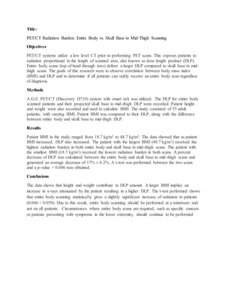
1. Title:
PET/CT Radiation Burden: Entire Body vs. Skull Base to Mid-Thigh Scanning
Objectives
PET/CT systems utilize a low level CT prior to performing PET scans. This exposes patients to
radiation proportional to the length of scanned area, also known as dose length product (DLP).
Entire body scans (top of head through toes) deliver a larger DLP compared to skull base to mid-
thigh scans. The goals of this research were to observe correlation between body mass index
(BMI) and DLP and to determine if all patients can safely receive an entire body scan regardless
of diagnosis.
Methods
A G.E. PET/CT (Discovery D710) system with smart mA was utilized. The DLP for entire body
scanned patients and their projected skull base to mid-thigh DLP were recorded. Patient height
and weight were also recorded and used to calculate BMI. The study consisted of 55 adult
patients, with varying BMI. Patient BMI was compared to their DLP, along with the difference
between entire body and skull base to mid-thigh DLP.
Results
Patient BMI in the study ranged from 18.7 kg/m2 to 48.7 kg/m2. The data showed that as patient
BMI increased, DLP also increased. The patient with the largest BMI (48.7 kg/m2) received the
highest radiation burden in both entire body and skull base to mid-thigh scans. The patient with
the smallest BMI (18.7 kg/m2) received the lowest radiation burden in both scans. A percent
decrease of DLP was calculated between the entire body and skull base to mid-thigh scans and
generated an average decrease of 17% ± 6.2%. A t-test was performed between the two scans
and a p-value = 0.046 was obtained.
Conclusions
The data shows that height and weight contribute to DLP amount. A larger BMI implies an
increase in x-rays attenuated by the patient resulting in a higher DLP. The t-test performed shows
that entire body scanning statistically gives a slightly significant increase of radiation to patients
(0.046 < 0.050). Due to this result, entire body scanning should be performed at a minimum and
not on all patients. Circumstances where entire body scans are required, such as specific cancers
or patients presenting with symptoms in the lower extremity, should continue to be performed