ABSTRACT:
Objective: This study purports to answer the question: Does a dual-energy CT scan of the chest using reduced radiation result in images of equal or better quality compared to those produced by the gold standard of care?
Methods: With the agreement of the Ethical Review Committee and written informed consent from 32 patients, who received dual-energy CT (DECT) scan of the chest in a dual-source scanner, a second set of images was taken at a reduced radiation dose. On virtual monochromatic images at 40 and 60 keV, three thoracic radiologists evaluated image quality, normal thoracic structures, and pulmonary and mediastinal aberrations. Students analyzed the data using analysis of variance, Kappa statistics, and Wilcoxon signed-rank tests.
Results: No irregularities in the scans were missed in the virtual monochrome photographs of all patients at a lower radiation dose, and the images were found to be of sufficient quality. At 40 and 60 keV, standard-of-care pictures produced equal contrast enhancement and lesion detection. Observers were entirely consistent with one another. Among other characteristics, reduced-dose DECT had a CTDIvol of 3.0 ±0.6 mGy, and a size specified dose estimate (SSDE) of 4.0 ±0.6 mGy, a dose-length product (DLP) of 107 ±30 mgy.cm, and an effective dose (ED) of 1.15 ±0.4 mSv.
Conclusion: Dual-energy computed tomography of the chest allows for the administration of lower radiation doses (CTDIvol <3 mGy).
Thesis / Doctoral Project / Dissertation Proposal
Student Information:
Student GUID Number:
833168318
Student Name: (As it appears on your transcript)
Abdullatif Abdullah
Address:
1850 Columbia Pike Apt 406, Arlington, Virginia, 22204
E-Mail Address:
[email protected]
Phone Number:
571-340-6065
Degree:
Masters in Health Physics
Expected Graduation Month/Year
05 / 2022
Dept./Major:
Health Physics
I. Title:
Estimation of Peak Skin Dose and Its Relation to the Size Specific Dose Estimate
II. Problem or Hypothesis:
The CT Dose Index (CTDIvol) was originally designed as an index of dose associated with various CT diagnostic procedures not as a direct dosimetry method for individual patient dose assessments. There is no current method for calculating peak skin dose (PSD) using the key metrics provided from the radiation dose structure report of a CT scanner. Every CT study is required to output the kVp and mAs that were used, the dose length product and CT dose index volume which will all be shown on the CT console, but there is no direct method to go straight to the PSD. This project will test the hypothesis that the SSDE has a sufficiently strong linear relationship with PSD to allow direct calculation of the PSD directly from the SSDE.
III. Review of Related Literature:
The highest radiation dose accruing at a single site on a patient’s skin is referred to as the peak skin dose (PSD) which is related to the Computed Tomography dose index (CTDIvol) that is displayed on the console of CT scanners. However, the CT Dose Index was originally designed as an index not as a direct dosimetry method for patient dose assessment. More recently, modifications to original CTDI concept have attempted to convert it into to patient dosimetry method, but have with mixed results in terms of accuracy. Nonetheless, CTDI-based dosimetry is the current worldwide standard for estimation of patient dose in CT. Therefore, CTDIvol is often used to enable medical physicists to compare the dose output between different CT scanners.
Fearon, Thomas (2011) explained that current estimation of radiation dose from CT scans on patients has relied on the measurement of Computed Tomography Dose Index (CTDI) in standard cylindrical phantoms, and calculations based on mathematical representations of “standard man.” The purpose of this study was to investigate the feasibility of adapting a radiation treatment planning system (RTPS) to provide patient-specific CT dosimetry. A radiation treatment planning system was modified to calculate patient-specific CT dose distributions, which can be represented by dose at specific points within an organ of interest, as well as organ dose-volume (after image segmentation) for a GE Light Speed Ultra Plus CT scanner. Digital representations of the phantoms (virtual phantom) were acquired with the GE CT scanner in axial mode. Thermoluminescent dosimeter (TLDs) measurements in pediatric anthropomorphic phantoms were utilized t ...
CT scans still play a critical role in managing COVID-19. Patients with a severe coronavirus infection show different features on their computed tomography
The Computed Tomography (CT) dose output of some selected hospitals in the Federal capital Territory, Abuja, Nigeria have been determined by calculating the Effective doses of CT Chest and Abdomen-Pelvis of selected hospitals and compared its average with the Mean Reference Dose of CT Chest and Abdomen-Pelvis from four hospitals in the Federal Capital Territory, Abuja, Nigeria. Effective Dose and Scan type were extracted from the CT Chest and Abdomen-Pelvis examinations recorded. The Effective Dose of each patient undergoing the Chest and Abdomen-Pelvis examinations were calculated using the coefficient factor and the DLP values. Patients’ CT dose data from the ages of 18 to 60years from each of the 4 centres for each study type from January, 2013 to December, 2014 was extracted. A total of 112 patients’ CT dose data was extracted. Chest CT Effective Dose ranged from 9.0 to 34.0mSv, while Abdomen-Pelvis CT Effective Dose ranged from 15.9 to 61.0 for all the Centres in Federal Capital Territory, Abuja. This is higher than the recommended Reference Effective Dose range for CT Chest which is from 5 – 7mSv. and for CT Abdomen-Pelvis is from 8 – 14mSv. The mean effective dose from the Chest CT is 21.8mSv and from the Abdomen-Pelvis is 31.9mSv.
Using Distance Measure based Classification in Automatic Extraction of Lungs ...sipij
We introduce in this paper a reliable method for automatic extraction of lungs nodules from CT chest
images and shed the light on the details of using the Weighted Euclidean Distance (WED) for classifying
lungs connected components into nodule and not-nodule. We explain also using Connected Component
Labeling (CCL) in an effective and flexible method for extraction of lungs area from chest CT images with
a wide variety of shapes and sizes. This lungs extraction method makes use of, as well as CCL, some
morphological operations. Our tests have shown that the performance of the introduce method is high.
Finally, in order to check whether the method works correctly or not for healthy and patient CT images, we
tested the method by some images of healthy persons and demonstrated that the overall performance of the
method is satisfactory.
Breast conserving surgery followed by adjuvant radiotherapy is adopted in the early detected cases and mastectomy followed by radiotherapy or chemotherapy in the advanced cases are the general practices.
USING DISTANCE MEASURE BASED CLASSIFICATION IN AUTOMATIC EXTRACTION OF LUNGS ...sipij
We introduce in this paper a reliable method for automatic extraction of lungs nodules from CT chest
images and shed the light on the details of using the Weighted Euclidean Distance (WED) for classifying
lungs connected components into nodule and not-nodule. We explain also using Connected Component
Labeling (CCL) in an effective and flexible method for extraction of lungs area from chest CT images with
a wide variety of shapes and sizes. This lungs extraction method makes use of, as well as CCL, some
morphological operations. Our tests have shown that the performance of the introduce method is high.
Finally, in order to check whether the method works correctly or not for healthy and patient CT images, we
tested the method by some images of healthy persons and demonstrated that the overall performance of the
method is satisfactory.
Ionizing radiation makes invasive cardiology procedures such as coronary angiography, percutaneous coronary intervention (PCI), and electrophysiologic diagnostics and therapeutics possible .
Radiation risks can be thought of as deterministic (effects after exceeding certain threshold, e.g., skin burns) or stochastic (a risk of an outcome is proportional to the dose received, e.g., malignancy or teratogenicity) .
Reducing the radiation exposure in the cardiac catheterization laboratory is important, especially as procedures are becoming more complex .
Image segmentation is still an active reason of research, a relevant research area
in computer vision and hundreds of image segmentation techniques have been proposed by
the researchers. All proposed techniques have their own usability and accuracy. In this paper
we are going present a review of some best lung nodule existing detection and segmentation
techniques. Finally, we conclude by focusing one of the best methods that may have high
level accuracy and can be used in detection of lung very small nodules accurately.
Deep learning method for lung cancer identification and classificationIAESIJAI
Lung cancer (LC) is calming many lives and is becoming a serious cause of concern. The detection of LC at an early stage assists the chances of recovery. Accuracy of detection of LC at an early stage can be improved with the help of a convolutional neural network (CNN) based deep learning approach. In this paper, we present two methodologies for Lung cancer detection (LCD) applied on Lung image database consortium (LIDC) and image database resource initiative (IDRI) data sets. Classification of these LC images is carried out using support vector machine (SVM), and deep CNN. The CNN is trained with i) multiple batches and ii) single batch for LC image classification as non cancer and cancer image. All these methods are being implemented in MATLAB. The accuracy of classification obtained by SVM is 65%, whereas deep CNN produced detection accuracy of 80% and 100% respectively for multiple and single batch training. The novelty of our experimentation is near 100% classification accuracy obtained by our deep CNN model when tested on 25 Lung computed tomography (CT) test images each of size 512×512 pixels in less than 20 iterations as compared to the research work carried out by other researchers using cropped LC nodule images.
Study on the Impact of FOCUS-PDCA Management Model on the Disinfection Qualit...MehranMouzam
To analyze the impact of FOCUS-PDCA management model on the disinfection quality of flexible endoscopes. Method: A study was conducted on 128 flexible endoscopes in our hospital. According to different management plans, flexible endoscopes were divided into a control group (conventional management model) and an experimental group (FOCUS-PDCA management model). The flexible endoscopes evaluated in both control group and the experimental group were 64 each. The ATP values, management quality, and bacterial colony exceeding standards were observed in two groups. Results: Before management, there was no significant difference in ATP values between the two groups, with P>0.05; after management, compared with the control group (106.25 ± 6.812), the ATP value in the experimental group was lower, with P<0.05. The scores of disinfection standards (4.39 ± 0.49), cleaning standards (4.22 ± 0.45), management systems (4.13 ± 0.34), and management assessment (4.97 ± 0.25) in the experimental group were higher than those in the control group (3.89 ± 0.31, 3.20 ± 0.41, 3.12 ± 0.13, 3.95 ± 0.21), with P<0.05. In the experimental group, the bacterial colony exceeding standard rate of gastroscopy was 3.13%, bacterial colony exceeding standard rate of colonoscopy was 0.00%, bacterial colony exceeding standard rate of bronchoscopy was 1.56%, and the total bacterial colony exceeding standard rate of total colonies was 6.25%, which were significantly lower than the control group's 12.50%, 7.81%, 12.50%, and 32.81%, respectively with P<0.05. Conclusion: The FOCUS-PDCA management model is more conducive to reducing the ATP values of flexible endoscopes, improving the disinfection qualification rate, and improving management quality. This model is worthy of further promotion.
The Evolving Nature of Mother-Representations Across the Waves of FeminismMehranMouzam
Whether they’ve occupied the foreground or the background of literary works, mothers as primary subjects or as their shadows - have forever been weaved into the vital, in stories told either about them and/or, about their children. Motherhood and the matrifocal narrative, on the whole, have undergone various conceptual reconstructions that have been both a direct and indirect result of the different waves of feminism across the globe. Feminist concerns over ideas of motherhood and their related representations in literary texts, popular culture, and media, etc. have sought to understand the dichotomy between biological ideas of being a mother and its social and cultural constructions, which essentially shape the gendered expectations of mothers, especially because such socio-cultural constructions carry the cis-gendered heteronormative expectation of what it necessarily means to be a ‘socially accepted’ mother. The ''maternal'' representations in literature and other artistic mediums have evolved to accommodate the ever-changing, dynamism that the term ''mother'' brings forth. The mother figure is no longer only nurturing, ever-suffering and sappy but also loud, angry, and articulate.
More Related Content
Similar to Reduced Radiation Exposure in Dual-Energy Computed Tomography of the Chest: Impact on Image Quality
The Computed Tomography (CT) dose output of some selected hospitals in the Federal capital Territory, Abuja, Nigeria have been determined by calculating the Effective doses of CT Chest and Abdomen-Pelvis of selected hospitals and compared its average with the Mean Reference Dose of CT Chest and Abdomen-Pelvis from four hospitals in the Federal Capital Territory, Abuja, Nigeria. Effective Dose and Scan type were extracted from the CT Chest and Abdomen-Pelvis examinations recorded. The Effective Dose of each patient undergoing the Chest and Abdomen-Pelvis examinations were calculated using the coefficient factor and the DLP values. Patients’ CT dose data from the ages of 18 to 60years from each of the 4 centres for each study type from January, 2013 to December, 2014 was extracted. A total of 112 patients’ CT dose data was extracted. Chest CT Effective Dose ranged from 9.0 to 34.0mSv, while Abdomen-Pelvis CT Effective Dose ranged from 15.9 to 61.0 for all the Centres in Federal Capital Territory, Abuja. This is higher than the recommended Reference Effective Dose range for CT Chest which is from 5 – 7mSv. and for CT Abdomen-Pelvis is from 8 – 14mSv. The mean effective dose from the Chest CT is 21.8mSv and from the Abdomen-Pelvis is 31.9mSv.
Using Distance Measure based Classification in Automatic Extraction of Lungs ...sipij
We introduce in this paper a reliable method for automatic extraction of lungs nodules from CT chest
images and shed the light on the details of using the Weighted Euclidean Distance (WED) for classifying
lungs connected components into nodule and not-nodule. We explain also using Connected Component
Labeling (CCL) in an effective and flexible method for extraction of lungs area from chest CT images with
a wide variety of shapes and sizes. This lungs extraction method makes use of, as well as CCL, some
morphological operations. Our tests have shown that the performance of the introduce method is high.
Finally, in order to check whether the method works correctly or not for healthy and patient CT images, we
tested the method by some images of healthy persons and demonstrated that the overall performance of the
method is satisfactory.
Using Distance Measure based Classification in Automatic Extraction of Lungs ...sipij
We introduce in this paper a reliable method for automatic extraction of lungs nodules from CT chest
images and shed the light on the details of using the Weighted Euclidean Distance (WED) for classifying
lungs connected components into nodule and not-nodule. We explain also using Connected Component
Labeling (CCL) in an effective and flexible method for extraction of lungs area from chest CT images with
a wide variety of shapes and sizes. This lungs extraction method makes use of, as well as CCL, some
morphological operations. Our tests have shown that the performance of the introduce method is high.
Finally, in order to check whether the method works correctly or not for healthy and patient CT images, we
tested the method by some images of healthy persons and demonstrated that the overall performance of the
method is satisfactory.
Using Distance Measure based Classification in Automatic Extraction of Lungs ...sipij
We introduce in this paper a reliable method for automatic extraction of lungs nodules from CT chest
images and shed the light on the details of using the Weighted Euclidean Distance (WED) for classifying
lungs connected components into nodule and not-nodule. We explain also using Connected Component
Labeling (CCL) in an effective and flexible method for extraction of lungs area from chest CT images with
a wide variety of shapes and sizes. This lungs extraction method makes use of, as well as CCL, some
morphological operations. Our tests have shown that the performance of the introduce method is high.
Finally, in order to check whether the method works correctly or not for healthy and patient CT images, we
tested the method by some images of healthy persons and demonstrated that the overall performance of the
method is satisfactory.
Breast conserving surgery followed by adjuvant radiotherapy is adopted in the early detected cases and mastectomy followed by radiotherapy or chemotherapy in the advanced cases are the general practices.
USING DISTANCE MEASURE BASED CLASSIFICATION IN AUTOMATIC EXTRACTION OF LUNGS ...sipij
We introduce in this paper a reliable method for automatic extraction of lungs nodules from CT chest
images and shed the light on the details of using the Weighted Euclidean Distance (WED) for classifying
lungs connected components into nodule and not-nodule. We explain also using Connected Component
Labeling (CCL) in an effective and flexible method for extraction of lungs area from chest CT images with
a wide variety of shapes and sizes. This lungs extraction method makes use of, as well as CCL, some
morphological operations. Our tests have shown that the performance of the introduce method is high.
Finally, in order to check whether the method works correctly or not for healthy and patient CT images, we
tested the method by some images of healthy persons and demonstrated that the overall performance of the
method is satisfactory.
Ionizing radiation makes invasive cardiology procedures such as coronary angiography, percutaneous coronary intervention (PCI), and electrophysiologic diagnostics and therapeutics possible .
Radiation risks can be thought of as deterministic (effects after exceeding certain threshold, e.g., skin burns) or stochastic (a risk of an outcome is proportional to the dose received, e.g., malignancy or teratogenicity) .
Reducing the radiation exposure in the cardiac catheterization laboratory is important, especially as procedures are becoming more complex .
Image segmentation is still an active reason of research, a relevant research area
in computer vision and hundreds of image segmentation techniques have been proposed by
the researchers. All proposed techniques have their own usability and accuracy. In this paper
we are going present a review of some best lung nodule existing detection and segmentation
techniques. Finally, we conclude by focusing one of the best methods that may have high
level accuracy and can be used in detection of lung very small nodules accurately.
Deep learning method for lung cancer identification and classificationIAESIJAI
Lung cancer (LC) is calming many lives and is becoming a serious cause of concern. The detection of LC at an early stage assists the chances of recovery. Accuracy of detection of LC at an early stage can be improved with the help of a convolutional neural network (CNN) based deep learning approach. In this paper, we present two methodologies for Lung cancer detection (LCD) applied on Lung image database consortium (LIDC) and image database resource initiative (IDRI) data sets. Classification of these LC images is carried out using support vector machine (SVM), and deep CNN. The CNN is trained with i) multiple batches and ii) single batch for LC image classification as non cancer and cancer image. All these methods are being implemented in MATLAB. The accuracy of classification obtained by SVM is 65%, whereas deep CNN produced detection accuracy of 80% and 100% respectively for multiple and single batch training. The novelty of our experimentation is near 100% classification accuracy obtained by our deep CNN model when tested on 25 Lung computed tomography (CT) test images each of size 512×512 pixels in less than 20 iterations as compared to the research work carried out by other researchers using cropped LC nodule images.
Study on the Impact of FOCUS-PDCA Management Model on the Disinfection Qualit...MehranMouzam
To analyze the impact of FOCUS-PDCA management model on the disinfection quality of flexible endoscopes. Method: A study was conducted on 128 flexible endoscopes in our hospital. According to different management plans, flexible endoscopes were divided into a control group (conventional management model) and an experimental group (FOCUS-PDCA management model). The flexible endoscopes evaluated in both control group and the experimental group were 64 each. The ATP values, management quality, and bacterial colony exceeding standards were observed in two groups. Results: Before management, there was no significant difference in ATP values between the two groups, with P>0.05; after management, compared with the control group (106.25 ± 6.812), the ATP value in the experimental group was lower, with P<0.05. The scores of disinfection standards (4.39 ± 0.49), cleaning standards (4.22 ± 0.45), management systems (4.13 ± 0.34), and management assessment (4.97 ± 0.25) in the experimental group were higher than those in the control group (3.89 ± 0.31, 3.20 ± 0.41, 3.12 ± 0.13, 3.95 ± 0.21), with P<0.05. In the experimental group, the bacterial colony exceeding standard rate of gastroscopy was 3.13%, bacterial colony exceeding standard rate of colonoscopy was 0.00%, bacterial colony exceeding standard rate of bronchoscopy was 1.56%, and the total bacterial colony exceeding standard rate of total colonies was 6.25%, which were significantly lower than the control group's 12.50%, 7.81%, 12.50%, and 32.81%, respectively with P<0.05. Conclusion: The FOCUS-PDCA management model is more conducive to reducing the ATP values of flexible endoscopes, improving the disinfection qualification rate, and improving management quality. This model is worthy of further promotion.
The Evolving Nature of Mother-Representations Across the Waves of FeminismMehranMouzam
Whether they’ve occupied the foreground or the background of literary works, mothers as primary subjects or as their shadows - have forever been weaved into the vital, in stories told either about them and/or, about their children. Motherhood and the matrifocal narrative, on the whole, have undergone various conceptual reconstructions that have been both a direct and indirect result of the different waves of feminism across the globe. Feminist concerns over ideas of motherhood and their related representations in literary texts, popular culture, and media, etc. have sought to understand the dichotomy between biological ideas of being a mother and its social and cultural constructions, which essentially shape the gendered expectations of mothers, especially because such socio-cultural constructions carry the cis-gendered heteronormative expectation of what it necessarily means to be a ‘socially accepted’ mother. The ''maternal'' representations in literature and other artistic mediums have evolved to accommodate the ever-changing, dynamism that the term ''mother'' brings forth. The mother figure is no longer only nurturing, ever-suffering and sappy but also loud, angry, and articulate.
Investigating the Challenges Faced by Iraqi Secondary School Students in Engl...MehranMouzam
The aim of the current study is to uncover the challenges encountering Iraqi students in the secondary school classrooms. Five students in a public secondary school located in Misan province participate in this investigational study. The study explores the challenges the students face in the learning process; particularly in the acquisition of oral proficiency. However, oral proficiency, even as used by the teacher, hardly ever functions as a means for students to acquire knowledge and explore new ideas. This paper attempts to identify the challenges or problems that students encounter in teaching English oral proficiency.
Data collection methods used in this study include students’ interviews and classroom observations. After collecting information and taking notes on the students’ oral proficiency. Data collected demonstrates that the acquisition of the students’ oral proficiency is associated with several challenges and problems that inhibit their pursuit to interact and express themselves in real-life situations. In sum, the study concludes that when learning English oral proficiency, several challenges prevent the students’ oral performance or progress such as improperly trained teacher, government policy, assessment systems, exposure to English, and less use of audio-visual aids etc.
RIGHT TO DIE: A STUDY OF DIFFERENT JURISDICTIONSMehranMouzam
1. INTRODUCTIONThe right to die is the concept based on the opinion that a human being is entitled to end their life.
Euthanasia, or mercy killing, means the deliberate killing of a patient who is terminally ill and/or
in severe and chronic pain. The word ‘Euthanasia’ is a derivative from the Greek words ‘eu’ and
‘thanotos’ which literally mean “good death”.1 The death of a terminally ill patient is accelerated
through active or passive means in order to relieve such patient of pain or suffering. However, the
issue of euthanasia is not as simple as the literal translation of the term. The issue is complex and
involves several moral, ethical, societal and economic aspects.2 Those who are in favor of
euthanasia argue on the right to self-determination and futility of prolonging a life without meaning
and dignity and those who are against the practice believe that emphasizes must be given to
palliative care, and that legalizing euthanasia would be violate of the principle of sanctity of life.3
It is because of this that most of the States allow only passive euthanasia and to check the misuse
enacted laws on the subject.
Appointment of non muslim ruler in muslim countryMehranMouzam
The modern world has turned multicultural and the socio-political changes have created a new code of conduct at the global level. Multireligion societies are getting developed. Now the nature of modern politics creates new questions. It has been said that Muslim countries have also to choose a new democratic system of politics in which it is not inevitable now to appoint only Muslim rulers in Muslim states. Traditional and liberal narratives have got into the clash in Muslim countries in this regard. This study deals with the logic of both above-said categories of thinkers. The shreds of evidence from the Quran and Hadith, which prohibit the appointment of non-Muslim in Muslim countries, are presented first along with the logics of Muslim scholars who take them into consideration. The later liberal narrative has been explained followed by ending remarks through conclusion.
A study on urdu speakers’ use of english stress patterns phonological variationMehranMouzam
The aim of this research paper is to study Urdu Speakers’ use of English Stress Patterns and their phonological variation from native speakers of Pakistani EFL learners. The stress patterns of English language are affected by the influence of L1Urdu speakers’ perception in Pakistan which ultimately influences English pronunciation and sometimes its meanings as well. It also results difficulties faced by learners in our class rooms. Based on phonological differences between two languages, the researchers assume that there is a wide discrepancy in stress patterns among those spoken and used by native speakers and read and perceived by Pakistani students in our classrooms using English as second language. It carries a tangible impact of Urdu stress pattern with almost equal stress on all the syllables which is quite problematic both for teachers and learners of English whether it is as Second Language Learning or as Foreign Language Learning. To find out concrete results quantitative analysis of stress patterns was made on the selected sample taking from public sector university students. Findings of the research provide a useful pedagogical insight into the perspective of English language teaching with particular emphasis on spoken proficiency of English among students whose L1 is Urdu. The findings of the research suggest invariably the wrong placement of lexical stress in English words in Pakistan by Urdu speakers who have Urdu as L1 because they either place the stress on the syllable preceding the actual syllable or following it. Finally, it is suggested to follow the native speakers tone as a final remedy.
Forensic discourse analysis of legal and courtroom interaction dr arshad aliMehranMouzam
The primary objective of this study is to look into the complexities and complications of legal discourse and how they manifest themselves in the courtroom. The research looks at the dynamics in a courtroom and the jury room in the film 12 Angry Men. The study aims to show how language acts as a source of agency and power in a legal setting, as well as to look into how speakers cooperate in a legal setting. The researcher devised a framework based on Heffer's (2013) legal and forensic discourse model as well as Grice's (1975) Cooperative Principle and its maxims. The data for the study comes from the film 12 Angry Men, which is based on a true story. Forensic discourse analysis was used to analyse the data. This method analyses the utterances and other features present in the legal discourse, as well as its implications. The main findings of the study show that the judge's voice is projected in the court with a significant amount of dominance. Similarly, there is a lack of direct communication that affects the trial by making it difficult for the jury to fully comprehend the facts of the case. Furthermore, the agency is frequently removed from the jury, resulting in a misunderstanding of the case. The majority of the jury members are bored and sleepy, while others have an unhealthy fondness for the prosecution. The final finding concerns the jury members' power play in the jury trial, as evidenced by the jury members' failure to project their voices effectively, and their lack of cooperation. The forensic discourse analysis reveals that all of the maxims were repeatedly violated by the jury members. The most frequently flouted maxims, however, were those of quantity and relevance. This demonstrates how the desire for authority and the lack of agency can have far-reaching implications for the final decision.
IMPACT OF PATERNALISTIC LEADERSHIP ON EMPLOYEE COMMITMENT AND INNOVATIVE WOR...MehranMouzam
1. Description of Research Work
Innovation is a key concern of HR now a days. It is the key requirement for organizational success (Akram,
Lei, Haider, & Hussain, 2018). It gives new ways to do work, to overcome situations, to solve problems
and to make decisions. Dedahanov et al. (2019) claims that PLB and EE promotes innovative work
behaviour of employees. This positive impact can be enhanced by I-deals that are now trending in
manipulating job behaviours of employees. Recent studies conducted by Microsoft Japan on a four-day
working week claims that, not only the employees became happier and satisfied, they became productive
at workplace. These shortened working weeks and flexibility made employees more efficient and
productive at workplace (Paul, 2019). Leadership styles are now playing major roles in organizations and
are proved to be crucial factors for their success. PLB is a practical approach to get maximum out of
employees. According to Hornung, Rouseau, Glaser, Angerer, and Weigh (2011), loyalty and commitment
of employees are major outcomes of PLB. When employees are committed at workplace, their performance
boost ups which ultimately effects the organizational performance positively. This study is conducted to
observe and to describe the role of PLB on employee’s commitment level and how I-deals can mediate this
relationship in pharmaceutical sector. According to PPMA (2017), Pakistan’s pharmaceutical industry is
progressive, vibrant and future oriented. It consists of more than 700 manufacturing units, having 25-
multinational pharmaceutical units working in country. 70% of country’s demand of finished medicines is
been met by this sector and have shown a remarkable growth over last ten years and has export turnover of
more than 12 million and accounts for less than one percent of country’s GDP. To observe this productive
phenomenon of PLB, I-deals, EE, IWB and commitment in such a fast-growing sector can help to develop
positive outcomes for them.
Antibiotic Stewardship by Anushri Srivastava.pptxAnushriSrivastav
Stewardship is the act of taking good care of something.
Antimicrobial stewardship is a coordinated program that promotes the appropriate use of antimicrobials (including antibiotics), improves patient outcomes, reduces microbial resistance, and decreases the spread of infections caused by multidrug-resistant organisms.
WHO launched the Global Antimicrobial Resistance and Use Surveillance System (GLASS) in 2015 to fill knowledge gaps and inform strategies at all levels.
ACCORDING TO apic.org,
Antimicrobial stewardship is a coordinated program that promotes the appropriate use of antimicrobials (including antibiotics), improves patient outcomes, reduces microbial resistance, and decreases the spread of infections caused by multidrug-resistant organisms.
ACCORDING TO pewtrusts.org,
Antibiotic stewardship refers to efforts in doctors’ offices, hospitals, long term care facilities, and other health care settings to ensure that antibiotics are used only when necessary and appropriate
According to WHO,
Antimicrobial stewardship is a systematic approach to educate and support health care professionals to follow evidence-based guidelines for prescribing and administering antimicrobials
In 1996, John McGowan and Dale Gerding first applied the term antimicrobial stewardship, where they suggested a causal association between antimicrobial agent use and resistance. They also focused on the urgency of large-scale controlled trials of antimicrobial-use regulation employing sophisticated epidemiologic methods, molecular typing, and precise resistance mechanism analysis.
Antimicrobial Stewardship(AMS) refers to the optimal selection, dosing, and duration of antimicrobial treatment resulting in the best clinical outcome with minimal side effects to the patients and minimal impact on subsequent resistance.
According to the 2019 report, in the US, more than 2.8 million antibiotic-resistant infections occur each year, and more than 35000 people die. In addition to this, it also mentioned that 223,900 cases of Clostridoides difficile occurred in 2017, of which 12800 people died. The report did not include viruses or parasites
VISION
Being proactive
Supporting optimal animal and human health
Exploring ways to reduce overall use of antimicrobials
Using the drugs that prevent and treat disease by killing microscopic organisms in a responsible way
GOAL
to prevent the generation and spread of antimicrobial resistance (AMR). Doing so will preserve the effectiveness of these drugs in animals and humans for years to come.
being to preserve human and animal health and the effectiveness of antimicrobial medications.
to implement a multidisciplinary approach in assembling a stewardship team to include an infectious disease physician, a clinical pharmacist with infectious diseases training, infection preventionist, and a close collaboration with the staff in the clinical microbiology laboratory
to prevent antimicrobial overuse, misuse and abuse.
to minimize the developme
Navigating the Health Insurance Market_ Understanding Trends and Options.pdfEnterprise Wired
From navigating policy options to staying informed about industry trends, this comprehensive guide explores everything you need to know about the health insurance market.
R3 Stem Cells and Kidney Repair A New Horizon in Nephrology.pptxR3 Stem Cell
R3 Stem Cells and Kidney Repair: A New Horizon in Nephrology" explores groundbreaking advancements in the use of R3 stem cells for kidney disease treatment. This insightful piece delves into the potential of these cells to regenerate damaged kidney tissue, offering new hope for patients and reshaping the future of nephrology.
Reduced Radiation Exposure in Dual-Energy Computed Tomography of the Chest: Impact on Image Quality
1. 1
Reduced Radiation Exposure in Dual-Energy
Computed Tomography of the Chest: Impact on Image Quality
ABSTRACT:
Objective: This study purports to answer the question: Does a dual-energy CT
scan of the chest using reduced radiation result in images of equal or better quality
compared to those produced bythe gold standard of care?
Methods:With the agreement of the Ethical Review Committee and written
informed consent from 32 patients, who received dual-energy CT (DECT) scanof
the chest in a dual-source scanner, a second set of images was taken at a reduced
radiation dose. On virtual monochromatic images at 40 and 60 keV, three thoracic
radiologists evaluated image quality, normal thoracic structures, and pulmonary
and mediastinal aberrations. Students analyzed the data using analysis of variance,
Kappa statistics, and Wilcoxon signed-rank tests.
Results:No irregularities in the scans were missed in the virtual monochrome
photographs of all patients at a lower radiation dose, and the images were found to
be of sufficient quality. At 40 and 60 keV, standard-of-care pictures produced
equal contrast enhancement and lesion detection. Observers were entirely
consistent with one another. Among other characteristics, reduced-doseDECT had
a CTDIvol of 3.0 ±0.6 mGy, and a size specified doseestimate (SSDE) of 4.0 ±0.6
mGy, a dose-length product(DLP) of 107 ±30 mgy.cm, and an effective dose(ED)
of 1.15 ±0.4 mSv.
Conclusion: Dual-energy computed tomography of the chest allows for the
administration of lower radiation doses (CTDIvol <3 mGy).
INTRODUCTION:
Dual-energy computed tomography (DECT) was originally a potential alternative
that enabled more accurate images, but it required a high doseof radiation and had
issues with the two kV image files collected during two consecutive DECT
acquisitions not being in the same location at the same time (1). In the 1990s,
numerous studies demonstrated that DECT was superior to single-energy CT for
2. 2
detecting lung nodules (2). However, according to a study financed by the
Fleischner Society, DECT still proved to be unable to distinguish lung nodules
toward the decade's end (3).
As public awareness of the dangers of CT scan radiation has grown, additional
clinical research and technological advancements have been made to minimise CT
scan radiation levels (4). According to some experts, DECT can be performed with
doses comparable to those used in single-energy CT in multidetector CT (5). In
fact, doctors have employed both single-phase and dual-phase DECT scans to
detect benign and malignant mediastinal tumours. These tests have identified
aneurysms of the aorta, chronic pulmonary embolism, lung emboli, and pulmonary
nodules (6,7). Few persons who use chest DECT have considered reducing their
daily radiation exposure. Recent studies have demonstrated that DECT scans are
effective for examining lung parenchyma and pulmonary embolisms. This is
accomplished through the use of material separation techniques and the creation of
virtual monochrome images (8,9).
As mentioned above, the goal of this study is to determine whether chest dual-
energy CT can be administered at lower doses than the current gold standard of
care and still produceviable images.
METHODOLOGY:
The voxel standard deviation was used to assess the number of CT counts and the
amount of noise in DECT images (defined as the standard deviation of voxel
values). Chest phantoms were used in this study. Double-sided tape was applied to
the surface of test tubes containing iopamidol 370 mg/mL diluted contrast material
at 1:20 and 1:40 concentrations. Two scans of the phantom were performed using
standard-of-care and low-dose DECT procedures. Changing this has no effect on
the amount of time required to scan a file or on the size of the file scanned. CT
scans and standard deviations (SDs) were calculated for each region at ten
locations on the right upper lung and chest wall of the phantom. The diluted
contrast solution test tubes were scanned using CT scanners and their standard
deviations were recorded twice.
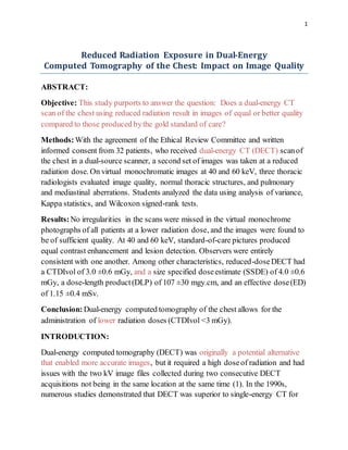
3. 3
Figure1: Imageof the anthropomorphicphantom'schest in the transverse plane
from a CT scan.
Participants in the Study
Our Institutional Review Board's Human Research Committee approved this study
after everyone signed an informed consent form to participate. To ensure that the
research was conducted properly, HIPPAA regulations were strictly adhered to.
4. 4
The researchers searched the Radiology Information System (RIS) for patients who
needed a conventional chest CT with contrast enhancement. They discovered that
there were individuals. To participate in the study, participants needed to be alert,
have stable blood pressure, and be at least 56 years old. A CT scan was not
performed if the patient could not hold their breath for more than 10 seconds orif
the patient was allergic to the contrast used. Certain individuals were unable to
participate in the study due to their BMI being greater than 32 kg/m2. Individuals
who had previously undergone two CT scans at our clinic — one in the prone
position and another in the supine position — were also denied participation due to
a history of interstitial lung disease. Forty-five individuals participated in the study.
Eight patients stated that they did not wish to be treated after signing an informed
consent form. Five persons were forced to abandon the experiment for health
reasons after an accident involving a single-energy CT scanner. As a result, there
were finally 32 adults participating: 12 men and 20 women. The average age was
71 ±6 years in general. On average, the men were 70 ±5 years old, and the women
were 72 ±7 years old, with a range of 56 to 87 years. The 32 individuals who
underwent clinical CT scans included those with lung cancer, pneumonia, and
other non-pulmonary ailments.
For each CT scan performed, the patient received 65mL of IV contrast material.
This was achievable in this circumstance because the CT scanner was a
multidetector CT scanner with a zooming focus point. It took 35 seconds for the
individual to be scanned in preparation for the contrastmedia injection. They were
scanned at a rate of 2.5 mL per second. Each scan lasted nearly the same length of
time (approximately 3 s).
Patients could see how much of their bodywas covered by both standard-of-care
and low-dose imaging because they would eventually get a chest CT scan. CT
scans with a standard dosetook longer than scans with a low dose. The decreased-
dosage technique reduced the quality reference mAs to approximately half that of
the standard of care (10). Between the two scans, all other scanning parameters
remained constant. It was not included in the set of low-dose images. Each photo
set's CTDIvol and dose-length products (DLP) were determined. We were able to
determine the water-equivalent diameter (WED) and size-specific dose estimate
(SSDE) for each person by utilising software called the Radmetrics Enterprise
5. 5
Platform (11). This meant that each chest CT image was estimated because it took
0.14 of a second to convert each image to an EDT. This was the DLP's operation. It
resembled a map (12).
Images of normal and lower doses were created using Healthcare's proprietary
technology. The images were created with the industry-standard medium-smooth
soft-tissue reconstruction kernel, a proprietary iterative reconstruction technique
(I30f). There were no patients with interstitial lung disease in this research. As a
result, we used a soft-tissue kernel for all of the images in order to improve spatial
resolution and the ability to see edges (which provides optimal image contrast at
the costof spatial resolution).
This is how transversely orientated pictures (80/Sn140 kV) were created. Each
slice was separated by a 2mm gap. Virtual monochromatic images at 40 and 60
keV, as well as images of blood flow and images with no contrast, were created
using dedicated computers. Because virtual monochromatic low-energy images
had the same or less noise and additionally might aid in making iodine more
visible, they were the best choice (13–16). The images taken at 40 keV were
chosen for the investigation becausethey best fit the K-edge of iodine (33 keV). A
60 keV resolution image has better contrastthan a 65-70 keV resolution image due
to the absenceof noise. This is why they were chosen for investigation (8, 17).
Before the investigators examined the photographs, no scan settings nor patient
information were associated with individual participants.
Board-certified thoracic radiologists viewed and analyzed the chest CT images
using a DICOM-compliant image viewer. Radiologists were unaware of how much
radiation was emitted during each imaging series, since they did not administer the
tests. Each radiologist examined standard-of-care and low-dose images privately
without collaboration. A side-by-side comparison was required to determine
whether the image quality was the same for both treatments and whether noise
obscured the lesions. Radiologists graded the appearance of images of mediastinal
and lung lesions, as well as normal bodycomponents, using a two-point scale. The
lesions included lung fissures and the sub-segmental bronchial wall, the
pericardium, and sub-centimeter mediastinal lymph nodes. This study examined a
variety of factors that influence the appearance of an image (18).
6. 6
Two radiologists conducted this research examining the effect of low-dose DECT
images on diagnostic information, but they were unable to view what was
occurring in the lung parenchyma or mediastinum due to their “blindness.” At 40
and 60 keV, two monochromatic pictures were created (images illustrating
standard-of-care methods). An outstanding grade indicated that anomalous
structures could be observed clearly in this example due to excellent delineation. If
images received a high score, they were perceived as having some blurriness or
ambiguity over how to assess them. If they received a low grade, they were
considered invisible. Monochromatic 40 keV images were superior to standard-of-
care images due to the reduced doseof DECT utilized to increase contrast.
The image noise and signal data for the tracheal lumen, mid-thoracic vertebral
body, and paraspinal muscle ROIs were gathered concurrently, with CT numbers
expressed in HUs. Additionally, CT scans of the right pulmonary artery were
performed to determine its appearance on the CT. One researcher created circular
ROIs on virtual monochromatic images at 60 and 40 keV using standard-of-care
datasets. These took up a lot of space. They occupied an area of between 0.5 and
0.8 cm2 , respectively. The signal to noise ratio (SNR) and contrast to noise ratio
(CNR) were also calculated (20). The CNR was determined using tracheal ROIs.
Due to the ten-second delay between the standard-of-care and reduced-dose
photographs, it was difficult to determine the amount of iodine in blood flow
photographs.
StatisticalAnalysis:
SPSS Statistics, version 25.0, was used to examine the data and determine an
appropriate method of analysis. Students employed a t-test to assess the quantity of
image noise and the number of CT scans performed in the right pulmonary artery
between two groups of individuals. The study used the Wilcoxon signed-rank test
to evaluate the subjective image quality of standard of care with low-dose DECT.
Cohen's kappa statistic was used to determine the degree to which two radiologists
might agree. According to Cohen, those with a scoreof less than 0.20 could claim
only “slight agreement,” whereas those individuals with a scoreof more than 0.60
could claim "substantial agreement." As illustrated in the image, a one-way
analysis of variance was used to compare HU values between persons who
7. 7
received standard of care and those who received less care in a phantom study. We
considered this to be significant because our p value was 0.05.
RESULTS:
The demographics of our patients are summarized in the table below (Table 1).
Between standard-of-care and reduced-doseDECT scans, soft tissue and lung
parenchyma CT values did not differ substantially (p >0.05, respectively).
According to our findings, there was no significant difference in picture noise
between standard-of-care and reduced-dosetreatments (p >0.05). (Table 2)
Table 1:
Characteristics Males Females
No. of participants 12 20
Age (years)(Mean ±SD) 70 ± 5 72 ± 7
Weight (kilogram) 80 ± 12 66 ± 10
BMI (Mean ±SD) 25.5 ± 3.7 24.7 ± 3.5
BMI (According to sub categories)
≤ 20 kilogram/meter2
1 2
9. 9
Lung parenchyma
(40 keV)
-780.2 ±18.1 -777.8 ±21.5
Contrastmedia
1:20 (40 keV)
1495.1 ±49.2 1487.6 ±53.4
Contrastmedia
1:20 (60 keV)
673.5 ±20.7 667.3 ±23.6
Contrastmedia
1:40 (40 keV)
780.6 ±35.6 767.4 ±34.3
Contrastmedia
1:40 (60 keV)
346.5 ±13.3 339.3 ±19.4
The scans were carried out at an energy level of 60 keV in order to comparetwo
monochromatic pictures side by side. DECT images taken at standard and
decreased doses indicated normal lung fissures, small lymph nodes, and the
pericardium. There were no visible distinctions between these two sorts of
photographs. All 39 lesions were identified on virtual monochromatic images in
the standard-of-care DECT, but were also visible in the reduced-doseDECT.
Non-calcified solid nodules with a diameter of less than one centimeter were found
in 27 of the participants. Only one patient had solid nodules that were non-calcified
and measured more than one centimetre in diameter. The overall subjective image
quality was assessed to be satisfactory in all 32 cases (kappa = 1) when
monochromatic 60 keV monochrome images were used in the reduced-doseDECT
approach. The detection of lesions can be aided by monochromatic monochrome
10. 10
images with a resolution of 40 keV. Despite the dosage reduction, the number and
kind of mediastinal lesions were identical in the standard-of-care (60 keV) and
reduced-dose(40 keV) pictures. Because of the perfect interobserver agreement
(kappa = 1), the reduced-dosepicture sequence inspired a lot of trust.
Lung nodules as small as 2 mm in diameter can be detected using this approach.
Both intraobserver (kappa = 0.73) and interobserver (kappa = 0.66) agreement
were rated as acceptable. There was a considerable improvement in interobserver
agreement when noncalcified lung nodules less than 5 mm in diameter were
omitted from the analysis. Three patients received four points for having a high
level of diagnostic confidence. There were 28 patients who had a high level of
diagnostic certainty.
Table 3 summarizes the results of the objective image quality evaluation. Noise in
the tracheal lumen was significantly reduced (12%) in the reduced-doseimaging
series, whereas noise in muscle and bone tissues increased up to 44 percent.
Regardless, the CNR increased as a result to 20 percent.
Table 3:
Standard-of-Care
DECT
Reduced-Dose
DECT
%
Difference
Variable 60 keV 40 keV 60 keV 40 keV 60
keV
40
keV
Image Noise (mean ±SD)
Trachea 8.2 ±2.5 8.7 ±3.2 7.0 ±3.0 8.0±5.7 -11% -7%
11. 11
Muscle 13.6 ±3.1 23.5 ±5.1 18.3±3.4 34.1±6.3 34% 43%
Bone 24.7 ±8.2 39.7 ±14.9 28.7±6.3 48.7±13.3 17% 21%
Signal-to-NoiseRatio (mean ±SD)
Trachea 131.5 ±7.1 125.2
±40.3
157.4 ±
51.2
151.6 ±
53.4
18% 20%
Muscle 3.8 ± 1.1 2.2 ± 0.8 3.2 ± 0.8 1.9 ± 0.9 -20% -21%
Bone 8.2 ± 4.1 8.7 ± 4.1 6.7 ± 2.6 7.4 ± 3.3 -18% -16%
Contrast-to-NoiseRatio (mean±SD)
Muscle 124.9
±38
118.6
±38.7
148.7
±48.9
142.6 ±
51.2
18% 19%
Bone 108.3
±33
86.7 ± 27.2 128.3
±41.4
101.6
±35.6
18% 16%
Reducing the dosageto 60 keV for virtual monochrome photographs resulted in an
average attenuation of 228 ±72 Hu for images of the right pulmonary artery taken
concurrently with standard-of-care images. Attenuation values of virtual
12. 12
monochromatic images at 40 keV were greater than those at 60 keV. Despite the
10-second delay between treatments, reduced-doseprotocolphotos revealed
somewhat greater attenuation in the right pulmonary vein (452-102 HU) than 60-
keV images collected with conventional treatment (403-118 HU).
Medicine that is considered to be standard-of-care CTDIvol had a higher
concentration of CTDIvol, SSDE, DLP, and ED than the reduced-dosage
medication's lower dose. The following table summarizes these figures (p <0.001).
The p-value is <0.0001 in this situation. Other participants who underwent DECT
of the chest did not report any abnormal CT results. The average doseadministered
in the emergency department (ED) for chest CT scans was less than seven
millisieverts (mSv) for each patient in our analysis (21).
DISCUSSION:
Doctors can do DECT on the chest at lower doses without sacrificing diagnostic
information in patients with BMIs less than 32 kg/m2. For the lower dosages, a
CTDIvol of no more than 3.0 and a DLP of no more than 30 mGy.cm may be
employed down to a CTDIvol of 3.0 ±0.7 mGy and a DLP of 107 ±30 mGy.cm.
Despite the reduction in radiation exposure, small nodules, bronchial fissures,
pulmonary arteries, and bronchial walls could still be detected in the photographs.
As a result, we proved the feasibility of using low-dose virtual monochromatic
images to detect mediastinal and pulmonary abnormalities.
Our findings contradict previous research on DECT treatments, which concludes
that the scanning device exposes patients to high radiation doses during use (22,
23). CT angiography and urography are two clinical scenarios in which recent
research have considered low-radiation-dose protocols and the possibility of
eliminating one or more scanning steps (24, 25). DECT scanners may be able to
drastically reduce the amount of radiation they create compared to other scanners.
Our DECT study used a lower radiation dosethan previous studies (8). According
to the researchers, chest DECT was performed in the lab with a CTDIvol of 5.44
milliGy and 2.26 milliSv. This means that they were able to obtain the required
results with extremely low doses. Additionally, this study establishes that DECT
has a higher CNR at 120 kV than single-energy CT. A CT angiography
13. 13
examination was performed with the assistance of a dual-source DECT scanner.
They determined the average DLP to be 403.4 mGy.cm. According to Hwang et al.
(26), the ED was 1.78mSv when a dual-source DECT scanner with single-energy
collection was used for low-dose chest CT (120 kV).
For this purpose, virtual monochromatic pictures of the pulmonary artery with a
lower energy level, namely 40 keV rather than 60 keV, are preferable. In most of
the patients we evaluated, attenuation in the lung was more obvious on 40 keV
(reduced-doseprotocol) images than on 60 keV images, even after a 10-second
delay. This is because iodine's K-edge (33 keV) is closer to 40 keV than to 60 keV.
Due to DECT's ability to boostcontrast, it may be able to lower the amount of
contrast medium injected during CT scans. This may be beneficial for people who
are at risk of contrast-induced nephropathy
Patients who had previously gotten standard-of-care DECT were exposed to very
little radiation when we photographed them using low-dose DECT. This resulted in
a reduction in the size of our study sample. We were able to mitigate the risks
associated with the higher radiation exposure, however, by including only
individuals above the age of 56. Due to the fact that the rapid kV-switching
technology on our other DECT scanners did not allow us to reduce the radiation
doseby 50%, we used a single DECT scanner from a single vendor. We
recommend utilizing the lowest feasible doseof DECT chest treatment when
delivering it. This is because the rapid kV-switching approachis only effective
with pre-set doses and tube currents. However, because the SAFIRE S3 iterative
reconstruction setting has been shown to be beneficial for all patients and is
regularly used in our practice, it was chosen. Even so, we do not know how
reduced-doseDECT of the chest will effect individuals with a bodymass index
greater than 32 kg/m2 or who have interstitial lung disease. When low-dose DECT
is applied on the chest and other areas of the body, it is necessary to verify
quantitative iodine levels detected in material degradation images. Our clinical
standard of care demands us to examine all images of the lungs and mediastinum
with a single kernel (I30f). As a result, we made no attempt to determine if
reduced-doseDECT made it more difficult to understand data with a high spatial
frequency or with small kernels in the lung.
14. 14
With a patient’s BMI of less than 32 kg/m2, chest DECT can be used at a lower
dose, i.e., down to 3.0 mGy. By using low-dose monochromatic images at 40 keV
and 60 keV, radiologists can analyze normal as well as diseased thoracic findings.
REFERENCES:
1. Kan WC, Wiley Jr AL, Wirtanen GW, et al. High Z elements in human
sarcomata: assessmentby multienergy CT and neutron activation analysis. AJR
Am J Roentgenol. 1980;135:123–129. [PubMed] [Google Scholar]
2. Bhalla M, Shepard JA, Nakamura K, et al. Dual kV CT to detect calcification in
solitary pulmonary nodule. J Comput Assist Tomogr. 1995;19:44–
47. [PubMed] [Google Scholar]
3. Swensen SJ, Yamashita K, McCollough CH, et al. Lung nodules: dual-kilovolt
peak analysis with CT-multicenter study. Radiology. 2000;214:81–
85. [PubMed] [Google Scholar]
4. Kalra MK, Maher MM, Toth TL, et al. Strategies for CT radiation dose
optimization. Radiology. 2004;230:619–628. [PubMed] [Google Scholar]
5. Schenzle JC, Sommer WH, Neumaier K, et al. Dual energy CT of the chest: how
about the dose?Invest Radiol. 2010;45:347–353. [PubMed] [Google Scholar]
6. Lu GM, Zhao Y, Zhang LJ, et al. Dual-energy CT of the lung. AJR Am J
Roentgenol. 2012;199(5) Suppl:S40–S53. [PubMed] [Google Scholar]
7. Otrakji A, Digumarthy SR, Lo Gullo R, et al. Dual-energy CT:spectrum of
thoracic abnormalities. Radiographics. 2016;36:38–52. [PubMed] [Google
Scholar]
8. Ohana M, Labani A, Severac F, et al. Single source dual energy CT: what is the
optimal monochromatic energy level for the analysis of the lung parenchyma? Eur
J Radiol. 2017; 88:163–170. [PubMed] [Google Scholar]
9. Apfaltrer P, Sudarski S, Schneider D, et al. Value of monoenergetic low-kV dual
energy CT datasets for improved image quality of CT pulmonary angiography. Eur
J Radiol. 2014;83:322–328. [PubMed] [Google Scholar]
10. Kalra MK, Maher MM, TothTL, et al. Techniques and applications of
automatic tube current modulation for CT. Radiology. 2004;233:649–
657. [PubMed] [Google Scholar]
15. 15
11. McCollough C, Bakalyar DM, Bostani M, et al. Use of water equivalent
diameter for calculating patient size and size-specific doseestimates (SSDE) in
CT: the report of AAPM Task Group 220. AAPM Rep. 2014;2014:6–23. [PMC
free article] [PubMed] [Google Scholar]
12. Christner JA, Kofler JM, McCollough CH. Estimating effective dosefor CT
using dose-length productcompared with using organ doses:consequences of
adopting International Commission on Radiological Protection publication 103 or
dual-energy scanning. AJR Am J Roentgenol. 2010;194:881–
889. [PubMed] [Google Scholar]
13. Yu L, Christner JA, Leng S, et al. Virtual monochromatic imaging in dual-
sourcedual-energy CT: radiation doseand image quality. Med
Phys. 2011;38:6371–6379. [PMC free article] [PubMed] [Google Scholar]
14. Yu L, Leng S, McCollough CH. Dual-energy CT-based monochromatic
imaging. AJR Am J Roentgenol. 2012;199(5) Suppl:S9–S15. [PubMed] [Google
Scholar]
15. Mileto A, Barina A, Marin D, et al. Virtual monochromatic images from dual-
energy multidetector CT:variance in CT numbers from the same lesion between
single-source projection-based and dual-source image-based
implementations. Radiology. 2016;279:269–277. [PubMed] [Google Scholar]
16. Tamm EP, Le O, Liu X, et al. Abdom Radiol. Vol. 42. NY: 2017. "How to"
incorporate dual-energy imaging into a high volume abdominal imaging practice;
pp. 688–701. [PMC free article] [PubMed] [Google Scholar]
17. Cheng J, Yin Y, Wu H, et al. Optimal monochromatic energy levels in spectral
CT pulmonary angiography for the evaluation of pulmonary embolism. PLoS
One. 2013;8:e63140. [PMC free article] [PubMed] [Google Scholar]
18. Singh S, Kalra MK, Gilman MD, et al. Adaptive statistical iterative
reconstruction technique for radiation dosereduction in chest CT:a pilot
study. Radiology. 2011;259:565–573. [PubMed] [Google Scholar]
19. Neroladaki A, Botsikas D, Boudabbous S, et al. Computed tomography of the
chest with model-based iterative reconstruction using a radiation exposure similar
to chest X-ray examination: preliminary observations. Eur Radiol. 2013;23:360–
366. [PubMed] [Google Scholar]
20. Miéville FA, Gudinchet F, Brunelle F, et al. Iterative reconstruction methods in
two different MDCT scanners: physical metrics and 4-alternative forced-choice
16. 16
detectability experiments-a phantom approach. Phys Med. 2013;29:99–
110. [PubMed] [Google Scholar]
21. Mettler Jr FA, Huda W, Yoshizumi TT, et al. Effective doses in radiology and
diagnostic nuclear medicine: a catalog. Radiology. 2008;248:254–
263. [PubMed] [Google Scholar]
22. Pourjabbar S, Singh S, Kulkarni N, et al. Dose reduction for chest CT:
comparison of two iterative reconstruction techniques. Acta Radiol. 2015;56:688–
695. [PubMed] [Google Scholar]
23. de Broucker T, Pontana F, Santangelo T, et al. Single- and dual-source chest
CT protocols:levels of radiation dosein routine clinical practice. Diagn Interv
Imaging. 2012;93:852–858. [PubMed] [Google Scholar]
24. Javor D, Wressnegger A, Unterhumer S, et al. Endoleak detection using single-
acquisition split-bolus dual-energy computer tomography (DECT) Eur
Radiol. 2017;27:1622–1630. [PMC free article] [PubMed] [Google Scholar]
25. Chen CY, Hsu JS, Jaw TS, et al. Split-bolus portal venous phase dual-energy
CT urography: protocoldesign, image quality, and dosereduction. AJR Am J
Roentgenol. 2015;205:W492–W501. [PubMed] [Google Scholar]
26. Hwang HJ, Seo JB, Lee JS, et al. Radiation dosereduction of chest CT with
iterative reconstruction in image space - Part I: studies on image quality using dual
sourceCT. Korean J Radiol. 2012;13:711–719. [PMC free
article] [PubMed] [Google Scholar]
27. Yuan R, Shuman WP, Earls JP, et al. Reduced iodine load at CT pulmonary
angiography with dual-energy monochromatic imaging: comparisonwith standard
CT pulmonary angiography—a prospective randomized
trial. Radiology. 2012;262:290–297. [PubMed] [Google Scholar]