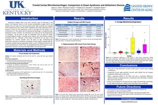
1. DS MC DS/AD OC AD
Groups
MicrohemorrhageCount
YC
Frontal Cortex Microhemorrhages: Comparison in Down Syndrome and Alzheimers Disease
James C. Fitch1, Paulina R. Davis1,2, Frederick A. Schmitt1,3, Elizabeth Head1,2
University of Kentucky, Sanders Brown Center on Aging1, Dept. of Pharmacology & Nutritional Sciences2,
Department of Neurology3, Lexington, KY, United States; 2 , Lexington, KY, United States;
Subjects: 46 samples individuals with DS, AD, DS with AD, along with their age matched
controls.
Brain Tissue Preparation:
• Frontal cortex brain tissue was used from each case.
• Tissue was sectioned 50 um thick using a vibratome.
Prussian Blue Staining and MH Quantification:
• A solution of potassium ferrocyanide in acidic solution was used to stain iron rich
hemoglobin blue in the frontal cortex tissue
• Only iron in the extracellular matrix is colorized (indicative of MH).
• Must be within 2 cell diameters of a blood vessel to be considered an MH.
Sample Analysis:
• 6 images were captured at 20X magnification (3 superficial layer and 3 of corresponding
deep layer) and MH counted for each tissue
• Data is represented as total MH count per case.
Alzheimer’s disease (AD) is the most common cause of dementia in the
elderly. The hallmark lesions of AD include neurofibrillary tangles (NFTs) and
plaques that are made up of the β-amyloid protein (Aβ), resulting from cleavage
of the amyloid precursor protein (APP). Individuals with Down Syndrome (DS) or
trisomy 21 have an over expression of APP as the gene for this protein is on
chromosome 21. Thus adults with DS develop AD pathology in a progressive age-
dependent manner. Therfore, DS individuals are at high risk for the development
of dementia. By 40 years of age, all individuals with DS have full blown
neuropathological changes, including senile plaques and neurofibrillary tangles,
consistent with AD. However, clinical signs of dementia are not seen in DS
individuals for another decade, over 50 years of age. Cerebrovascular
dysfunction may be an important contributor to brain aging in people with DS.
Increased cerebral amyloid angiopathy (CAA) is seen in individuals with DS due
to the overexpression of APP. Thus, we hypothesized that there may be an age-
dependent increase in microhemorrhages (MH) in DS and due to AD.
Materials and Methods
• There was no age dependent increase in MH count in control cases ranging
2 to 88 years.
• Compared to their aged matched controls, both DS/AD and AD showed
noteworthy differences in MH count.
• When comparing DS/AD to AD there was also a important difference
between groups, showing that DS in conjunction with AD further increases
MH count more than AD.
• Clinical trials designed for DS with AD may target this pathology specifically,
or that adverse responses related to MH may need to be considered
Introduction Results
Conclusions
Acknowledgements
Funded by NIH/DHHS xxx/xxx
Results
Future Directions
• Future directions of this study will investigate the association between Aβ
accumulation in CAA to the frequency of MH.
• By looking for IgG in different brain tissue samples, the type and strength of the
immune response can be learned to gain a better understanding of the response to
diseases such as DS and AD.
2. Representation MH Count From Each Group
Table 1: Grouping used in study. Average ages, post mortem interval, and average bleed
count per frame. *() indicates standard deviation.
Group N Age* PMI* MH Count*
Young controls (YC) 6 17.5 (16.2) 14.0 (7.4) 0.3 (0.5)
Middle aged controls (MC) 9 50.3 (9.1) 15.5 (6.1) 2.9 (5.9)
Old controls (OC) 8 81.0 (4.7) 3.4 (3.5) 2.0 (2.3)
Young DS (DS) 7 16.9 (16.9) 20.8 (12.8) 0.7 (1.3)
DS with AD (DS/AD) 8 53.4 (7.7) 9.3 (7.6) 17.0 (11.0)
Sporadic AD (AD) 7 83.3 (5.5) 5.0 (2.4) 10.9 (9.3)
Figure 1: Prussian Blue stained tissue representing average MH counts for each study
group. YC)-Young Controls, MC)-Middle Aged Controls, OC)-Old Controls, DS)-Young Down
syndrome, DS/AD) Down syndrome with Alzheimer’s disease, AD)-Sporadic Alzheimer’s
disease. Arrows indicate MH , arrow head indicate the location of blood vessels.
Subject Groups and MH Counts
Figure 2: A significant difference in MH counts is seen when comparing, DS/AD
(p=<0.05), to all groups. Both DS/AD and AD showed a significant difference in MH count
from their aged matched controls (p=<0.001) and (p=<0.018) respectively. Most notably,
there was a significant difference between DS/AD and AD (p=<0.029).
Identifying Microhemorrhages
Figure 1: Illustration of MH labeled by Prussian Blue. It is within appropriate proximity of blood
vessels (two cell diameter) and is punctate in nature. Arrows indicate MH, Arrowheads indicate
blood vessels
DS/AD
YC
MC
OC
DS
AD
Tissue Sample and Preparation
DS/AD
4. Average Microhemorrhage Count