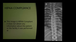
This document provides an evaluation of an AP thoracic spine x-ray image based on several criteria:
1. The image meets HIPAA compliance standards but is missing patient and facility information.
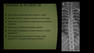
2. The anatomical marker is correctly placed but post-processing was likely used.
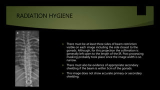
3. Radiation hygiene standards for beam restriction and shielding are not met.
4. The image shows the correct position/projection for a thoracic spine x-ray.
5. There are no artifacts but the part is slightly rotated and not optimally centered.
6. Image quality could be improved with better positioning, centering, and use of a compensating filter.
7. The image meets minimum acceptance standards