2. homeostasis all depend on cell-ECM interactions (34). Such in-
teractions involve adhesion receptors (e.g., integrins [35]), inter-
cellular adhesion complexes (e.g., tight junctions [36]), and extra-
cellular polysaccharides (e.g., glycosaminoglycans [37]), which
activate cellular receptors. In contrast, the bacterial ECM has long
been thought to function merely as a passive extracellular scaffold
that holds the biofilm together and protects resident cells from
environmental stresses.
In this review, we first describe the structural roles of different
exopolymeric substances that constitute the ECM of various bio-
films. Then, we focus on studies that demonstrate the role of some
of these ECM components in regulating genetic programs of the
biofilm cells (Fig. 1). The examples presented in this review are
divided according to the three stages of biofilm formation (38),
i.e., (i) attachment, exploration of the surface, monolayer forma-
tion, and aggregation, (ii) 3D structure development and pattern-
ing, and (iii) dispersal.
ECM COMPONENTS IN BACTERIAL BIOFILMS
The most extensively studied biofilm ECM components are car-
bohydrate-rich polymers (i.e., extracellular polysaccharides or ex-
opolysaccharides), proteins, and nucleic acids (25).
Exopolysaccharides. Exopolysaccharides generated by bacte-
ria have been recognized to significantly impact bacterial viru-
lence and promote capsule formation. Various genetic analyses
have provided strong evidence that biofilm exopolysaccharides
play a fundamental structural role in different bacterial species.
Mutants defective in the production of exopolysaccharides display
severe defects in biofilm formation and in achieving complex bio-
film architecture. Common bacterial exopolysaccharides include
cellulose (39–43) and the staphylococcal polysaccharide intercel-
lular adhesin (PIA) (44). PIA-related polymers are produced by
Staphylococcus epidermidis and S. aureus (45, 46) and by several
Gram-negative bacterial species (47–52). In S. epidermidis and S.
aureus, PIA synthesis is mediated by the icaADBC operon (53–55).
The PIA molecule contains both positive and negative charges,
which are important for its adhesive properties (44). Many staph-
ylococcal isolates lack ica and are still capable of producing bio-
films, indicating alternative routes of biofilm formation (see be-
low).
Many bacterial species are capable of producing several differ-
ent exopolysaccharides, simultaneously or differentially, as a
function of environmental factors or the genetic background of
the specific strain. For instance, the ECM of P. aeruginosa biofilms
can contain three exopolysaccharides: alginate, Psl, and Pel (56).
Alginate is a polymer of manuronic acid and guluronic acid,
and its synthesis is mediated by the alg operon, as well as by 12
additional genes (57). P. aeruginosa isolates from lungs of cystic
fibrosis (CF) patients overproduce alginate, resulting in a mucoid
colony phenotype. Alginate is not essential for biofilm formation,
as the laboratory strains of P. aeruginosa PAO1 and PA14 do not
produce alginate and are capable of forming a submerged biofilm
in a microtiter plate assay (58). However, mutants in alginate bio-
synthesis form biofilms with an altered architecture compared to
the parental strains (59, 60).
Pel and Psl, the other two exopolysaccharides produced by P.
aeruginosa, play redundant roles in defining ECM structure (61).
Psl is composed of mannose, rhamnose, and glucose and is syn-
thesized by the gene products of the pslA-O operon (62–65). Pel is
FIG 1 Signals from the ECM during biofilm development. (Bottom panel) Scheme of the different stages of biofilm development. (I) Attachment, monolayer
formation, and aggregation. (II) 3D structure development and patterning. (III) Dispersal. (Top panel) (A) P. aeruginosa PAO1 deposits trails of high local
concentrations of ECM (in green) that attract other cells and induce further ECM production (114, 128). (B) In B. subtilis, inhibition of the rotation of the flagella,
e.g., by the viscous environment of the ECM, induces ECM production (150, 151). (C) TasA (brown), a structural amyloid in B. subtilis ECM, can be toxic to
vegetatively growing B. subtilis cells (80). (D) During B. subtilis biofilm development, ECM induces emergence of different cell subpopulations: motile cells,
ECM-producing cells, and sporulating cells (31). (E) In S. aureus PSMs, peptides (purple) can create structural amyloid fibers (85) but have a destabilizing effect
on the biofilm in their monomeric form (176). (F) In C. crescentus biofilms, eDNA induces dispersal by inhibiting the reattachment of the mature stalked cell by
binding to the exopolysaccharides of the holdfast (dark brown) (175).
Meeting Review
July 2015 Volume 197 Number 13 jb.asm.org 2093Journal of Bacteriology
onJuly26,2015byWEIZMANNINSTOFSCIENCEhttp://jb.asm.org/Downloadedfrom
3. a glucose-rich polymer, and its synthesis is mediated by the pelA-G
operon (66, 67). As the PAO1 and PA14 laboratory strains do not
produce alginate, they rely on Pel and Psl production for their
biofilm formation (58). However, the ECM of PAO1 contains
mostly Psl, while PA14 cannot produce Psl and its primary exopo-
lysaccharide is Pel (61, 67–69). In addition to its structural role, Psl
is specifically important for surface attachment (61, 70).
The biofilm of the B. subtilis soil bacterium is strengthened by
several exopolysaccharide polymers as well. These exopolysaccha-
rides are produced by the epsA-O operon and are composed of
glucose, galactose, and N-acetylgalactosamine (71). Colonies of
mutants in the epsA-O operon are not as structured and wrinkled
as the wild-type colonies (72). When B. subtilis is grown in su-
crose-rich growth medium, it is also capable of producing levan, a
fructan biopolymer, via the sacB-yveB-yveA levansucrase tricis-
tronic operon (73).
Proteinaceous components. The proteinaceous components
of the biofilm ECM are especially intriguing. We first focus on
bacterial functional amyloids. Amyloids are insoluble fibrous ag-
gregates of proteins that contain parallel beta sheets, first identi-
fied in human neurodegenerative diseases (74). In bacteria, how-
ever, amyloid fibers were found to be the major proteinaceous
component of the microbial ECM and are produced by both
Gram-negative and Gram-positive bacterial species. In Gram-
positive bacteria, the best-characterized functional amyloids are
the TasA amyloid fibers, produced by B. subtilis (75–77). The TasA
amyloid fibers are attached to the cell wall and mediate cell-to-cell
adhesion in conjunction with other extracellular components
(76). TapA, an additional protein encoded by the tapA-sipW-tasA
operon, is a minor extracellular component incorporated within
the TasA fibers (78). SipW, the product of the third gene in the
tapA-sipW-tasA operon, is a signal peptidase that processes both
TasA and TapA to their mature forms (79–82). SipW also pro-
motes adherence of B. subtilis to surfaces (75, 83, 84). This second
function of SipW is independent of its function in TasA and TapA
maturation and involves a distinct signal peptidase domain (84).
Functional amyloids are surprisingly common structural ECM
components in a variety of bacterial biofilms, including S. aureus,
Pseudomonas species, Salmonella enterica serovar enteritidis, and
E. coli (85–89).
Amyloid fiber proteins, like TasA, show self-assembly proper-
ties. BslA, another protein component in the ECM of B. subtilis,
also self-polymerizes into a structural element (90, 91). BslA is a
cell surface-associated amphiphilic protein which is reminiscent
of hydrophobin of filamentous fungi (90). BslA forms a hydro-
phobic coat on the surface of B. subtilis biofilms that may enhance
protection against environmental insults (90).
However, not all proteinaceous components of a biofilm are
functional amyloids. Cell-cell and cell-host tissue contacts within
a biofilm can be mediated by surface adhesins. Adhesins are sur-
face proteins of bacteria that promote the adhesion of cells to the
host tissues. Among them are S. aureus biofilm-associated pro-
teins Bap and SasG (92) and fibronectin binding proteins FnBPA
and FnBPB (93). Intriguingly, FnBPA and FnBPB have been
shown to stimulate integrin signaling and actin rearrangements in
host cells, supporting the notion of ECM-cell signaling between
bacteria and their host (94). In V. cholerae, the ECM is composed
of three structural proteins—RbmA, RbmC, and Bap1 (95–97)—
and the exopolysaccharide Vibrio polysaccharide (VPS). Berk et al.
(98) simultaneously visualized these proteins during biofilm de-
velopment in a chambered cover glass. Interestingly, the proteins
exhibited distinct localization patterns and were not distributed
uniformly throughout the biofilm. During the development of the
biofilm structure, Berk et al. noted that the cells arranged in de-
fined clusters and were encapsulated by Bap1 and RmbC, which
interacted with the VPS exopolysaccharide. In contrast, RmbA
was found only within the cell clusters and was essential for their
formation. Taken together, these observations indicate that each
structural biofilm protein in V. cholerae plays a specific role.
eDNA. Extracellular genomic DNA (eDNA) was found to be
an important structural component in many bacterial biofilms
(99, 100). Addition of DNase to growing or mature biofilms of
various bacterial species results in inhibition of biofilm formation
or disruption of the established biofilms (101). In S. aureus, S.
epidermidis, V. cholerae, and P. aeruginosa PAO1, the DNase-
driven disruption of established biofilms was dependent on bio-
film age; young biofilms were more sensitive to DNase than older
biofilms (102–105). Thus, eDNA is important for the structure of
the young biofilm but its role is later taken over by other exopo-
lymers (101). The chemical nature of the long, charged DNA mol-
ecule is thought to modulate the cell surface properties and to
promote cell-to-cell and cell-to-surface adhesion (100). In addi-
tion, eDNA was shown to interact with other ECM components,
such as exopolysaccharide and protein components, which has
been suggested to add to structure stability (101). For instance,
type IV pili (T4P) in P. aeruginosa can bind to eDNA and in this
manner may mediate cell attachment to the eDNA scaffold (106).
eDNA is released to the ECM from lysed cells or via eDNA secre-
tion mechanisms suggested to exist in certain species (99). In S.
aureus, the mechanism that controls cell lysis was shown to be
analogous to the bacteriophage holin-antiholin system (107). Ho-
lins are membrane proteins that facilitate access of endolysins to
the cell wall, leading to peptidoglycan cleavage and to cell lysis
(108). Antiholins are proteins that inhibit holin activity. The S.
aureus CidA and LrgA proteins act as holin and antiholin, respec-
tively (109–113). Deletion of the cidA gene or the lrgAB operon
results in altered cell lysis and disrupted biofilm structure, medi-
ated by a change in the amount of released eDNA (102, 113).
The different components of the ECM, i.e., exopolysaccha-
rides, proteins, and eDNA, were recently demonstrated to partic-
ipate in several nonstructural roles, which are discussed in length
in the following sections.
ECM SIGNALING DURING BACTERIAL EXPLORATION OF THE
SURFACE
In many cases, bacterial biofilms are surface associated and are sug-
gested to require a stimulus-response mechanism to coordinate be-
tween surface attachment and ECM production. Thus, it is not sur-
prising that bacteria developed numerous ECM-derived signaling
mechanisms that induce biofilm initiation (Fig. 1, panel I).
In the P. aeruginosa PAO1 strain, the presence of exopolysac-
charide Psl was found to act as a signal that activates the produc-
tion of bis-(3=-5=)-cyclic dimeric GMP (c-di-GMP) (114). c-di-
GMP is a second messenger prevalent in numerous bacterial
species (115). It is a key regulator in the planktonic-to-biofilm
switch, mediated by activation of biofilm production and repres-
sion of motility (115–117). Intracellular c-di-GMP concentra-
tions are tightly regulated by the counteractive action of diguany-
late cyclases that produce c-di-GMP from two GTP molecules and
of phosphodiesterases that break it down to 5=-phosphoguanylyl-
Meeting Review
2094 jb.asm.org July 2015 Volume 197 Number 13Journal of Bacteriology
onJuly26,2015byWEIZMANNINSTOFSCIENCEhttp://jb.asm.org/Downloadedfrom
4. (3=-5=)-guanosine (116). There are 41 predicted diguanylate cy-
clase and phosphodiesterase genes in P. aeruginosa PAO1, many of
which contain domains known to participate in signal sensing and
transduction (118). Two important c-di-GMP receptors are in-
volved in biofilm formation: FleQ and PelD (119, 120). FleQ is a
repressor of the pelA-G and pslA-O operons, which drive exopo-
lysaccharide synthesis, and of cdrA, which encodes an adhesin
(119, 121, 122). Upon c-di-GMP binding, FleQ repression is re-
lieved, leading to induction of the pelA-G, pslA-O, and cdrA oper-
ons. PelD is part of the biosynthetic pathway of the Pel polysac-
charide (120). Its binding to c-di-GMP was found to be essential
for Pel production (120). Thus, through binding to FleQ and
PelD, high levels of c-di-GMP lead to induction of ECM produc-
tion, while low levels lead to repression (123, 124). Interestingly,
Irie et al. (114) found that the presence of Psl leads to increased
intracellular c-di-GMP levels. Both overexpression of Psl, under
the control of an arabinose-inducible promoter, and addition of
purified Psl from another biofilm enhanced c-di-GMP levels by
up to 2-fold (114). Irie et al. showed that Psl-induced c-di-GMP
elevation depended on the activity of SadC and SiaD, two digua-
nylate cyclases, which have been previously established as critical
for biofilm maturation (125, 126). As mentioned above, high c-di-
GMP levels lead to activation of ECM production. Thus, these
findings present a positive-feedback loop in which sensing of the
newly produced ECM components leads to activation of further
ECM production. The fact that Psl can act when applied exoge-
nously demonstrates that it can amplify ECM production in
neighbor cells that are not yet producing ECM.
Apart from its amplificatory function, Psl was found to play a
critical role in directing the cells at the forefront of the P. aerugi-
nosa PAO1 biofilm. During the initial stages of microcolony for-
mation in flow cells, P. aeruginosa PAO1 cells deposit trails of Psl
as they move along the surface, using T4P twitching motility (127,
128). In twitching motility, T4P extend, attach to the surface, and
retract, leading to cell progression in the retraction direction (129,
130). Using cell-tracking algorithms, Zhao et al. (128) demon-
strated that these Psl trails influence the surface motility of cells
that later encounter these trails, encouraging them to follow the
same routes. ⌬pslD mutant cells explore much more of the surface
than wild-type cells, as they lack Psl traffic signs. On the other
hand, a Psl-overproducing mutant is limited to fewer trajectories
as a result of the higher local concentration of Psl. Furthermore,
Zhao et al. showed that, after cell division, the tendency of daugh-
ter cells to stay near the mother cell depends on the presence of Psl.
Cells of the ⌬pslD mutant strain left the mother cell more fre-
quently than wild-type strain cells, while cells of the ECM-over-
producing mutant left the mother cell less frequently. Deletion of
pilA, the gene that encodes the T4P structural protein, resulted in
a reduction of the surface area that was explored. This behavior
indicates that bacterial microcolony initiation is self-organizing
and that local concentrations of Psl are used to induce positive-
feedback loops. Cells then tend to reside in areas with significant
Psl accumulation. Together with the aforementioned evidence
that high Psl concentrations serve as a signal to produce more Psl,
those results imply that, during the initial phase of biofilm forma-
tion, a group of pioneer cells locally increases ECM concentrations
to attract more cells that later contribute to the ECM production
and serve as the founding population of the microcolony. Note
that both studies used the PAO1 P. aeruginosa laboratory strain,
which produces mainly Psl. As mentioned above, PA14, the other
well-studied P. aeruginosa laboratory strain, does not produce Psl,
and its primary exopolysaccharide is Pel. It will be interesting to
study whether PA14 shows similar behavior given the suggested
structural redundancy between the two exopolysaccharides (61)
or, alternatively, whether cell guidance is a unique property of Psl.
In a different study, Gloag et al. (131) showed that eDNA pro-
motes cell migration through interconnected channels in P.
aeruginosa biofilms. In this experimental system, P. aeruginosa
cells created channels on the surface of the semisolid substrate,
which was solidified by the addition of gellan gum. The channels
assembled into a network that facilitated colony expansion. The
formation of the channel network was achieved by the coordi-
nated movement of leader groups followed by cells that traced the
same trails. eDNA was found to be necessary for this tracing, and
addition of DNase caused the leader groups to remain isolated and
hampered the construction of the channel network and colony
expansion. Moreover, DNA strands were found to be aligned with
the direction of movement of the cells, possibly aiming the cells in
the correct direction. The cells moved using T4P twitching motil-
ity. T4P were shown to bind DNA, and it was suggested that T4P
eDNA binding was responsible for the directed movement of cells.
Importantly, the involvement of T4P-eDNA interaction in P.
aeruginosa PAO1 biofilm structure in flow cells was demonstrated
previously (132–134). When grown in flow cells with glucose min-
imal medium, P. aeruginosa creates typical mushroom-shaped
structures, with a stalk and a cap (132). In these structures, eDNA
was found to be located at the stalk (135). The formation of the cap
required T4P-dependent migration of a subpopulation of cells
(133), which was also shown to depend on the presence of eDNA
at the stalk (134). In these studies, eDNA plays a role in guiding
cell migration within the biofilm structure. Similarly to the Psl
example mentioned above, ECM signals, laid down by the cells in
the colony frontline, direct the cells in the back, allowing the col-
ony to expand to new territories.
ECM-guided cell migration resembling that of P. aeruginosa
was observed in the bacterium Myxococcus xanthus (136). M. xan-
thus is a Gram-negative soil bacterium, demonstrating multiple
types of social behaviors, and thus is used as an important model
of bacterial development (137). One of these social behaviors is
social motility (S-motility), which occurs during growth on nutri-
ent-rich medium, on which M. xanthus cells collectively migrate,
creating a swarm. S-motility was found to be driven by T4P
twitching motility (138, 139). Interestingly, during swarming, M.
xanthus cells seem to follow the same routes as precedent cells,
resembling P. aeruginosa (140). M. xanthus T4P participate in
S-motility by binding exopolysaccharides that are produced dur-
ing swarming, causing retraction of the pili (136). These exopoly-
saccharides cover the cell surface, and in mutants lacking them,
S-motility is inhibited (136, 141). Thus, movement is possible
only when T4P attach to adjacent cell surface exopolysaccharides.
It was suggested that a portion of the surface exopolysaccharide is
shed during cell migration (142), possibly creating trails that sub-
sequent cells follow. In the biofilm state of M. xanthus, also termed
the fruiting body, the exopolysaccharides may create a network
between cells, which might facilitate T4P twitching motility (143,
144). Interestingly, production of exopolysaccharides depends on
the presence of assembled T4P (145, 146). This might imply a
positive-feedback loop similar to Psl-induced ECM production in
P. aeruginosa (147).
Recently, an example of posttranslational self-amplification of
Meeting Review
July 2015 Volume 197 Number 13 jb.asm.org 2095Journal of Bacteriology
onJuly26,2015byWEIZMANNINSTOFSCIENCEhttp://jb.asm.org/Downloadedfrom
5. exopolysaccharide production was discovered in B. subtilis (148)
(Fig. 2). EpsA and EpsB constitute a tyrosine kinase that was found
to undergo autophosphorylation and remain inactive in the ab-
sence of a signal. However, in the presence of B. subtilis exopoly-
saccharides, autophosphorylation was repressed and the EpsAB
kinase phosphorylated EpsE, a glycosyltransferase in the B. subtilis
exopolysaccharide biosynthetic pathway. EpsE phosphorylation
was suggested to promote production of exopolysaccharides. The
binding of the EpsA extracellular domain to exopolysaccharides
was found to be specific to B. subtilis exopolysaccharides. Interest-
ingly, the extracellular domain of the S. aureus CapA (an EpsA
homolog) bound specifically the S. aureus exopolysaccharide PIA.
Similarly to the P. aeruginosa Psl, these findings present a mecha-
nism for positive-feedback loop regulation of exopolysaccharide
production, enabling amplification of the signal and the construc-
tion of the biofilm. The species specificity of the signal further
emphasizes the idea of directed regulation.
Such regulation of glycosyltransferases is only one example of
ECM signaling during biofilm development in B. subtilis. The pro-
duction of exopolysaccharides creates a highly viscous environ-
ment (149), which can be sensed by the bacterial cell flagella. In B.
subtilis, the attachment step was suggested to be governed and
amplified by mechanosensing (Fig. 2). B. subtilis biofilm develop-
ment requires the activation of three transcriptional regulators:
ComA, Spo0A, and DegU (38). Recent studies showed that the
disruption of flagellar rotation increased the DegUϳP level (150,
151). Phosphorylated DegU triggers the biosynthesis of ␥-poly-
glutamic acid (␥-PGA), a unique ECM polymer composed of glu-
tamic acid and produced by the pgs operon (152–154). Deletion of
either MotA or MotB, which together form the stator part of the
flagellar motor, resulted in overproduction of ␥-PGA and a mu-
coid colony phenotype. The same phenotype was achieved by
other methods that hampered flagellar rotation, such as (i) muta-
tions in MotB that disrupted proton flux through the motor, (ii)
addition of specific antibodies against the flagellar filament, and
(iii) overexpression of EpsE, which can function as a molecular
clutch that separates the flagellar stator and rotor compartments
(155, 156). Overproduction of ␥-PGA required the presence of
DegU and DegS (150), as well as of proteins involved in flagellar
filament assembly (151). The results of the studies presented here
suggest that hampering the rotation of the flagella during initial
attachment results in altered gene expression. We propose that the
flagella can sense the viscous ECM environment in later stages of
microcolony formation by the same mechanism. Viscous envi-
ronments are thought to increase flagellar motor torque (157),
and a high viscosity level was shown to affect gene expression in V.
cholerae (158, 159), P. aeruginosa (125, 160), and Proteus mirabilis
(161, 162). In order to examine this hypothesis in B. subtilis, it will
be interesting to measure the levels of ␥-PGA production in cells
grown in environments of various viscosities, perhaps by supply-
ing different concentrations of ECM exopolysaccharides. This sig-
naling pathway could constitute a positive-feedback loop, as the
highly viscous environment of the ECM can halt the flagella. Halt-
ing the flagella then promotes production of more ECM, which
further amplifies viscosity. Several mechanosensory mechanisms
leading to initiation of biofilm formation have been described in
numerous bacterial species, including P. aeruginosa, C. crescentus,
and V. cholerae. A recent review summarizes the current knowl-
edge of these mechanisms (157).
During the initial stages of biofilm formation, the population is
still divided between motile planktonic cells and sessile surface or
ECM-associated cells. As the biofilm matures, the motile cell pop-
FIG 2 ECM-cell signaling induces positive-feedback in ECM production. There are three ECM-derived signals that induce ECM production in B. subtilis
biofilms as follows. (A) Disruption of flagellar rotation, which can occur in the viscous ECM environment, causing DegS-dependent phosphorylation of DegU
and induction of the pgs operon, yielding ␥-PGA production, and of bslA, which forms the biofilm hydrophobic coat (150, 151). (B) The KinD kinase senses high
osmotic pressure and phosphorylates Spo0A. Phosphorylated Spo0A leads to the activation (low Spo0AϳP) or deactivation (high Spo0AϳP) of the epsA-O
operon for production of exopolysaccharides and of the tapA-sipW-tasA operon, inducing TasA amyloid fiber production (163–165). (C) EpsA-specific binding
to B. subtilis exopolysaccharides causes inhibition of EpsAB autophosphorylation and phosphorylation of EpsE, which may then lead to increased ECM
production (148).
Meeting Review
2096 jb.asm.org July 2015 Volume 197 Number 13Journal of Bacteriology
onJuly26,2015byWEIZMANNINSTOFSCIENCEhttp://jb.asm.org/Downloadedfrom
6. ulation gradually diminishes and most of the cells become sessile.
During the initial stages of B. subtilis biofilm formation, an un-
usual selection for ECM producers occurs. The amyloid fibers
forming TasA, a structural component of the B. subtilis biofilm
(76), also serve as a secreted antibacterial protein (80). Intrigu-
ingly, when B. subtilis cells were grown planktonically, they dis-
played sensitivity to TasA (80). Thus, TasA production may se-
verely retard growth of planktonic motile cells in the initial stages
of biofilm formation, shifting the population toward immobility
and ECM association, which are crucial for further biofilm devel-
opment. While this is a tempting speculation, the sensitivity of
planktonic cells to TasA needs to be tested in the biofilm environ-
ment.
Overall, the examples described here present different and
complementary mechanisms for ECM sensing. The sensing of P.
aeruginosa Psl, and of B. subtilis exopolysaccharides by EpsAB,
relies on a species-specific signal. The cells recognize and respond
to the exact chemical properties of specific ECM components. In
contrast, eDNA sensing by P. aeruginosa and the possible flagellar
mechanosensing of the viscous environment in B. subtilis present
signals that are more global. General signals may provide an ad-
vantage, as they can be sensed by many types of bacteria. The
importance of such universal physical cues is prominent in mul-
tispecies biofilms in which various bacterial species must coordi-
nate their behavior.
ECM SIGNALING DURING 3D BIOFILM STRUCTURE
DEVELOPMENT
The effects of ECM signaling during biofilm development can be
quite dramatic. Analysis of the spatiotemporal gene expression
profiles of a B. subtilis ECM mutant lacking both exopolysaccha-
rides and TasA fibers demonstrated alterations in the number and
localization of motile cells, ECM producers, and sporulating cells
within the mature colony (31) (Fig. 1, panel II, and Fig. 2). Spe-
cifically, reduced expression of the motility reporter was noted.
The few cells that expressed the motility reporter were not local-
ized at the base of the colony as observed in wild-type biofilms,
suggesting that the ECM plays a critical role in regulating existence
and localization of motile cells (31; N. Steinberg and I. Kolodkin-
Gal, unpublished data). Interestingly, transcription of the two
ECM operons, epsA-O and tapA-sipW-tasA, was dramatically in-
creased in the ECM mutant (31), suggesting that the B. subtilis
ECM serves a dual signaling role; it positively posttranslationally
regulates ECM production, as mentioned in the previous section
(148), and negatively regulates ECM production at the transcrip-
tional level. ECM-driven negative feedback on its own production
may play an important role in adaptation to the biofilm environ-
ment. In this manner, cells do not waste resources by constantly
producing matrix, as a positive-feedback loop would predict. It
would be interesting to test whether accumulation of sufficient
matrix suppresses further matrix expression, thus overcoming the
effects of the positive-feedback loop.
Strikingly, virtually no sporulating cells were observed in the B.
subtilis ECM mutant colony, at a time point at which wild-type
biofilm had a high percentage of sporulating cells (31, 163). A
mixture of the ECM mutant cells with sporulation mutant cells
was capable of producing intact ECM, resulting in colonies with
wild-type architecture and restored sporulation and suggesting
that the ECM induces sporulation as a trans-acting signal.
What type of signal does the ECM convey that causes the ob-
served effects? One attractive hypothesis claims that reduced ECM
production supports increased cell growth. If the energy required
to synthesize the ECM components is instead channeled toward
other metabolic pathways, the ECM mutant cells might continue
growing for longer periods of time, delaying the initiation of spo-
rulation. However, when the total number of cells in the biofilm of
the ECM mutant was compared to the number of cells present in
the wild-type biofilm, over the course of development, there were
significantly more cells in the biofilms formed by the wild-type
strain than in those formed by the ECM mutant (163). When
ECM mutant cells were visually compared to wild-type cells under
the microscope, no differences in cell size were observed. There-
fore, the delay observed in sporulation could not be explained by a
prolonged period of growth of the ECM mutant cells (31). Can an
ECM-generated chemical cue promote B. subtilis development
and, more specifically, sporulation? Mutants with mutations in
two different ECM components, the TasA protein and the exopo-
lysaccharides, were both defective in sporulation when grown un-
der biofilm-inducing conditions but not when grown in dispersed
cultures (163). However, TasA fibers and exopolysaccharides have
strikingly different chemistries; thus, it is not plausible that the
ECM generates a chemical signal that regulates sporulation.
Another hypothesis as to the lack of specificity in the signal
source is that a nonspecific physical cue, for instance, increasing
osmotic pressure during the accumulation of ECM, might trigger
sporulation. Rubinstein et al. (164) exploited unique properties of
polymeric solutions to differentiate between the effects of multiple
physical parameters. They tested the effects of both polyethylene
glycol (PEG) and dextran over a range of molecular sizes and
concentrations as well as various concentrations of the dextran
subunit dextrose. The expression levels of ECM and sporulation
genes strongly correlated with the osmotic pressure exerted by the
various polymer solutions. Polymer solutions that exerted suffi-
cient osmotic pressure complemented the sporulation defect of an
ECM mutant in trans, as did purified polymers from P. aeruginosa
ECM, added exogenously to the B. subtilis ECM mutants. Both
sporulation and ECM production are regulated by the master reg-
ulator Spo0A. While low levels of Spo0AϳP trigger ECM produc-
tion, high levels trigger sporulation. In this case, the osmotic pres-
sure exerted by both exopolysaccharides and amyloid fibers was
shown to be sensed by KinD, as a kinD mutant did not respond to
increased osmotic pressure (164). KinD is a membrane histidine
kinase that phosphorylates Spo0A and was shown to affect both
sporulation and matrix production, with activity redundant with
that of KinC (163, 165). A deletion of kinD suppresses the sporu-
lation defect of the ECM mutants (163), suggesting that KinD is
the sensor of the ECM. Importantly, KinD has also been shown to
specifically respond to a broad range of plant exopolysaccharides
(166), as well as to several small molecules (167, 168). It is highly
plausible that the specificity of this membrane kinase dramatically
changes as a function of the mediator that it binds, such as the Med
lipoprotein (169). Alternatively, it is plausible that KinD senses
somewhat generic signals such as membrane stress, caused by in-
creased osmotic pressure, antibiotics, or membrane interactions
with high concentrations of polymers.
Taken together, the findings in B. subtilis biofilms present a
case for several arguments. First, sensing the ECM may initiate a
local positive-feedback loop by inducing a minor increase in the
level of Spo0AϳP, thereby promoting further ECM production.
The gradual increase in ECM-driven local osmotic pressure can
Meeting Review
July 2015 Volume 197 Number 13 jb.asm.org 2097Journal of Bacteriology
onJuly26,2015byWEIZMANNINSTOFSCIENCEhttp://jb.asm.org/Downloadedfrom
7. drive neighboring bacteria to induce their own ECM production
in mixed communities. When sufficient ECM is accumulated, fur-
ther phosphorylation of Spo0A by KinD promotes sporulation
and shuts off ECM production. Second, aside from sporulation,
several severe defects in other developmental programs (e.g.,
competence and motility) were observed in these ECM mutants
(31, 32). It is unlikely that these defects can be solely attributed to
Spo0A-phosphorylation or to the structural role of the ECM. We
suggest that several independent signaling pathways are activated
by the ECM during the 3D patterning of B. subtilis biofilms.
Spatial arrangements of different cell types in the biofilm 3D
structure were also demonstrated in E. coli and P. aeruginosa (170–
172). Specifically, flagellated cells were observed at the bottom and
exterior parts of E. coli biofilms grown on agar (170), as was ob-
served in B. subtilis biofilms. In addition, ECM-producing cells,
which produce Curli, the amyloid fiber of E. coli, were located at
the top of the colony (170). In the context of this review, it will be
interesting to examine the influence of the presence of each of the
ECM components of E. coli and P. aeruginosa on the quantity and
spatial organization of the different cell types in the biofilm.
ECM SIGNALING DURING THE LATE STAGES OF THE
BIOFILM CYCLE
As the biofilm matures, resources become limited and waste prod-
ucts accumulate. Bacteria incapable of escaping the biofilm be-
come trapped in what evolves into a death trap (173). Therefore, at
a certain time, it is beneficial for the constituent cells of the biofilm
to disperse (174). Effective probing of ECM maintenance level and
density enables cells to determine the ideal time for dispersal (Fig.
1, panel III).
Dispersal triggered by an ECM polymer was demonstrated in
C. crescentus biofilms, which are composed of swarmer cells and
sessile stalked cells. The stalked cells have a polar adhesin com-
posed of polysaccharides, called the holdfast, which is required
both for permanent adhesion to surfaces and for biofilm forma-
tion. As cell death within the biofilm commences, eDNA accumu-
lates. Berne et al. (175) have shown that eDNA inhibits submerged
C. crescentus biofilm formation, in static cultures and in flow cells.
When applied externally, the purified eDNA effectively blocked
biofilm formation. This robust hindrance occurred both when the
eDNA was purified from the spent medium and when genomic
DNA was purified from C. crescentus cells. eDNA was found to
specifically bind the C. crescentus holdfast. This ligand-receptor
interaction specifically prevented the attachment of the newly
produced stalked cells to one another and to the surface. When
eDNA was added to established biofilms grown in flow cells, it did
not cause dispersal. However, the biomass of the biofilm remained
constant, indicating that the progeny of the attached cells dis-
persed. Specific targeting of nonattached cells in the mature bio-
film stimulates dispersal without the simultaneous destruction of
the biofilm. While eDNA-bound dispersing cells fail to efficiently
attach to a surface, they can produce progeny swarmer eDNA-free
cells that are capable of settling in a new environment.
In S. aureus, surfactant-like small peptides called phenol-solu-
ble modulins (PSMs) were recently found to form functional am-
yloid fibers that have a structural role in the S. aureus biofilm (85).
However, in their monomeric form, they were found to promote
biofilm disassembly by reducing the surface tension (176). Distin-
guishing between the structural role of the PSMs and their role in
dispersal of biofilms is challenging. Aggregation and disaggrega-
tion of PSMs are probably dynamic events occurring in parallel
during biofilm formation. Periasamy et al. (176) suggested a pos-
sible role of PSMs in local biofilm dispersal in flow cells, where
mutants incapable of producing PSMs formed thicker, denser,
and smoother biofilms. In addition, after 72 h in flow cells, the
wild-type strain showed waves of detachment and growth, while
the biofilm of the PSM mutants remained thick. PSM expression
was localized to specific areas of the biofilm, suggesting a signaling
role in pattern formation. Together, these findings suggest regu-
lation of biofilm dispersal via ECM components, where a struc-
tural element in the biofilm acts as a dispersal signal as well. PSMs
were also suggested to play a role in regulating their own expres-
sion, though the underlying mechanism is still unknown (85).
CONCLUSIONS AND FUTURE DIRECTIONS: HOW CELL-ECM
INTERACTIONS SHAPE MULTICELLULAR MICROBIAL
COMMUNITIES
This review presents evidence from a number of studies demon-
strating the pivotal role of the ECM in controlling gene expression
and cell behavior in bacterial biofilms, throughout the various
stages of biofilm formation and maturation.
During the initial stages of attachment, it is important for the
pioneer cell population to create a positive-feedback loop that will
lead to ECM and cell accumulation. As a biofilm is a multicellular
structure, a bacterial cell that approaches a surface must signal
other cells, or its daughter cells, to attach at locations close to its
attachment site and to start producing ECM components. As local
concentrations of ECM in the attachment area of the pioneer cells
rise, they can be used as a signal for other cells.
This was demonstrated in P. aeruginosa, as Psl and eDNA trails
being laid by the pioneer cells direct cell movement to these spe-
cific areas (128, 131). Psl in P. aeruginosa, and the B. subtilis ex-
opolysaccharide, are specifically sensed by each of these bacteria
and are used as a signal to increase ECM production, demonstrat-
ing a positive-feedback loop (114, 148). Another example is B.
subtilis, which uses its flagella as mechanosensory organelles that
might sense the ECM viscous environment; halting the flagellum
leads to the induction of ECM production (150, 151).
When the B. subtilis biofilm develops, ECM probing is crucial
for coordinating cell behaviors over large scales and to obtain a
proper structure formation. When ECM components are absent,
biofilm development is disrupted (31, 128, 131). Specifically, the
size and the spatial organization of different cell type subpopula-
tions in the biofilm are modified in ECM mutants compared to
wild-type cells (31). The defects in cell behavior can be explained
as a lack of ECM-derived regulatory cues. It will be interesting to
examine the possible role of the ECM in regulation of cell local-
ization and genetic program activation in other species (170–172).
As the biofilm reaches its last stages, ECM biochemistry, struc-
ture, and integrity serve as ideal probes of the physiological state of
the biofilm. ECM structural components that either collapse or
undergo active destruction during the dispersal stage may act as
dispersing signals. PSMs that create structural amyloid fibers in S.
aureus ECM (85) were shown to trigger dispersal in their mono-
meric form (176), suggesting that when the ECM is dismantled to
its building blocks, the breakdown products may induce dispersal.
eDNA, which functions as a structural component in many bac-
terial species (99, 100), was found to inhibit attachment of C.
crescentus cells by binding to their attachment organelle, the stalk
(175). Thus, eDNA from lysed cells can induce dispersal of the
Meeting Review
2098 jb.asm.org July 2015 Volume 197 Number 13Journal of Bacteriology
onJuly26,2015byWEIZMANNINSTOFSCIENCEhttp://jb.asm.org/Downloadedfrom
8. new progeny of the biofilm cells, releasing them to search for new
colonization sites.
In multicellular eukaryotes, numerous examples of ECM-
driven signals crucial for determination of both cell behavior and
correct tissue morphogenesis have been described. There is no
apparent reason to think that bacterial ECM would differ in this
aspect. ECM-derived signals may be a common feature of cell
communities surrounded by an extracellular matrix, as such an
arrangement allows probing of the local environment and dy-
namic adjustment of signals. The studies presented in this review
are undoubtedly just the tip of the iceberg—many more examples
of bacterial ECM signaling are sure to be discovered in the near
future.
ACKNOWLEDGMENTS
The Kolodkin-Gal laboratory is supported by ISF-icore grant 152/1, by a
research grant from Ayala Benjamin-Mashat, KAMIN program for R & D
of the Israeli chamber of commerce, and by the Angel-Fiavovich fund for
ecological research. I.K.-G. is a recipient of the Rowland and Sylvia Career
Development Chair.
REFERENCES
1. Kolter R, Greenberg EP. 2006. Microbial sciences: the superficial life of
microbes. Nature 441:300–302. http://dx.doi.org/10.1038/441300a.
2. Aguilar C, Vlamakis H, Losick R, Kolter R. 2007. Thinking about
Bacillus subtilis as a multicellular organism. Curr Opin Microbiol 10:
638–643. http://dx.doi.org/10.1016/j.mib.2007.09.006.
3. Costerton JW, Stewart PS, Greenberg EP. 1999. Bacterial biofilms: a
common cause of persistent infections. Science 284:1318–1322. http:
//dx.doi.org/10.1126/science.284.5418.1318.
4. Entcheva-Dimitrov P, Spormann AM. 2004. Dynamics and control of
biofilms of the oligotrophic bacterium Caulobacter crescentus. J Bacteriol
186:8254–8266. http://dx.doi.org/10.1128/JB.186.24.8254-8266.2004.
5. Stewart PS, Franklin MJ. 2008. Physiological heterogeneity in biofilms.
Nat Rev Microbiol 6:199–210. http://dx.doi.org/10.1038/nrmicro1838.
6. Xu KD, Stewart PS, Xia F, Huang CT, McFeters GA. 1998. Spatial
physiological heterogeneity in Pseudomonas aeruginosa biofilm is deter-
mined by oxygen availability. Appl Environ Microbiol 64:4035–4039.
7. Rani SA, Pitts B, Beyenal H, Veluchamy RA, Lewandowski Z, Davison
WM, Buckingham-Meyer K, Stewart PS. 2007. Spatial patterns of DNA
replication, protein synthesis, and oxygen concentration within bacterial
biofilms reveal diverse physiological states. J Bacteriol 189:4223–4233.
http://dx.doi.org/10.1128/JB.00107-07.
8. Wilking JN, Zaburdaev V, De Volder M, Losick R, Brenner MP, Weitz
DA. 2013. Liquid transport facilitated by channels in Bacillus subtilis
biofilms. Proc Natl Acad Sci U S A 110:848–852. http://dx.doi.org/10
.1073/pnas.1216376110.
9. Dietrich LE, Okegbe C, Price-Whelan A, Sakhtah H, Hunter RC,
Newman DK. 2013. Bacterial community morphogenesis is intimately
linked to the intracellular redox state. J Bacteriol 195:1371–1380. http:
//dx.doi.org/10.1128/JB.02273-12.
10. Kolodkin-Gal I, Elsholz AK, Muth C, Girguis PR, Kolter R, Losick R.
2013. Respiration control of multicellularity in Bacillus subtilis by a com-
plex of the cytochrome chain with a membrane-embedded histidine ki-
nase. Genes Dev 27:887–899. http://dx.doi.org/10.1101/gad.215244.113.
11. Kempes CP, Okegbe C, Mears-Clarke Z, Follows MJ, Dietrich LE.
2014. Morphological optimization for access to dual oxidants in bio-
films. Proc Natl Acad Sci U S A 111:208–213. http://dx.doi.org/10.1073
/pnas.1315521110.
12. Monds RD, O’Toole GA. 2009. The developmental model of microbial
biofilms: ten years of a paradigm up for review. Trends Microbiol 17:73–
87. http://dx.doi.org/10.1016/j.tim.2008.11.001.
13. Serra DO, Hengge R. 2014. Stress responses go three dimensional—the
spatial order of physiological differentiation in bacterial macrocolony
biofilms. Environ Microbiol 16:1455–1471. http://dx.doi.org/10.1111
/1462-2920.12483.
14. Davies D. 2003. Understanding biofilm resistance to antibacterial
agents. Nat Rev Drug Discov 2:114–122. http://dx.doi.org/10.1038
/nrd1008.
15. Fux CA, Costerton JW, Stewart PS, Stoodley P. 2005. Survival strate-
gies of infectious biofilms. Trends Microbiol 13:34–40. http://dx.doi.org
/10.1016/j.tim.2004.11.010.
16. Mah TF, Pitts B, Pellock B, Walker GC, Stewart PS, O’Toole GA. 2003.
A genetic basis for Pseudomonas aeruginosa biofilm antibiotic resis-
tance. Nature 426:306–310. http://dx.doi.org/10.1038/nature02122.
17. Stewart PS. 1994. Biofilm accumulation model that predicts antibiotic
resistance of Pseudomonas aeruginosa biofilms. Antimicrob Agents
Chemother 38:1052–1058. http://dx.doi.org/10.1128/AAC.38.5.1052.
18. Stewart PS. 2002. Mechanisms of antibiotic resistance in bacterial bio-
films. Int J Med Microbiol 292:107–113. http://dx.doi.org/10.1078/1438
-4221-00196.
19. Stewart PS, Costerton JW. 2001. Antibiotic resistance of bacteria in
biofilms.Lancet358:135–138.http://dx.doi.org/10.1016/S0140-6736(01)
05321-1.
20. Bryers JD. 2008. Medical biofilms. Biotechnol Bioeng 100:1–18. http:
//dx.doi.org/10.1002/bit.21838.
21. Heithoff DM, Mahan MJ. 2004. Vibrio cholerae biofilms: stuck between
a rock and a hard place. J Bacteriol 186:4835–4837. http://dx.doi.org/10
.1128/JB.186.15.4835-4837.2004.
22. Yildiz FH, Visick KL. 2009. Vibrio biofilms: so much the same yet so
different. Trends Microbiol 17:109–118. http://dx.doi.org/10.1016/j.tim
.2008.12.004.
23. Chen Y, Yan F, Chai Y, Liu H, Kolter R, Losick R, Guo JH. 2013.
Biocontrol of tomato wilt disease by Bacillus subtilis isolates from natural
environments depends on conserved genes mediating biofilm formation.
Environ Microbiol 15:848–864. http://dx.doi.org/10.1111/j.1462-2920
.2012.02860.x.
24. Rudrappa T, Czymmek KJ, Pare PW, Bais HP. 2008. Root-secreted
malic acid recruits beneficial soil bacteria. Plant Physiol 148:1547–1556.
http://dx.doi.org/10.1104/pp.108.127613.
25. Branda SS, Vik S, Friedman L, Kolter R. 2005. Biofilms: the matrix
revisited. Trends Microbiol 13:20–26. http://dx.doi.org/10.1016/j.tim
.2004.11.006.
26. O’Toole G, Kaplan HB, Kolter R. 2000. Biofilm formation as microbial
development. Annu Rev Microbiol 54:49–79. http://dx.doi.org/10.1146
/annurev.micro.54.1.49.
27. Sauer K, Camper AK, Ehrlich GD, Costerton JW, Davies DG. 2002.
Pseudomonas aeruginosa displays multiple phenotypes during develop-
ment as a biofilm. J Bacteriol 184:1140–1154. http://dx.doi.org/10.1128
/jb.184.4.1140-1154.2002.
28. Southey-Pillig CJ, Davies DG, Sauer K. 2005. Characterization of
temporal protein production in Pseudomonas aeruginosa biofilms. J
Bacteriol 187:8114–8126. http://dx.doi.org/10.1128/JB.187.23.8114
-8126.2005.
29. Lopez D, Vlamakis H, Kolter R. 2009. Generation of multiple cell types
in Bacillus subtilis. FEMS Microbiol Rev 33:152–163. http://dx.doi.org
/10.1111/j.1574-6976.2008.00148.x.
30. Chai Y, Chu F, Kolter R, Losick R. 2008. Bistability and biofilm for-
mation in Bacillus subtilis. Mol Microbiol 67:254–263. http://dx.doi.org
/10.1111/j.1365-2958.2007.06040.x.
31. Vlamakis H, Aguilar C, Losick R, Kolter R. 2008. Control of cell fate by
the formation of an architecturally complex bacterial community. Genes
Dev 22:945–953. http://dx.doi.org/10.1101/gad.1645008.
32. López D, Vlamakis H, Losick R, Kolter R. 2009. Paracrine signaling in
a bacterium. Genes Dev 23:1631–1638. http://dx.doi.org/10.1101/gad
.1813709.
33. López D, Vlamakis H, Losick R, Kolter R. 2009. Cannibalism enhances
biofilm development in Bacillus subtilis. Mol Microbiol 74:609–618.
http://dx.doi.org/10.1111/j.1365-2958.2009.06882.x.
34. Adams JC, Watt FM. 1993. Regulation of development and differenti-
ation by the extracellular matrix. Development 117:1183–1198.
35. Schwartz MA. 2001. Integrin signaling revisited. Trends Cell Biol 11:
466–470. http://dx.doi.org/10.1016/S0962-8924(01)02152-3.
36. Matter K, Balda MS. 2003. Signalling to and from tight junctions. Nat
Rev Mol Cell Biol 4:225–236. http://dx.doi.org/10.1038/nrm1055.
37. Entwistle J, Hall CL, Turley EA. 1996. HA receptors: regulators of
signalling to the cytoskeleton. J Cell Biochem 61:569–577. http://dx.doi
.org/10.1002/(SICI)1097-4644(19960616)61:4Ͻ569::AID-JCB10Ͼ3.0
.CO;2-B.
38. Vlamakis H, Chai Y, Beauregard P, Losick R, Kolter R. 2013. Sticking
together: building a biofilm the Bacillus subtilis way. Nat Rev Microbiol
11:157–168. http://dx.doi.org/10.1038/nrmicro2960.
Meeting Review
July 2015 Volume 197 Number 13 jb.asm.org 2099Journal of Bacteriology
onJuly26,2015byWEIZMANNINSTOFSCIENCEhttp://jb.asm.org/Downloadedfrom
9. 39. Ross P, Mayer R, Benziman M. 1991. Cellulose biosynthesis and func-
tion in bacteria. Microbiol Rev 55:35–58.
40. Ude S, Arnold DL, Moon CD, Timms-Wilson T, Spiers AJ. 2006.
Biofilm formation and cellulose expression among diverse environmen-
tal Pseudomonas isolates. Environ Microbiol 8:1997–2011. http://dx.doi
.org/10.1111/j.1462-2920.2006.01080.x.
41. Serra DO, Richter AM, Hengge R. 2013. Cellulose as an architectural
element in spatially structured Escherichia coli biofilms. J Bacteriol 195:
5540–5554. http://dx.doi.org/10.1128/JB.00946-13.
42. Jonas K, Tomenius H, Kader A, Normark S, Romling U, Belova LM,
Melefors O. 2007. Roles of curli, cellulose and BapA in Salmonella bio-
film morphology studied by atomic force microscopy. BMC Microbiol
7:70. http://dx.doi.org/10.1186/1471-2180-7-70.
43. Solano C, Garcia B, Valle J, Berasain C, Ghigo JM, Gamazo C, Lasa I.
2002. Genetic analysis of Salmonella enteritidis biofilm formation: criti-
cal role of cellulose. Mol Microbiol 43:793–808. http://dx.doi.org/10
.1046/j.1365-2958.2002.02802.x.
44. Rohde H, Frankenberger S, Zahringer U, Mack D. 2010. Structure,
function and contribution of polysaccharide intercellular adhesin (PIA)
to Staphylococcus epidermidis biofilm formation and pathogenesis of
biomaterial-associated infections. Eur J Cell Biol 89:103–111. http://dx
.doi.org/10.1016/j.ejcb.2009.10.005.
45. Mack D, Fischer W, Krokotsch A, Leopold K, Hartmann R, Egge H,
Laufs R. 1996. The intercellular adhesin involved in biofilm accumula-
tion of Staphylococcus epidermidis is a linear beta-1,6-linked glucosami-
noglycan: purification and structural analysis. J Bacteriol 178:175–183.
46. Cramton SE, Gerke C, Schnell NF, Nichols WW, Gotz F. 1999. The
intercellular adhesion (ica) locus is present in Staphylococcus aureus and
is required for biofilm formation. Infect Immun 67:5427–5433.
47. Wang X, Preston JF 3rd, Romeo T. 2004. The pgaABCD locus of
Escherichia coli promotes the synthesis of a polysaccharide adhesin re-
quired for biofilm formation. J Bacteriol 186:2724–2734. http://dx.doi
.org/10.1128/JB.186.9.2724-2734.2004.
48. Darby C, Hsu JW, Ghori N, Falkow S. 2002. Caenorhabditis elegans:
plague bacteria biofilm blocks food intake. Nature 417:243–244. http:
//dx.doi.org/10.1038/417243a.
49. Bobrov AG, Kirillina O, Forman S, Mack D, Perry RD. 2008. Insights
into Yersinia pestis biofilm development: topology and co-interaction of
Hms inner membrane proteins involved in exopolysaccharide produc-
tion. Environ Microbiol 10:1419–1432. http://dx.doi.org/10.1111/j.1462
-2920.2007.01554.x.
50. Kaplan JB, Velliyagounder K, Ragunath C, Rohde H, Mack D, Kno-
bloch JK, Ramasubbu N. 2004. Genes involved in the synthesis and
degradation of matrix polysaccharide in Actinobacillus actinomycetem-
comitans and Actinobacillus pleuropneumoniae biofilms. J Bacteriol
186:8213–8220. http://dx.doi.org/10.1128/JB.186.24.8213-8220.2004.
51. Izano EA, Sadovskaya I, Vinogradov E, Mulks MH, Velliyagounder K,
Ragunath C, Kher WB, Ramasubbu N, Jabbouri S, Perry MB, Kaplan
JB. 2007. Poly-N-acetylglucosamine mediates biofilm formation and an-
tibiotic resistance in Actinobacillus pleuropneumoniae. Microb Pathog
43:1–9. http://dx.doi.org/10.1016/j.micpath.2007.02.004.
52. Parise G, Mishra M, Itoh Y, Romeo T, Deora R. 2007. Role of a putative
polysaccharide locus in Bordetella biofilm development. J Bacteriol 189:
750–760. http://dx.doi.org/10.1128/JB.00953-06.
53. Mack D, Nedelmann M, Krokotsch A, Schwarzkopf A, Heesemann J,
Laufs R. 1994. Characterization of transposon mutants of biofilm-
producing Staphylococcus epidermidis impaired in the accumulative
phase of biofilm production: genetic identification of a hexosamine-
containing polysaccharide intercellular adhesin. Infect Immun 62:3244–
3253.
54. Heilmann C, Gerke C, Perdreau-Remington F, Gotz F. 1996. Charac-
terization of Tn917 insertion mutants of Staphylococcus epidermidis
affected in biofilm formation. Infect Immun 64:277–282.
55. Heilmann C, Schweitzer O, Gerke C, Vanittanakom N, Mack D, Gotz
F. 1996. Molecular basis of intercellular adhesion in the biofilm-forming
Staphylococcus epidermidis. Mol Microbiol 20:1083–1091. http://dx.doi
.org/10.1111/j.1365-2958.1996.tb02548.x.
56. Franklin MJ, Nivens DE, Weadge JT, Howell PL. 2011. Biosynthesis of
the Pseudomonas aeruginosa extracellular polysaccharides, alginate, Pel,
and Psl. Front Microbiol 2:167. http://dx.doi.org/10.3389/fmicb.2011
.00167.
57. Hay ID, Ur Rehman Z, Ghafoor A, Rehm BHA. 2010. Bacterial bio-
synthesis of alginates. J Chem Technol Biotechnol 85:752–759. http://dx
.doi.org/10.1002/jctb.2372.
58. Wozniak DJ, Wyckoff TJ, Starkey M, Keyser R, Azadi P, O’Toole GA,
Parsek MR. 2003. Alginate is not a significant component of the extra-
cellular polysaccharide matrix of PA14 and PAO1 Pseudomonas aerugi-
nosa biofilms. Proc Natl Acad Sci U S A 100:7907–7912. http://dx.doi.org
/10.1073/pnas.1231792100.
59. Hentzer M, Teitzel GM, Balzer GJ, Heydorn A, Molin S, Givskov M,
Parsek MR. 2001. Alginate overproduction affects Pseudomonas aerugi-
nosa biofilm structure and function. J Bacteriol 183:5395–5401. http://dx
.doi.org/10.1128/JB.183.18.5395-5401.2001.
60. Stapper AP, Narasimhan G, Ohman DE, Barakat J, Hentzer M, Molin
S, Kharazmi A, Hoiby N, Mathee K. 2004. Alginate production affects
Pseudomonas aeruginosa biofilm development and architecture, but is
not essential for biofilm formation. J Med Microbiol 53:679–690. http:
//dx.doi.org/10.1099/jmm.0.45539-0.
61. Colvin KM, Irie Y, Tart CS, Urbano R, Whitney JC, Ryder C, Howell
PL, Wozniak DJ, Parsek MR. 2012. The Pel and Psl polysaccharides
provide Pseudomonas aeruginosa structural redundancy within the bio-
film matrix. Environ Microbiol 14:1913–1928. http://dx.doi.org/10.1111
/j.1462-2920.2011.02657.x.
62. Friedman L, Kolter R. 2004. Two genetic loci produce distinct carbo-
hydrate-rich structural components of the Pseudomonas aeruginosa
biofilm matrix. J Bacteriol 186:4457–4465. http://dx.doi.org/10.1128/JB
.186.14.4457-4465.2004.
63. Matsukawa M, Greenberg EP. 2004. Putative exopolysaccharide syn-
thesis genes influence Pseudomonas aeruginosa biofilm develop-
ment. J Bacteriol 186:4449–4456. http://dx.doi.org/10.1128/JB.186
.14.4449-4456.2004.
64. Jackson KD, Starkey M, Kremer S, Parsek MR, Wozniak DJ. 2004.
Identification of psl, a locus encoding a potential exopolysaccharide
that is essential for Pseudomonas aeruginosa PAO1 biofilm forma-
tion. J Bacteriol 186:4466–4475. http://dx.doi.org/10.1128/JB.186.14
.4466-4475.2004.
65. Byrd MS, Sadovskaya I, Vinogradov E, Lu H, Sprinkle AB, Richardson
SH, Ma L, Ralston B, Parsek MR, Anderson EM, Lam JS, Wozniak DJ.
2009. Genetic and biochemical analyses of the Pseudomonas aeruginosa
Psl exopolysaccharide reveal overlapping roles for polysaccharide syn-
thesis enzymes in Psl and LPS production. Mol Microbiol 73:622–638.
http://dx.doi.org/10.1111/j.1365-2958.2009.06795.x.
66. Friedman L, Kolter R. 2004. Genes involved in matrix formation in
Pseudomonas aeruginosa PA14 biofilms. Mol Microbiol 51:675–690.
http://dx.doi.org/10.1046/j.1365-2958.2003.03877.x.
67. Colvin KM, Gordon VD, Murakami K, Borlee BR, Wozniak DJ, Wong
GC, Parsek MR. 2011. The pel polysaccharide can serve a structural and
protective role in the biofilm matrix of Pseudomonas aeruginosa. PLoS
Pathog 7:e1001264. http://dx.doi.org/10.1371/journal.ppat.1001264.
68. Yang L, Hu Y, Liu Y, Zhang J, Ulstrup J, Molin S. 2011. Distinct roles
of extracellular polymeric substances in Pseudomonas aeruginosa bio-
film development. Environ Microbiol 13:1705–1717. http://dx.doi.org
/10.1111/j.1462-2920.2011.02503.x.
69. Ghafoor A, Hay ID, Rehm BH. 2011. Role of exopolysaccharides in
Pseudomonas aeruginosa biofilm formation and architecture. Appl
Environ Microbiol 77:5238–5246. http://dx.doi.org/10.1128/AEM
.00637-11.
70. Ma L, Jackson KD, Landry RM, Parsek MR, Wozniak DJ. 2006.
Analysis of Pseudomonas aeruginosa conditional psl variants reveals
roles for the psl polysaccharide in adhesion and maintaining biofilm
structure postattachment. J Bacteriol 188:8213–8221. http://dx.doi.org
/10.1128/JB.01202-06.
71. Chai Y, Beauregard PB, Vlamakis H, Losick R, Kolter R. 2012. Galac-
tose metabolism plays a crucial role in biofilm formation by Bacillus
subtilis. mBio 3:e00184-12. http://dx.doi.org/10.1128/mBio.00184-12.
72. Branda SS, Gonzalez-Pastor JE, Ben-Yehuda S, Losick R, Kolter R.
2001. Fruiting body formation by Bacillus subtilis. Proc Natl Acad Sci
U S A 98:11621–11626. http://dx.doi.org/10.1073/pnas.191384198.
73. Dogsa I, Brloznik M, Stopar D, Mandic-Mulec I. 2013. Exopolymer
diversity and the role of levan in Bacillus subtilis biofilms. PLoS One
8:e62044. http://dx.doi.org/10.1371/journal.pone.0062044.
74. Fowler DM, Koulov AV, Balch WE, Kelly JW. 2007. Functional amy-
loid—from bacteria to humans. Trends Biochem Sci 32:217–224. http:
//dx.doi.org/10.1016/j.tibs.2007.03.003.
75. Branda SS, Chu F, Kearns DB, Losick R, Kolter R. 2006. A major
Meeting Review
2100 jb.asm.org July 2015 Volume 197 Number 13Journal of Bacteriology
onJuly26,2015byWEIZMANNINSTOFSCIENCEhttp://jb.asm.org/Downloadedfrom
10. protein component of the Bacillus subtilis biofilm matrix. Mol Microbiol
59:1229–1238. http://dx.doi.org/10.1111/j.1365-2958.2005.05020.x.
76. Romero D, Aguilar C, Losick R, Kolter R. 2010. Amyloid fibers provide
structural integrity to Bacillus subtilis biofilms. Proc Natl Acad Sci U S A
107:2230–2234. http://dx.doi.org/10.1073/pnas.0910560107.
77. Chai L, Romero D, Kayatekin C, Akabayov B, Vlamakis H, Losick R,
Kolter R. 2013. Isolation, characterization, and aggregation of a struc-
tured bacterial matrix precursor. J Biol Chem 288:17559–17568. http:
//dx.doi.org/10.1074/jbc.M113.453605.
78. Romero D, Vlamakis H, Losick R, Kolter R. 2011. An accessory protein
required for anchoring and assembly of amyloid fibres in B. subtilis bio-
films. Mol Microbiol 80:1155–1168. http://dx.doi.org/10.1111/j.1365
-2958.2011.07653.x.
79. Tjalsma H, Bolhuis A, van Roosmalen ML, Wiegert T, Schumann W,
Broekhuizen CP, Quax WJ, Venema G, Bron S, van Dijl JM. 1998.
Functional analysis of the secretory precursor processing machinery of
Bacillus subtilis: identification of a eubacterial homolog of archaeal and
eukaryotic signal peptidases. Genes Dev 12:2318–2331. http://dx.doi.org
/10.1101/gad.12.15.2318.
80. Stöver AG, Driks A. 1999. Secretion, localization, and antibacterial
activity of TasA, a Bacillus subtilis spore-associated protein. J Bacteriol
181:1664–1672.
81. Stöver AG, Driks A. 1999. Control of synthesis and secretion of the
Bacillus subtilis protein YqxM. J Bacteriol 181:7065–7069.
82. Tjalsma H, Stover AG, Driks A, Venema G, Bron S, van Dijl JM. 2000.
Conserved serine and histidine residues are critical for activity of the
ER-type signal peptidase SipW of Bacillus subtilis. J Biol Chem 275:
25102–25108. http://dx.doi.org/10.1074/jbc.M002676200.
83. Hamon MA, Stanley NR, Britton RA, Grossman AD, Lazazzera BA.
2004. Identification of AbrB-regulated genes involved in biofilm forma-
tion by Bacillus subtilis. Mol Microbiol 52:847–860. http://dx.doi.org/10
.1111/j.1365-2958.2004.04023.x.
84. Terra R, Stanley-Wall NR, Cao G, Lazazzera BA. 2012. Identification of
Bacillus subtilis SipW as a bifunctional signal peptidase that controls
surface-adhered biofilm formation. J Bacteriol 194:2781–2790. http://dx
.doi.org/10.1128/JB.06780-11.
85. Schwartz K, Syed AK, Stephenson RE, Rickard AH, Boles BR. 2012.
Functional amyloids composed of phenol soluble modulins stabilize
Staphylococcus aureus biofilms. PLoS Pathog 8:e1002744. http://dx.doi
.org/10.1371/journal.ppat.1002744.
86. Dueholm MS, Petersen SV, Sonderkaer M, Larsen P, Christiansen G,
Hein KL, Enghild JJ, Nielsen JL, Nielsen KL, Nielsen PH, Otzen DE.
2010. Functional amyloid in Pseudomonas. Mol Microbiol 77:1009–
1020. http://dx.doi.org/10.1111/j.1365-2958.2010.07269.x.
87. Dueholm MS, Sondergaard MT, Nilsson M, Christiansen G, Stensballe
A, Overgaard MT, Givskov M, Tolker-Nielsen T, Otzen DE, Nielsen
PH. 2013. Expression of Fap amyloids in Pseudomonas aeruginosa, P.
fluorescens, and P. putida results in aggregation and increased biofilm
formation. Microbiologyopen 2:365–382. http://dx.doi.org/10.1002
/mbo3.81.
88. Gibson DL, White AP, Rajotte CM, Kay WW. 2007. AgfC and AgfE
facilitate extracellular thin aggregative fimbriae synthesis in Salmonella
enteritidis. Microbiology 153:1131–1140. http://dx.doi.org/10.1099/mic
.0.2006/000935-0.
89. Chapman MR, Robinson LS, Pinkner JS, Roth R, Heuser J, Hammar
M, Normark S, Hultgren SJ. 2002. Role of Escherichia coli curli operons
in directing amyloid fiber formation. Science 295:851–855. http://dx.doi
.org/10.1126/science.1067484.
90. Kobayashi K, Iwano M. 2012. BslA(YuaB) forms a hydrophobic layer on
the surface of Bacillus subtilis biofilms. Mol Microbiol 85:51–66. http:
//dx.doi.org/10.1111/j.1365-2958.2012.08094.x.
91. Hobley L, Ostrowski A, Rao FV, Bromley KM, Porter M, Prescott AR,
MacPhee CE, van Aalten DM, Stanley-Wall NR. 2013. BslA is a self-
assembling bacterial hydrophobin that coats the Bacillus subtilis biofilm.
Proc Natl Acad Sci U S A 110:13600–13605. http://dx.doi.org/10.1073
/pnas.1306390110.
92. Roche FM, Meehan M, Foster TJ. 2003. The Staphylococcus aureus
surface protein SasG and its homologues promote bacterial adherence to
human desquamated nasal epithelial cells. Microbiology 149:2759–
2767. http://dx.doi.org/10.1099/mic.0.26412-0.
93. O’Neill E, Pozzi C, Houston P, Humphreys H, Robinson DA, Lough-
man A, Foster TJ, O’Gara JP. 2008. A novel Staphylococcus aureus
biofilm phenotype mediated by the fibronectin-binding proteins, FnBPA
and FnBPB. J Bacteriol 190:3835–3850. http://dx.doi.org/10.1128/JB
.00167-08.
94. Heying R, van de Gevel J, Que YA, Moreillon P, Beekhuizen H. 2007.
Fibronectin-binding proteins and clumping factor A in Staphylococcus
aureus experimental endocarditis: FnBPA is sufficient to activate human
endothelial cells. Thromb Haemost 97:617–626. http://dx.doi.org/10
.1160/TH06-11-0640.
95. Fong JC, Karplus K, Schoolnik GK, Yildiz FH. 2006. Identification and
characterization of RbmA, a novel protein required for the development
of rugose colony morphology and biofilm structure in Vibrio cholerae. J
Bacteriol 188:1049–1059. http://dx.doi.org/10.1128/JB.188.3.1049-1059
.2006.
96. Fong JC, Yildiz FH. 2007. The rbmBCDEF gene cluster modulates
development of rugose colony morphology and biofilm formation in
Vibrio cholerae. J Bacteriol 189:2319–2330. http://dx.doi.org/10.1128
/JB.01569-06.
97. Fong JC, Syed KA, Klose KE, Yildiz FH. 2010. Role of Vibrio polysac-
charide (vps) genes in VPS production, biofilm formation and Vibrio
cholerae pathogenesis. Microbiology 156:2757–2769. http://dx.doi.org
/10.1099/mic.0.040196-0.
98. Berk V, Fong JC, Dempsey GT, Develioglu ON, Zhuang X, Liphardt
J, Yildiz FH, Chu S. 2012. Molecular architecture and assembly princi-
ples of Vibrio cholerae biofilms. Science 337:236–239. http://dx.doi.org
/10.1126/science.1222981.
99. Okshevsky M, Meyer RL. 4 December 2013, posting date. The role of
extracellular DNA in the establishment, maintenance and perpetuation
of bacterial biofilms. Crit Rev Microbiol http://dx.doi.org/10.3109
/1040841X.2013.841639.
100. Das T, Sehar S, Manefield M. 2013. The roles of extracellular DNA in
the structural integrity of extracellular polymeric substance and bacterial
biofilm development. Environ Microbiol Rep 5:778–786. http://dx.doi
.org/10.1111/1758-2229.12085.
101. Okshevsky M, Regina VR, Meyer RL. 2014. Extracellular DNA as a
target for biofilm control. Curr Opin Biotechnol 33C:73–80. http://dx
.doi.org/10.1016/j.copbio.2014.12.002.
102. Mann EE, Rice KC, Boles BR, Endres JL, Ranjit D, Chandramohan L,
Tsang LH, Smeltzer MS, Horswill AR, Bayles KW. 2009. Modulation of
eDNA release and degradation affects Staphylococcus aureus biofilm
maturation. PLoS One 4:e5822. http://dx.doi.org/10.1371/journal.pone
.0005822.
103. Qin Z, Ou Y, Yang L, Zhu Y, Tolker-Nielsen T, Molin S, Qu D. 2007.
Role of autolysin-mediated DNA release in biofilm formation of Staph-
ylococcus epidermidis. Microbiology 153:2083–2092. http://dx.doi.org
/10.1099/mic.0.2007/006031-0.
104. Seper A, Fengler VH, Roier S, Wolinski H, Kohlwein SD, Bishop AL,
Camilli A, Reidl J, Schild S. 2011. Extracellular nucleases and extracellular
DNA play important roles in Vibrio cholerae biofilm formation. Mol Mi-
crobiol 82:1015–1037. http://dx.doi.org/10.1111/j.1365-2958.2011.07867.x.
105. Whitchurch CB, Tolker-Nielsen T, Ragas PC, Mattick JS. 2002. Extra-
cellular DNA required for bacterial biofilm formation. Science 295:1487.
http://dx.doi.org/10.1126/science.295.5559.1487.
106. van Schaik EJ, Giltner CL, Audette GF, Keizer DW, Bautista DL,
Slupsky CM, Sykes BD, Irvin RT. 2005. DNA binding: a novel function
of Pseudomonas aeruginosa type IV pili. J Bacteriol 187:1455–1464. http:
//dx.doi.org/10.1128/JB.187.4.1455-1464.2005.
107. Sadykov MR, Bayles KW. 2012. The control of death and lysis in staph-
ylococcal biofilms: a coordination of physiological signals. Curr Opin
Microbiol 15:211–215. http://dx.doi.org/10.1016/j.mib.2011.12.010.
108. Wang IN, Smith DL, Young R. 2000. Holins: the protein clocks of
bacteriophage infections. Annu Rev Microbiol 54:799–825. http://dx
.doi.org/10.1146/annurev.micro.54.1.799.
109. Brunskill EW, Bayles KW. 1996. Identification of LytSR-regulated genes
from Staphylococcus aureus. J Bacteriol 178:5810–5812.
110. Groicher KH, Firek BA, Fujimoto DF, Bayles KW. 2000. The Staphy-
lococcus aureus lrgAB operon modulates murein hydrolase activity
and penicillin tolerance. J Bacteriol 182:1794–1801. http://dx.doi.org/10
.1128/JB.182.7.1794-1801.2000.
111. Rice KC, Firek BA, Nelson JB, Yang SJ, Patton TG, Bayles KW. 2003.
The Staphylococcus aureus cidAB operon: evaluation of its role in regu-
lation of murein hydrolase activity and penicillin tolerance. J Bacteriol
185:2635–2643. http://dx.doi.org/10.1128/JB.185.8.2635-2643.2003.
112. Rice KC, Nelson JB, Patton TG, Yang SJ, Bayles KW. 2005. Acetic acid
induces expression of the Staphylococcus aureus cidABC and lrgAB
Meeting Review
July 2015 Volume 197 Number 13 jb.asm.org 2101Journal of Bacteriology
onJuly26,2015byWEIZMANNINSTOFSCIENCEhttp://jb.asm.org/Downloadedfrom
11. murein hydrolase regulator operons. J Bacteriol 187:813–821. http://dx
.doi.org/10.1128/JB.187.3.813-821.2005.
113. Rice KC, Mann EE, Endres JL, Weiss EC, Cassat JE, Smeltzer MS,
Bayles KW. 2007. The cidA murein hydrolase regulator contributes to
DNA release and biofilm development in Staphylococcus aureus. Proc
Natl Acad Sci U S A 104:8113–8118. http://dx.doi.org/10.1073/pnas
.0610226104.
114. Irie Y, Borlee BR, O’Connor JR, Hill PJ, Harwood CS, Wozniak DJ,
Parsek MR. 2012. Self-produced exopolysaccharide is a signal that stim-
ulates biofilm formation in Pseudomonas aeruginosa. Proc Natl Acad Sci
U S A 109:20632–20636. http://dx.doi.org/10.1073/pnas.1217993109.
115. Römling U, Galperin MY, Gomelsky M. 2013. Cyclic di-GMP: the first
25 years of a universal bacterial second messenger. Microbiol Mol Biol
Rev 77:1–52. http://dx.doi.org/10.1128/MMBR.00043-12.
116. Hengge R. 2009. Principles of c-di-GMP signalling in bacteria. Nat Rev
Microbiol 7:263–273. http://dx.doi.org/10.1038/nrmicro2109.
117. Boyd CD, O’Toole GA. 2012. Second messenger regulation of biofilm
formation: breakthroughs in understanding c-di-GMP effector systems.
Annu Rev Cell Dev Biol 28:439–462. http://dx.doi.org/10.1146/annurev
-cellbio-101011-155705.
118. Kulasakara H, Lee V, Brencic A, Liberati N, Urbach J, Miyata S, Lee
DG, Neely AN, Hyodo M, Hayakawa Y, Ausubel FM, Lory S. 2006.
Analysis of Pseudomonas aeruginosa diguanylate cyclases and phos-
phodiesterases reveals a role for bis-(3=-5=)-cyclic-GMP in virulence.
Proc Natl Acad Sci U S A 103:2839–2844. http://dx.doi.org/10.1073/pnas
.0511090103.
119. Hickman JW, Harwood CS. 2008. Identification of FleQ from Pseu-
domonas aeruginosa as a c-di-GMP-responsive transcription factor.
Mol Microbiol 69:376–389. http://dx.doi.org/10.1111/j.1365-2958
.2008.06281.x.
120. Lee VT, Matewish JM, Kessler JL, Hyodo M, Hayakawa Y, Lory S.
2007. A cyclic-di-GMP receptor required for bacterial exopolysaccharide
production. Mol Microbiol 65:1474–1484. http://dx.doi.org/10.1111/j
.1365-2958.2007.05879.x.
121. Baraquet C, Murakami K, Parsek MR, Harwood CS. 2012. The FleQ
protein from Pseudomonas aeruginosa functions as both a repressor and
an activator to control gene expression from the pel operon promoter in
response to c-di-GMP. Nucleic Acids Res 40:7207–7218. http://dx.doi
.org/10.1093/nar/gks384.
122. Borlee BR, Goldman AD, Murakami K, Samudrala R, Wozniak DJ,
ParsekMR.2010.Pseudomonasaeruginosausesacyclic-di-GMP-regulated
adhesintoreinforcethebiofilmextracellularmatrix.MolMicrobiol75:827–
842. http://dx.doi.org/10.1111/j.1365-2958.2009.06991.x.
123. Hickman JW, Tifrea DF, Harwood CS. 2005. A chemosensory system
that regulates biofilm formation through modulation of cyclic diguany-
late levels. Proc Natl Acad Sci U S A 102:14422–14427. http://dx.doi.org
/10.1073/pnas.0507170102.
124. Ueda A, Wood TK. 2009. Connecting quorum sensing, c-di-GMP, pel
polysaccharide, and biofilm formation in Pseudomonas aeruginosa
through tyrosine phosphatase TpbA (PA3885). PLoS Pathog 5:e1000483.
http://dx.doi.org/10.1371/journal.ppat.1000483.
125. Merritt JH, Brothers KM, Kuchma SL, O’Toole GA. 2007. SadC recip-
rocally influences biofilm formation and swarming motility via modula-
tion of exopolysaccharide production and flagellar function. J Bacteriol
189:8154–8164. http://dx.doi.org/10.1128/JB.00585-07.
126. Klebensberger J, Birkenmaier A, Geffers R, Kjelleberg S, Philipp B.
2009. SiaA and SiaD are essential for inducing autoaggregation as a spe-
cific response to detergent stress in Pseudomonas aeruginosa. Environ
Microbiol 11:3073–3086. http://dx.doi.org/10.1111/j.1462-2920.2009
.02012.x.
127. Wang S, Parsek MR, Wozniak DJ, Ma LZ. 2013. A spider web strategy
of type IV pili-mediated migration to build a fibre-like Psl polysaccharide
matrix in Pseudomonas aeruginosa biofilms. Environ Microbiol 15:
2238–2253. http://dx.doi.org/10.1111/1462-2920.12095.
128. Zhao K, Tseng BS, Beckerman B, Jin F, Gibiansky ML, Harrison JJ,
Luijten E, Parsek MR, Wong GC. 2013. Psl trails guide exploration and
microcolony formation in Pseudomonas aeruginosa biofilms. Nature
497:388–391. http://dx.doi.org/10.1038/nature12155.
129. Merz AJ, So M, Sheetz MP. 2000. Pilus retraction powers bacterial
twitching motility. Nature 407:98–102. http://dx.doi.org/10.1038
/35024105.
130. Skerker JM, Berg HC. 2001. Direct observation of extension and retrac-
tion of type IV pili. Proc Natl Acad Sci U S A 98:6901–6904. http://dx.doi
.org/10.1073/pnas.121171698.
131. Gloag ES, Turnbull L, Huang A, Vallotton P, Wang H, Nolan LM,
Mililli L, Hunt C, Lu J, Osvath SR, Monahan LG, Cavaliere R, Charles
IG, Wand MP, Gee ML, Prabhakar R, Whitchurch CB. 2013. Self-
organization of bacterial biofilms is facilitated by extracellular DNA.
Proc Natl Acad Sci U S A 110:11541–11546. http://dx.doi.org/10.1073
/pnas.1218898110.
132. Klausen M, Heydorn A, Ragas P, Lambertsen L, Aaes-Jorgensen A,
Molin S, Tolker-Nielsen T. 2003. Biofilm formation by Pseudomonas
aeruginosa wild type, flagella and type IV pili mutants. Mol Microbiol
48:1511–1524. http://dx.doi.org/10.1046/j.1365-2958.2003.03525.x.
133. Klausen M, Aaes-Jorgensen A, Molin S, Tolker-Nielsen T. 2003. In-
volvement of bacterial migration in the development of complex multi-
cellular structures in Pseudomonas aeruginosa biofilms. Mol Microbiol
50:61–68. http://dx.doi.org/10.1046/j.1365-2958.2003.03677.x.
134. Barken KB, Pamp SJ, Yang L, Gjermansen M, Bertrand JJ, Klausen M,
Givskov M, Whitchurch CB, Engel JN, Tolker-Nielsen T. 2008. Roles
of type IV pili, flagellum-mediated motility and extracellular DNA in the
formation of mature multicellular structures in Pseudomonas aerugi-
nosa biofilms. Environ Microbiol 10:2331–2343. http://dx.doi.org/10
.1111/j.1462-2920.2008.01658.x.
135. Allesen-Holm M, Barken KB, Yang L, Klausen M, Webb JS, Kjelleberg S,
Molin S, Givskov M, Tolker-Nielsen T. 2006. A characterization of DNA
release in Pseudomonas aeruginosa cultures and biofilms. Mol Microbiol
59:1114–1128. http://dx.doi.org/10.1111/j.1365-2958.2005.05008.x.
136. Li Y, Sun H, Ma X, Lu A, Lux R, Zusman D, Shi W. 2003. Extracellular
polysaccharides mediate pilus retraction during social motility of Myxo-
coccus xanthus. Proc Natl Acad Sci U S A 100:5443–5448. http://dx.doi
.org/10.1073/pnas.0836639100.
137. Zusman DR, Scott AE, Yang Z, Kirby JR. 2007. Chemosensory path-
ways, motility and development in Myxococcus xanthus. Nat Rev Micro-
biol 5:862–872. http://dx.doi.org/10.1038/nrmicro1770.
138. Wall D, Kaiser D. 1999. Type IV pili and cell motility. Mol Microbiol
32:1–10. http://dx.doi.org/10.1046/j.1365-2958.1999.01339.x.
139. Sun H, Zusman DR, Shi W. 2000. Type IV pilus of Myxococcus xanthus
is a motility apparatus controlled by the frz chemosensory system. Curr
Biol 10:1143–1146. http://dx.doi.org/10.1016/S0960-9822(00)00705-3.
140. Burchard RP. 1982. Trail following by gliding bacteria. J Bacteriol 152:
495–501.
141. Lu A, Cho K, Black WP, Duan XY, Lux R, Yang Z, Kaplan HB,
Zusman DR, Shi W. 2005. Exopolysaccharide biosynthesis genes re-
quired for social motility in Myxococcus xanthus. Mol Microbiol 55:
206–220. http://dx.doi.org/10.1111/j.1365-2958.2004.04369.x.
142. Hu W, Hossain M, Lux R, Wang J, Yang Z, Li Y, Shi W. 2011.
Exopolysaccharide-independent social motility of Myxococcus xanthus.
PLoS One 6:e16102. http://dx.doi.org/10.1371/journal.pone.0016102.
143. Kaiser D. 2008. Myxococcus-from single-cell polarity to complex mul-
ticellular patterns. Annu Rev Genet 42:109–130. http://dx.doi.org/10
.1146/annurev.genet.42.110807.091615.
144. Pathak DT, Wei X, Wall D. 2012. Myxobacterial tools for social inter-
actions. Res Microbiol 163:579–591. http://dx.doi.org/10.1016/j.resmic
.2012.10.022.
145. Black WP, Xu Q, Yang Z. 2006. Type IV pili function upstream of the
Dif chemotaxis pathway in Myxococcus xanthus EPS regulation. Mol
Microbiol 61:447–456. http://dx.doi.org/10.1111/j.1365-2958.2006
.05230.x.
146. Yang Z, Lux R, Hu W, Hu C, Shi W. 2010. PilA localization affects
extracellular polysaccharide production and fruiting body formation in
Myxococcus xanthus. Mol Microbiol 76:1500–1513. http://dx.doi.org
/10.1111/j.1365-2958.2010.07180.x.
147. Zhang Y, Ducret A, Shaevitz J, Mignot T. 2012. From individual cell
motility to collective behaviors: insights from a prokaryote, Myxococcus
xanthus. FEMS Microbiol Rev 36:149–164. http://dx.doi.org/10.1111/j
.1574-6976.2011.00307.x.
148. Elsholz AK, Wacker SA, Losick R. 2014. Self-regulation of exopolysac-
charide production in Bacillus subtilis by a tyrosine kinase. Genes Dev
28:1710–1720. http://dx.doi.org/10.1101/gad.246397.114.
149. Seminara A, Angelini TE, Wilking JN, Vlamakis H, Ebrahim S, Kolter
R, Weitz DA, Brenner MP. 2012. Osmotic spreading of Bacillus subtilis
biofilms driven by an extracellular matrix. Proc Natl Acad Sci U S A
109:1116–1121. http://dx.doi.org/10.1073/pnas.1109261108.
150. Cairns LS, Marlow VL, Bissett E, Ostrowski A, Stanley-Wall NR. 2013.
Meeting Review
2102 jb.asm.org July 2015 Volume 197 Number 13Journal of Bacteriology
onJuly26,2015byWEIZMANNINSTOFSCIENCEhttp://jb.asm.org/Downloadedfrom
12. A mechanical signal transmitted by the flagellum controls signalling in
Bacillus subtilis. Mol Microbiol 90:6–21. http://dx.doi.org/10.1111/mmi
.12342.
151. Chan JM, Guttenplan SB, Kearns DB. 2014. Defects in the flagellar
motor increase synthesis of poly-gamma-glutamate in Bacillus subtilis. J
Bacteriol 196:740–753. http://dx.doi.org/10.1128/JB.01217-13.
152. Ashiuchi M, Soda K, Misono H. 1999. A poly-gamma-glutamate syn-
thetic system of Bacillus subtilis IFO 3336: gene cloning and biochemical
analysis of poly-gamma-glutamate produced by Escherichia coli clone
cells. Biochem Biophys Res Commun 263:6–12. http://dx.doi.org/10
.1006/bbrc.1999.1298.
153. Urushibata Y, Tokuyama S, Tahara Y. 2002. Characterization of the
Bacillus subtilis ywsC gene, involved in gamma-polyglutamic acid pro-
duction. J Bacteriol 184:337–343. http://dx.doi.org/10.1128/JB.184.2
.337-343.2002.
154. Kimura K, Tran LS, Do TH, Itoh Y. 2009. Expression of the pgsB
encoding the poly-gamma-DL-glutamate synthetase of Bacillus subtilis
(natto). Biosci Biotechnol Biochem 73:1149–1155. http://dx.doi.org/10
.1271/bbb.80913.
155. Blair KM, Turner L, Winkelman JT, Berg HC, Kearns DB. 2008. A
molecular clutch disables flagella in the Bacillus subtilis biofilm. Science
320:1636–1638. http://dx.doi.org/10.1126/science.1157877.
156. Guttenplan SB, Blair KM, Kearns DB. 2010. The EpsE flagellar clutch is
bifunctional and synergizes with EPS biosynthesis to promote Bacillus
subtilis biofilm formation. PLoS Genet 6:e1001243. http://dx.doi.org/10
.1371/journal.pgen.1001243.
157. Belas R. 2014. Biofilms, flagella, and mechanosensing of surfaces by
bacteria. Trends Microbiol 22:517–527. http://dx.doi.org/10.1016/j.tim
.2014.05.002.
158. Häse CC, Mekalanos JJ. 1999. Effects of changes in membrane sodium
flux on virulence gene expression in Vibrio cholerae. Proc Natl Acad Sci
U S A 96:3183–3187. http://dx.doi.org/10.1073/pnas.96.6.3183.
159. Häse CC. 2001. Analysis of the role of flagellar activity in virulence gene
expression in Vibrio cholerae. Microbiology 147:831–837.
160. Caiazza NC, Merritt JH, Brothers KM, O’Toole GA. 2007. Inverse
regulation of biofilm formation and swarming motility by Pseudomonas
aeruginosa PA14. J Bacteriol 189:3603–3612. http://dx.doi.org/10.1128
/JB.01685-06.
161. Belas R, Suvanasuthi R. 2005. The ability of Proteus mirabilis to sense
surfaces and regulate virulence gene expression involves FliL, a flagellar
basal body protein. J Bacteriol 187:6789–6803. http://dx.doi.org/10
.1128/JB.187.19.6789-6803.2005.
162. Lee YY, Patellis J, Belas R. 2013. Activity of Proteus mirabilis FliL is
viscosity dependent and requires extragenic DNA. J Bacteriol 195:823–
832. http://dx.doi.org/10.1128/JB.02024-12.
163. Aguilar C, Vlamakis H, Guzman A, Losick R, Kolter R. 2010. KinD is
a checkpoint protein linking spore formation to extracellular-matrix
production in Bacillus subtilis biofilms. mBio 1:e00035-10. http://dx.doi
.org/10.1128/mBio.00035-10.
164. Rubinstein SM, Kolodkin-Gal I, McLoon A, Chai L, Kolter R, Losick
R, Weitz DA. 2012. Osmotic pressure can regulate matrix gene expres-
sion in Bacillus subtilis. Mol Microbiol 86:426–436. http://dx.doi.org/10
.1111/j.1365-2958.2012.08201.x.
165. McLoon AL, Kolodkin-Gal I, Rubinstein SM, Kolter R, Losick R. 2011.
Spatial regulation of histidine kinases governing biofilm formation in
Bacillus subtilis. J Bacteriol 193:679–685. http://dx.doi.org/10.1128/JB
.01186-10.
166. Beauregard PB, Chai Y, Vlamakis H, Losick R, Kolter R. 2013. Bacillus
subtilis biofilm induction by plant polysaccharides. Proc Natl Acad Sci
U S A 110:E1621–E1630. http://dx.doi.org/10.1073/pnas.1218984110.
167. Chen Y, Cao S, Chai Y, Clardy J, Kolter R, Guo JH, Losick R. 2012. A
Bacillus subtilis sensor kinase involved in triggering biofilm formation
on the roots of tomato plants. Mol Microbiol 85:418–430. http://dx.doi
.org/10.1111/j.1365-2958.2012.08109.x.
168. Bleich R, Watrous JD, Dorrestein PC, Bowers AA, Shank EA. 2015.
Thiopeptide antibiotics stimulate biofilm formation in Bacillus subtilis.
Proc Natl Acad Sci U S A 112:3086–3091. http://dx.doi.org/10.1073/pnas
.1414272112.
169. Banse AV, Hobbs EC, Losick R. 2011. Phosphorylation of Spo0A by the
histidine kinase KinD requires the lipoprotein med in Bacillus subtilis. J
Bacteriol 193:3949–3955. http://dx.doi.org/10.1128/JB.05199-11.
170. Serra DO, Richter AM, Klauck G, Mika F, Hengge R. 2013. Micro-
anatomy at cellular resolution and spatial order of physiological differ-
entiation in a bacterial biofilm. mBio 4:e00103-13. http://dx.doi.org/10
.1128/mBio.00103-13.
171. Lenz AP, Williamson KS, Pitts B, Stewart PS, Franklin MJ. 2008.
Localized gene expression in Pseudomonas aeruginosa biofilms. Appl
Environ Microbiol 74:4463–4471. http://dx.doi.org/10.1128/AEM
.00710-08.
172. Williamson KS, Richards LA, Perez-Osorio AC, Pitts B, McInnerney
K, Stewart PS, Franklin MJ. 2012. Heterogeneity in Pseudomonas
aeruginosa biofilms includes expression of ribosome hibernation factors
in the antibiotic-tolerant subpopulation and hypoxia-induced stress re-
sponse in the metabolically active population. J Bacteriol 194:2062–2073.
http://dx.doi.org/10.1128/JB.00022-12.
173. Oppenheimer-Shaanan Y, Steinberg N, Kolodkin-Gal I. 2013. Small
molecules are natural triggers for the disassembly of biofilms. Trends
Microbiol 21:594–601. http://dx.doi.org/10.1016/j.tim.2013.08.005.
174. Karatan E, Watnick P. 2009. Signals, regulatory networks, and materials
that build and break bacterial biofilms. Microbiol Mol Biol Rev 73:310–
347. http://dx.doi.org/10.1128/MMBR.00041-08.
175. Berne C, Kysela DT, Brun YV. 28 June 2010, posting date. A bacterial
extracellular DNA inhibits settling of motile progeny cells within a
biofilm. Mol Microbiol http://dx.doi.org/10.1111/j.1365-2958.2010
.07267.x.
176. Periasamy S, Joo HS, Duong AC, Bach TH, Tan VY, Chatterjee SS,
Cheung GY, Otto M. 2012. How Staphylococcus aureus biofilms de-
velop their characteristic structure. Proc Natl Acad Sci U S A 109:1281–
1286. http://dx.doi.org/10.1073/pnas.1115006109.
Meeting Review
July 2015 Volume 197 Number 13 jb.asm.org 2103Journal of Bacteriology
onJuly26,2015byWEIZMANNINSTOFSCIENCEhttp://jb.asm.org/Downloadedfrom
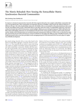
![homeostasis all depend on cell-ECM interactions (34). Such in-
teractions involve adhesion receptors (e.g., integrins [35]), inter-
cellular adhesion complexes (e.g., tight junctions [36]), and extra-
cellular polysaccharides (e.g., glycosaminoglycans [37]), which
activate cellular receptors. In contrast, the bacterial ECM has long
been thought to function merely as a passive extracellular scaffold
that holds the biofilm together and protects resident cells from
environmental stresses.
In this review, we first describe the structural roles of different
exopolymeric substances that constitute the ECM of various bio-
films. Then, we focus on studies that demonstrate the role of some
of these ECM components in regulating genetic programs of the
biofilm cells (Fig. 1). The examples presented in this review are
divided according to the three stages of biofilm formation (38),
i.e., (i) attachment, exploration of the surface, monolayer forma-
tion, and aggregation, (ii) 3D structure development and pattern-
ing, and (iii) dispersal.
ECM COMPONENTS IN BACTERIAL BIOFILMS
The most extensively studied biofilm ECM components are car-
bohydrate-rich polymers (i.e., extracellular polysaccharides or ex-
opolysaccharides), proteins, and nucleic acids (25).
Exopolysaccharides. Exopolysaccharides generated by bacte-
ria have been recognized to significantly impact bacterial viru-
lence and promote capsule formation. Various genetic analyses
have provided strong evidence that biofilm exopolysaccharides
play a fundamental structural role in different bacterial species.
Mutants defective in the production of exopolysaccharides display
severe defects in biofilm formation and in achieving complex bio-
film architecture. Common bacterial exopolysaccharides include
cellulose (39–43) and the staphylococcal polysaccharide intercel-
lular adhesin (PIA) (44). PIA-related polymers are produced by
Staphylococcus epidermidis and S. aureus (45, 46) and by several
Gram-negative bacterial species (47–52). In S. epidermidis and S.
aureus, PIA synthesis is mediated by the icaADBC operon (53–55).
The PIA molecule contains both positive and negative charges,
which are important for its adhesive properties (44). Many staph-
ylococcal isolates lack ica and are still capable of producing bio-
films, indicating alternative routes of biofilm formation (see be-
low).
Many bacterial species are capable of producing several differ-
ent exopolysaccharides, simultaneously or differentially, as a
function of environmental factors or the genetic background of
the specific strain. For instance, the ECM of P. aeruginosa biofilms
can contain three exopolysaccharides: alginate, Psl, and Pel (56).
Alginate is a polymer of manuronic acid and guluronic acid,
and its synthesis is mediated by the alg operon, as well as by 12
additional genes (57). P. aeruginosa isolates from lungs of cystic
fibrosis (CF) patients overproduce alginate, resulting in a mucoid
colony phenotype. Alginate is not essential for biofilm formation,
as the laboratory strains of P. aeruginosa PAO1 and PA14 do not
produce alginate and are capable of forming a submerged biofilm
in a microtiter plate assay (58). However, mutants in alginate bio-
synthesis form biofilms with an altered architecture compared to
the parental strains (59, 60).
Pel and Psl, the other two exopolysaccharides produced by P.
aeruginosa, play redundant roles in defining ECM structure (61).
Psl is composed of mannose, rhamnose, and glucose and is syn-
thesized by the gene products of the pslA-O operon (62–65). Pel is
FIG 1 Signals from the ECM during biofilm development. (Bottom panel) Scheme of the different stages of biofilm development. (I) Attachment, monolayer
formation, and aggregation. (II) 3D structure development and patterning. (III) Dispersal. (Top panel) (A) P. aeruginosa PAO1 deposits trails of high local
concentrations of ECM (in green) that attract other cells and induce further ECM production (114, 128). (B) In B. subtilis, inhibition of the rotation of the flagella,
e.g., by the viscous environment of the ECM, induces ECM production (150, 151). (C) TasA (brown), a structural amyloid in B. subtilis ECM, can be toxic to
vegetatively growing B. subtilis cells (80). (D) During B. subtilis biofilm development, ECM induces emergence of different cell subpopulations: motile cells,
ECM-producing cells, and sporulating cells (31). (E) In S. aureus PSMs, peptides (purple) can create structural amyloid fibers (85) but have a destabilizing effect
on the biofilm in their monomeric form (176). (F) In C. crescentus biofilms, eDNA induces dispersal by inhibiting the reattachment of the mature stalked cell by
binding to the exopolysaccharides of the holdfast (dark brown) (175).
Meeting Review
July 2015 Volume 197 Number 13 jb.asm.org 2093Journal of Bacteriology
onJuly26,2015byWEIZMANNINSTOFSCIENCEhttp://jb.asm.org/Downloadedfrom](data:image/gif;base64,R0lGODlhAQABAIAAAAAAAP///yH5BAEAAAAALAAAAAABAAEAAAIBRAA7)