
Recommended
More Related Content
What's hot
What's hot (20)
Similar to Biofilms
Similar to Biofilms (20)
Recently uploaded
Recently uploaded (19)
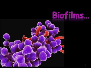
Biofilms
- 1. 1
- 3. Ultrastructure of biofilmsUltrastructure of biofilms Composition of biofilmsComposition of biofilms Stages of formation ofStages of formation of biofilmsbiofilms 3
- 4. Basic criteria for biofilmsBasic criteria for biofilms Characteristics of biofilmCharacteristics of biofilm Types of endodontic biofilmsTypes of endodontic biofilms -IntracanalIntracanal -ExtraradicularExtraradicular -PeriapicalPeriapical -BiomaterialBiomaterial 4
- 5. Microorganisms in biofilm formation Methods to isolate biofilm bacteria Biofilm detection Methods to eradicate biofilm 5
- 6. 6
- 7. In nature , bacteria 7
- 8. Microorganisms colonizing different sites in humans have been found to grow predominantly in complex structures known as biofilms. Biofilms Primordial multicellular protected mode organization of growth 8
- 9. Endodontic infections – biofilm model of growth TEM –dense aggregates of cocci and rods embedded in an extracellular matrix were observed along the walls 9
- 11. Individual microorganisms proliferating in a habitat – population Population – microcolonies Interaction of population – community Community- assembalge of populations Ecosystem – functional self supporting system 11
- 13. Population Perform functions – ecologic balance Functional role / niche in the community Limited no: of niches Competition b/n populations 13
- 14. Properties – greater than sum of the component populations Ability to withstand adverse conditions 14
- 16. Community profiling studies -Bacteria differed b/n individuals suffering from the same disease - Geographic locations -Different disease forms -Heterogenous etiology 16
- 17. DefinitionDefinition Biofilm is a mode of microbial growth where dynamic communities of interacting sessile cells are irreversibly attached to a solid substratum,as well as each other,and are embedded in a self-made matrix of extracellular polymeric substances. (Ingle 6th edition17
- 18. Biofilm can be defined as a sessile multicellular microbial community characterized by cells that are firmly attached to a surface and enmeshed in a self produced matrix of extracellular polymeric substance usually a polysaccharide - Stephan Cohen 18
- 19. Ability to form biofilm – virulence factor Biofilms -Microcolonies- towers and mushrooms -EPS matrix 19
- 20. Microcolonies - 300 cell thick Single bacterial species/ several different species Matrix Polysaccharide Nucleic acids, proteins Retain nutrients, water, enzymes 20
- 21. Microcolonies – separated by water channels Carry substrate, end products, signal molecules Formed by surface colonization by planktonic bacteria Co aggregate with other bacteria- Changes in gene expression, growth rate, protein production 21
- 22. Bacteria in biofilm – different phenotype, exposure to varying gradients of oxygen tension, ph, osmolarity Cell – cell communication – coordinate gene expression 22
- 23. Heterogenous arrangement of microbial cells on a solid surface Structural unit – microcolonies Int. Journal of Contemporary Dentistry 23
- 24. Three factors essential for biofilm are: 1.Microorganisms 2. Solid substrate 3. Fluid channels 24
- 25. 25
- 26. Composition Matrix material 85% volume 15% cells biopolymers such as polysaccharide, proteins, nucleic acids, and salts Glycocalyx matrix made of EPS surrounds microcolonies Anchors cells to substrate Haldal et al, Journal of International Oral Health 2016; 8(7):827-829 26
- 27. 27
- 28. Shape – determined by forces generated by flushing of fluid media 28
- 29. Stages of biofilm formation Stage I Stage II Stage III Stage IV Phase 1 Phase 2 Phase 3 Biofilm In Endodontics: New Understanding To An Old Problem, Usha H.L, Int. Journal of Contemporary Dentistry 2013(1) Biofilm In Endodontics: New Understanding To An Old Problem, Usha H.L, Int. Journal of Contemporary Dentistry 2013(1) 29
- 30. Stage I adsorption of inorganic and organic molecules, to the solid surface -formation of conditioning film Stage II Adhesion and colonization of planktonic microorganisms Attachment strengthened by polymer production and unfolding of cell surface structures 30
- 31. Factors affecting bacterial attachment pH Temp variations Flow rate of fluid Nutrients Surface energy of substrate Bacterial content 31
- 32. Bacterial growth stage Bacterial cell surface charge Surface hydrophobicity 32
- 33. Phase 1 Transport of the microbe to substrate surface and its attachment fimbriae, pili, flagella, EPS(glycocalyx) fimbriae, pili, flagella, EPS(glycocalyx) 33
- 34. Phase 2 Phase 2: Microbial and substrate adherence phase to form bridge. electrostatic attraction, covalent and hydrogen bonding ,dipole interaction and hydrophobic interaction. electrostatic attraction, covalent and hydrogen bonding ,dipole interaction and hydrophobic interaction. 34
- 35. Phase 3 specific microbial –substrate adherence phase adhesin or ligand 35
- 36. Stage III Bacterial growth and expansion monolayer of bacteria – 2* colonizers – microcolonies Towers – lateral and vertical growth 36
- 37. Two types of microbial interaction Co adhesion Co aggregation 37
- 38. Stage IV detachment of biofilm organisms seeding dispersal clumping dispersal 38
- 40. Stages of biofilm formation 40
- 41. Stages of biofilm formation Nov 2004, Endodontic Topics Gunnel Svensäter, Gunnar Bergenholtz 41
- 42. 42
- 43. Int. Journal of Contemporary Dentistry, 2010 1(3) 43
- 44. two main parts: (a) the initial interactions of cells with the substrate and (b) growth and development of the biofilm 44
- 45. 45
- 46. The phenotype of biofilm bacteria is distinct from planktonic bacteria due to following reasons: 1.EPS (extrapolymeric sacharide) protects the residing bacteria from environmental threats 2. Structure of biofilm permits trapping of nutrients and metabolites 3. Biofilm structures display organized internal compartmentalization 46
- 47. 4. Communication between bacterial cells and exchange of genetic materials. 47
- 48. BASIC CRITERIA FOR A BIOFILM Caldwell et al highlighted four characteristics of biofilm as follows: • Autopoiesis • Homeostasis • Synergy • Communality 48
- 49. Autopoiesis Ability to self organize Homeostasis Resist environmental pertubations Synergy Effective more in association than in isolation Communality Respond to changes as a group than an individual 49
- 50. Characteristics of biofilm Protection of Biofilm Bacteria from Environmental Threats Enhanced Tolerance to Antimicrobials Quorum Sensing 50
- 51. Cell surface structures -Capsule -Extracellular polysaccharide 51
- 52. Enhanced Tolerance to Antimicrobials -altered gene expression and -transfer of resistance genes -EPS matrix – barrier traps B lactamase -Bacteria – dormant state 52
- 53. - selective location of anaerobic niches - persisters 53
- 54. Quorum Sensing - bacterial cell-to-cell communication system -Chemical signals -characteristics of a specific strain of microbe determine its ability to co-exist 54
- 55. 55
- 56. 56
- 57. - E. fecalis, Streptococcus gordonii and Lactobacillus salivarius - Differential starvation endurance of E. Fecalis in mono-species and multi-species biofilms - protease production - co-existence between bacteria, as it is related to the virulence of bacteria 57
- 58. Specific quorum sensing genes -Biofilm forming ability of E feacalis -Eg: S-ribosylhomocysteine lyase [luxS]). -Future research- quorum-sensing inhibitors 58
- 59. Resistance to antimicrobials Physical - impaired penetration of antibiotics through the biofilm matrix 59
- 60. - Acquired differentiation of cells with low metabolic activity - differentiation of cells that actively respond to stress - differentiation of cells with a very high persistent phenotype 60
- 62. Anil Kishen 62
- 63. State of Nutrient Deprivation and Dormancy State of Nutrient Deprivation and Dormancy Cells – different physiological states 63
- 64. 64
- 65. Dormant state -Nutrient deprivation - general stress response and associated survival responses - resistance of biofilm cells to antimicrobials. 65
- 66. - E faecalis - Develop adaptive regulatory mechanisms - modify metabolic balance away from biosynthesis and reproduction Stringent response – global bac response – accumulation of p(ppGpp) 66
- 67. - Low nutrient survival of E feacalis - Form , develop and maintain stable biofilms - Dormant cells – ‘wake up’ n resume metabolic activity Strep anginosus & Lactobacill us salivarius Dormancy 24 hrs Inactive cells 67
- 68. Cell membrane integrity – maintained Reactivated – 96 hrs Undamaged cells – inactive Planktonic cells – active 2 hrs Biofilm cells – slow physiological response Strategy to resist stressful conditions 68
- 69. Formation of phenotypically different subpopulations evidence Difference in chemical conc gradient Adaptive variability 69
- 70. roo Mechanisms Root canal biofilms -Physiological differences Glucose deprivation Genetic alterations, mutations, genetic recombination, and stochastic gene expression. 70
- 71. roo Reorganization of subpopulations of cells In multispecies biofilms – imp for survival to stress from the envt Shapiro JA. Stud Hist Philos Biol Biomed Sci. 2007;38(4):807–19. 71
- 72. roo Development of persister cells in biofilms Survive even after exposure to lethal doses of antibiotics may represent (a)cells in some protected part of their cell cycle, (b)are capable of rapid adaptation, (c) are in a dormant state, or (d) are unable to initiate programmed cell death in response to the stimulus 72
- 73. Recalcitrant popn Produce cells – normal susceptibility Occur after exposure bac popn – high dose of single antimicrobial agent 73
- 74. E coli Expression of chromosomal toxin – antitoxin genes Operon Hip A – tolerance to ciprofloxacin And mitomycin Exposure to toxin --slow-growing, multiple drug- tolerant phenotypes by “shutting down” antibiotic targets 74
- 76. Types of endodontic biofilms 1.1. Intracanal biofilmIntracanal biofilm Seen on the root dentine of infected tooth Nair et al 1987 76
- 77. 77
- 78. cocci, rods, filaments and spirochetes 2. Extra radicular biofilm - root surface adjacent to the root apex of endodontically infected teeth -Fusobacterium nucleatum, - Porphyromonas gingivalis and - Tannellera forsythensis PCR based 16s rRNA gene assay 78
- 79. Scanning electron micrograph of bacterial biofilm on surface of root tip within periapical lesion of root-filled tooth with asymptomatic apical periodontitis. The biofilm is dominated by cocci and short rods in an extracellular matrix. 79
- 80. 3. Periapical microbial biofilms isolated biofilms in the periapical region of endodontically infected teeth seen even in the absence of root canal Infections Actinomyces species P. Propionicum 80
- 81. Scanning electron micrograph of bacterial biofilm adjacent to apical foramen of root-filled tooth with asymptomatic apical periodontitis. Bacterial colonies are recognized within smooth and structureless extracellular material. 81
- 82. Actinomyces – sulphur granules -aggregation of Actinomyces cells are influenced by pH, ionic strength and cell concentration 82
- 83. 4. Foreign body – centered biofilm bacteria adheres to an artificial biomaterial Surface major complication associated with prosthesis and also in an implant supported prosthesis 83
- 84. Opportunistic invasion by nosocomial organisms three phases- Transports of bacteria to biomaterial surface Initial, non-specific adhesion phase Specific adhesion phase 84
- 85. Coagulase –negative Staphylococcus, S. aureus , Streptococci, Enterococci, P.aeruginosa and fungi 85
- 86. Microorganisms in biofilm formation Microorganisms in biofilm formation 86
- 87. 87
- 88. -Fluorescent Microscopic Techniques with Super Resolution -Scanning Electron Microscopy (SEM) -Confocal Laser Scanning Microscopy 88
- 89. Microtiter plate based system -Closed system , no fluid movement -Envt in the experimental model changes -perform different tests at the same time 89
- 90. Biofilm quantification with microtiter plates may be categorized into biomass assays, viability assays, matrix quantification assays 90
- 91. Stains commonly used -crystal violet, -Nucleic acid stains such as Styo9, - non-fluorescent fluorescein diacetate, - tetrazolium salts such as XTT, - resazurin - dimethyl methylene blue 91
- 92. These methods show differential results for fungal and bacterial biofilms -Styo9 assay should not be used for CFU measurements in biofilms -crystal violet assay was non-repeatable for Pseudomonas aeruginosa biofilms -Styo9 assay is that it depends on microbial cell wall integrity, 92
- 93. - CFU counts only reproducing cells, and can over-quantify killed cells - these test methods measure different analytics to describe viability - it may be preferable to perform tests such as FDA or resazurin for quantification of biofilms with differentiation Between dead and live cells 93
- 94. Flow Displacement Biofilm Model Systems -the flow displacement system is open -Nutrients added at constant rate and waste products removed simultaneously -Concept - an initial film of macromolecular components needs to form on a surface to allow microbial adhesion - Fluid flow- microbial adhesion 94
- 95. - parallel plate flow chamber performed with controlled hydrodynamic conditions - Ideal flow rate - reduces technical variables, repeatability 95
- 96. Modified Robbins Device continuous formation of biofilm which is exposed to fluid flow Substrate - silicone or hydroxyapatite discs allows evaluation of more than one antibiofilm agent in the same experiment Device – can be modified, used along with flow devices 96
- 97. Microfluidic Device -Forming a biofilm under conditions similar to that physiologically - cell-to-fluid volume ratios and flow velocities - allows for a single cell resolution analysis of the biofilm under tightly controlled conditions 97
- 98. - chemical assays using small quantities of liquids on a small chip. Challenges - in terms of analysis of biofilms - with specific reference to quantification using methods such as fluorescent staining 98
- 99. - continuous optimizing confocal reflection microscopy, to quantitatively study the biovolume of biofilms 99
- 100. Fluorescent Microscopic Techniques Super Resolution STED (Stimulated Emission Depletion) PALM (Photo-Activated Localization Microscopy) and SIM (Structured-Illumination Microscopy) Biofilm should be labeled with fluorescent dyes 100
- 101. Scanning Electron Microscopy (SEM) allows scanning of microbial ecosystems qualitative information Detailed analysis of morphological structures Sample preparation – distortion of EPM Enviromental SEM 101
- 102. Laser scanning microscopy 1980 confocal laser scanning microscopy (CLSM) visualize multiple features in different channels that are spectrally resolved 102
- 103. structure, composition, microhabitats, activity, and processes Color probes Volumetric & structural quantification of multi channel signals in 4 dimensions 103
- 104. SRM – super resolution microscopy SRM + FISH – tracking of -Ribosome associated changes in activity levels -Subcellular localization at single cell level 104
- 105. 105
- 106. rRNA Fluorescence In Situ Hybridization (FISH) Visualize specific subpopulation of cells Maintaining 3 D structure Detection of biofilm in their natural envt No need for cultivation 106
- 107. Oligonucleotide probes + rRNA -Identification of single cells Species detection 107
- 108. Markers of cell viability Viability Culture methods Drawbacks • Under represent viable bacteria , injured • Media- lacking nutrients • Viable cells –losing ability to form colonies • Low metabolic activity 108
- 109. viability indicators LIVE DEAD kit Intact cells – SYTO 9 fluorescent green Dead- PI fluorescent red Fluorescent molecules - epifluorescence / LSM 109
- 110. Cell metabolic functions Tetrazolium salts INT CTC Correlation b/n INT / CTC positive cells and CFU count 110
- 111. Many therapeutic approaches Mechanical removal with instrumentation & irrigation 111
- 112. Challenges in root canal disinfection Anil Kishen 112
- 113. Anti biofilm strategies – Anil Kishen 113
- 114. Bettina Basrani 114
- 115. Cold Spring Harb Perspect Med. 2013 Apr; 3(4) 115
- 116. Surface coating 116
- 117. Root canal irrigants Proteolytic irrigants NaOCl – Sodium hypochlorite -potent disinfectant in endodontics -0.5- 6% -Irrigation regimen 117
- 118. Efficacy can be improved by various mechanisms Rosen et al – VNBC state 118
- 119. Antiseptics Chlorhexidine gluconate -Broad antimicrobial spectrum -Low toxicity , substantivity -Concentration 119
- 120. CHX+ cetrimide - alternating application - combined use - 2% CHX and 0.2% cetrimide – extended antimicrobial activity CHX plus 120
- 121. CHX – no role against biofilm Alexidine Greater affinity for lipoteichoic acids ALX 1% Octenedine hydrochloride positively charged bispyridinamine 121
- 122. Octenedine hydrochloride positively charged bispyridinamine Action against C albicans 122
- 123. Iodine potassium iodide 1829- Lugol – French physician To treat scrofula 1927- iodine products – root canal irrigants Broad antimicrobial action Used in combination with detergents No action against biofilms 123
- 124. Demineralizing agents Sequential use of EDTA and NaOCl – antibacterial action and biofilm disruption Maleic acid 0.88% for 30 seconds Altered cell permeability 124
- 125. 2.25% peracetic acid (PAA) More effective than CHX against E. Feacalis 125
- 126. Combination of solutions MTAD - 3% doxycycline, 4.25% citric acid and 0.5% Tween 80. Complete inhibition of bacterial growth by MTAD in a 3 week old biofilm Qmix CHX, EDTA and a detergent 126
- 127. 5 % NaOCl + 18% etidronic acid – continuous Chelation Excellent antibiofilm activity against biofilms of E. fecalis 127
- 128. Natural Agents (Phytotherapeutic or Ethnopharmacological Approaches) Berberine - antimicrobial plant alkaloid Morinda citrifolia Curcumin- Curcuma longa 128
- 129. Berberine + 1% chlorhexidine = 5.25% sodium hypochlorite and 2% chlorhexidine + miconazole – against C.albicans biofilms 129
- 130. Nanoparticles Based Disinfection Chitosan , Zinc oxide Silver (Ag-np) nanoparticles possess a broad spectrum of antimicrobial Activity- altering cell wall Permeability 130
- 131. Rose bengal-functionalized CS-np- effective in the presence of tissue inhibitors Rose bengal –light – cytotoxic Chitin – polymer 131
- 132. Chitosan + rose bengal - enhance the degradation resistance of collagen Silver nanoparticles sized 10–100 nm mesoporous bioactive calcium silicate nanoparticles and bioactive glass powder loaded with AgNp- reduction in adhesion of E. fecalis biofilms 132
- 133. Endodontic Topics 2012, 22, 99–123, Anil Kishen Endodontic Topics 2012, 22, 99–123, Anil Kishen 133
- 134. Miscellaneous Interventions -Enzymatic irrigation- Niazi et al - 1% trypsin and 1% proteinase K, with or -without ultrasonic activation 134
- 135. D-amino acids, specifically D-leucine 135
- 136. 136
- 137. Irrigant activation systems -Sonics and ultrasonics -Light: Non-Coherent (Photoactivated Disinfection) and Coherent (Laser Activated Disinfection) - Microbubble emulsion 137
- 138. Sonics and ultrasonics Dis-agglomeration of the bacterial biofilm Planktonic form – susceptible to antimicrobials Cell membrane permeability – altered 138
- 139. Shear stresses – disruption of biofilms Better penetration of irrigant into the dentinal tubules 139
- 141. - Er:YAG laser activation of 5 percent NaOCl and 17 percent EDTA was more effective than conventional irrigation for eradicating E. faecalis and preventing new bacterial growth ex vivo. Olivi G, et al J Am Dent Assoc. 2014 Aug;145(8):843-8 141
- 142. sub-ablative photoacoustic technique penetration of the irrigating solution into inaccessible areas of the root canal system, bacterial elimination by antimicrobial irrigants activated by the photomechanical effects of the PIPS-tapered and stripped laser tip (Jaramillo et al. 2012). 142
- 143. 143
- 144. 144
- 145. 145
- 146. Dentin surface with thermal damage and charring Er,Cr:YSGG laser radial firing tip Ledging and thermal damage Clean canal surface 146
- 147. PIPS 147
- 148. PIPS 148
- 149. Previous studies have shown that PIPS in combination with 5.25 and 6 % sodium hypochlorite is an effective means of eliminating resistant bacteria such as E. faecalis from the root canal System (Jaramillo et al. 2012; Olivi et al. 2014). 149
- 150. Comparison between (A) the classic PIPS protocol (adapted from Jamarillo ) and (B) the modified PIPS protocol. Barbara Skrlj Golob et al, (J Endod 2017;-:1–6) 150
- 151. Buffered 0.5 % sodium hypochlorite delivered by conventional method was effective in Removing E. faecalis from contaminated root canals; however,activation of a buffered 0.5 % sodium hypochlorite solution by PIPS significantly increased its antimicrobial capacity. Jaramillo et al. Evidence-Based Endodontics (2016) 1:6 151
- 152. PDT involves the use of a nontoxic dye or photosensitizer (PS) in combination with visible light, which in the presence of molecular oxygen leads to the production of cytotoxic oxygen radicals such as singlet oxygen. 152
- 153. 153
- 154. Microbubble emulsion -Halford et al - employs unstable gas-filled microbubbles that expand when exposed to ultrasonic waves : biofilm detachment -generate reactive oxygen species to exhibit an antibacterial effect. 154
- 155. Intracanal Medicaments Calcium hydroxide - ineffective against E. feacalis- 24 hours of treatment Even combining with chitosan nanoparticles – Cannot penetrate the EPS matrix of biofilms 155
- 156. Antibiotics TAP is significantly better than calcium hydroxide and chlorhexidine in disrupting biofilms of E. feacalis polymer nanofibers with TAP has been shown to bring about significant bacterial killing resistance to antibiotics 156
- 157. Dentin pretreatment for 4 weeks with 5, 50 or 500 mg/mL of DAP demonstrated significantly higher residual antibiofilm effects and complete eradication of E. faecalis biofilms in comparison to a 1 week pretreatment with similar concentrations. No sig antibiofilm effects with 1mg/ml irrespective of time Jenks, D et al ,Archives of Oral Biology, 70, 88–93 157
- 158. Biofilm mode of growth Structure and composition of biofilms Stages of formation of biofilm SummarySummary 158
- 159. Mechanisms of resistance to antimicrobials Biofilm detection Antibiofilm strategies 159
- 160. Surface coating Root canal irrigants Irrigant activation systems Intracanal medicaments 160
- 161. Conclusion It is clear that endodontic infections are caused by multispecies biofilms and that the interactions between different organisms can contribute to apical periodontitis progress and clinical outcome. 161
- 162. Further research in basic microbiological processes such as the molecular basis and biological effect of these host–bacterial connections may lead to an improvement of treatment regimens and also may identify new objectives and strategies for disease control. 162
- 163. References 1. Shapiro JA. Bacteria are small but not stupid: cognition, natural genetic engineering and sociobacteriology. Stud Hist Philos Biol Biomed Sci. 2007;38(4):807–19. 2. Cold Spring Harb Perspect Med. 2013 Apr; 3(4): a010306. 3. Chavez de Paz LE, Bergenholtz G, Dahlen G, Svensater G. Response to alkaline stress by root canal bacteria in biofi lms. Int Endod J. 2007;40(5):344–55 4. Bettina Basrani 5. Pathways of pulp 6. Ingles endodontics 7. Endodontic microbiology 163