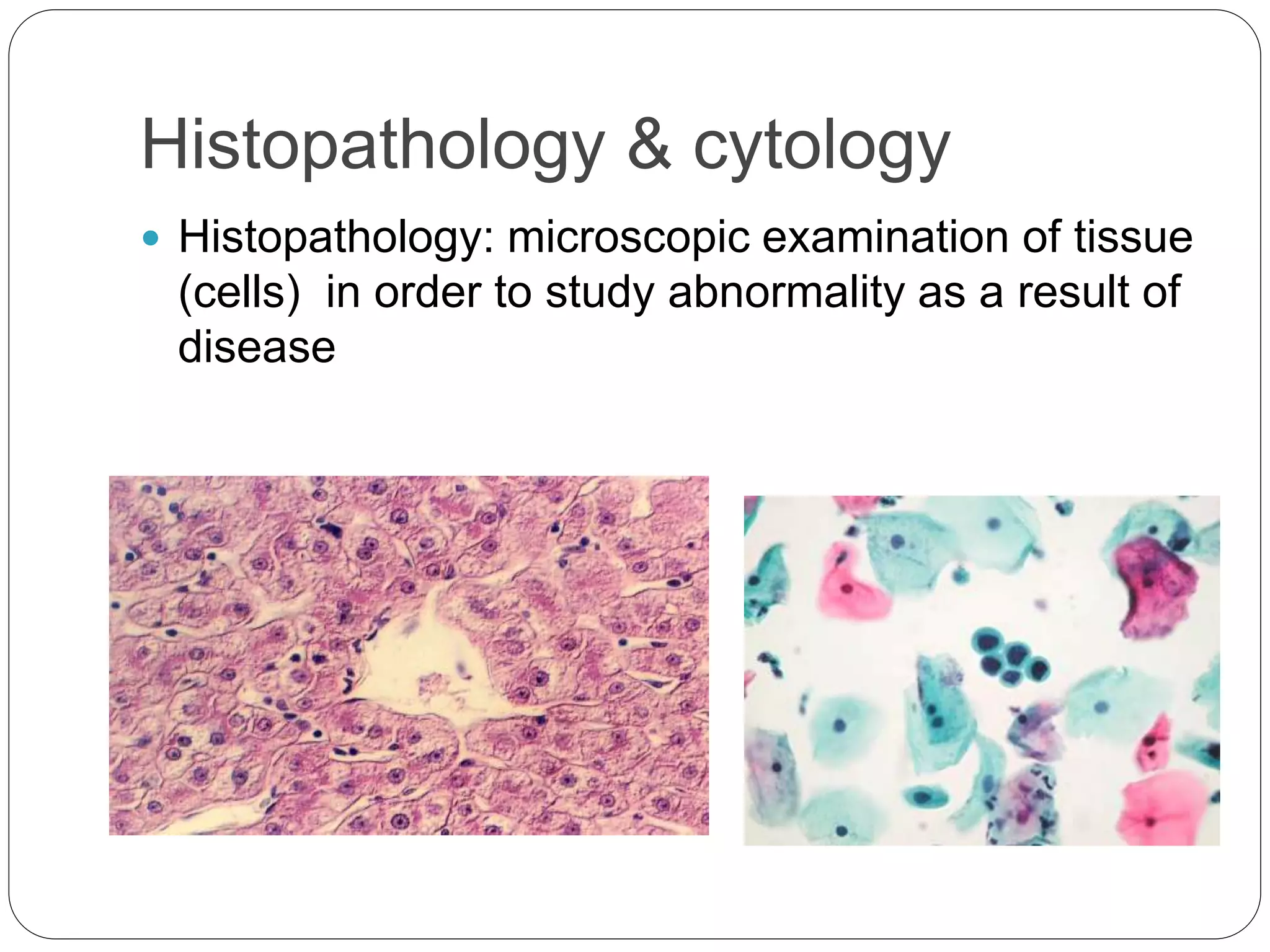
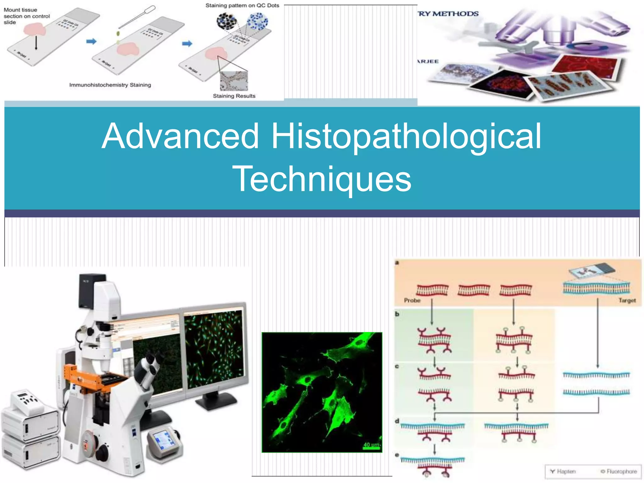
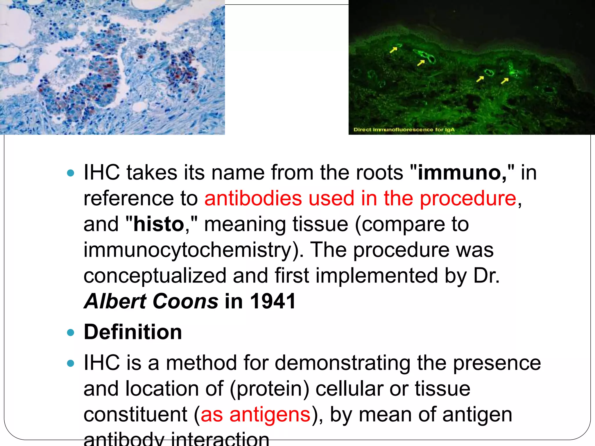
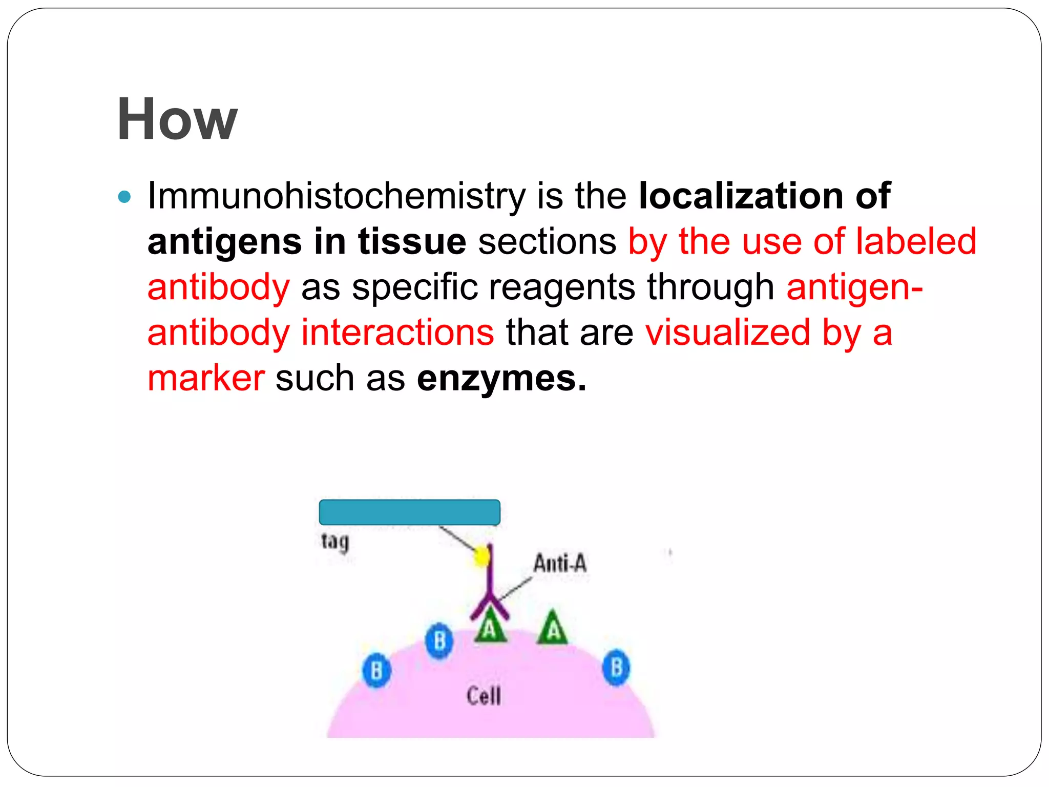
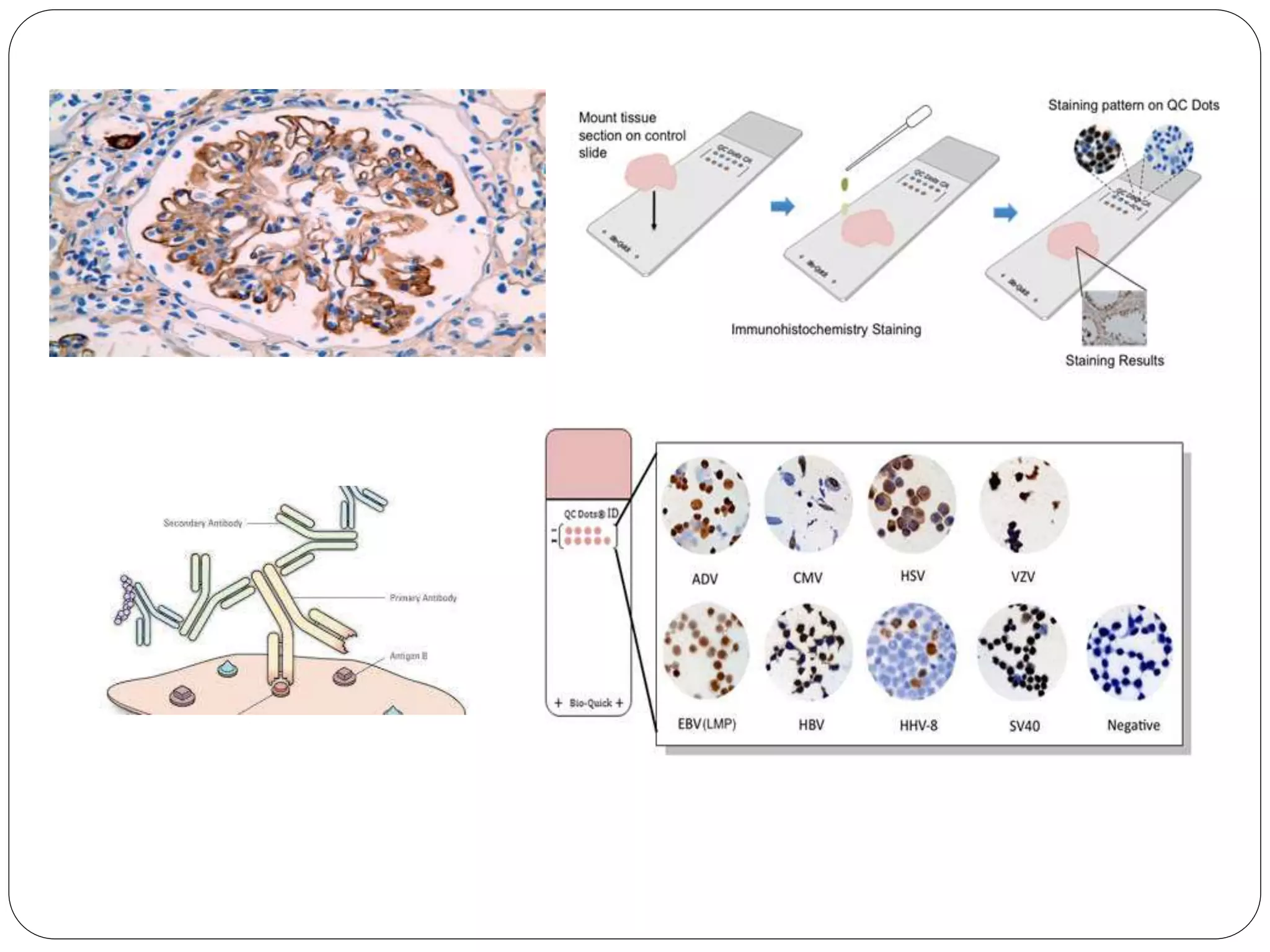
Advanced Histopathological
Techniques
Histological techniques
of preparation
Histopathology & cytology
 Histopathology: microscopic examination of tissue
(cells) in order to study abnormality as a result of
disease
Advanced Histopathological
Techniques
Prerequisites
 Cell biology- Normal histology
 Histopathological techniques
 Pathology
 Cytology
Topics:
 1: Introduction (course out lines, concepts in IHC)
 2: definitions, Production of antibodies, sample preparation, antigen retrieval
 3: Antibody labeling, IHC methods (1),
 4: IHC methods (2), detection of low level antigens , controls in immunohistochemical
staining,
 5: Applications of IHC , tumor markers (intermediate filaments)
 6: tumor markers (Epithelial related antibodies,Germ cell tumor marker,
Neuroendocrine marker )
 7: tumor markers (hormone receptor, oncogenes, bassment membrane,
mesenchaymel tissue marker)
 8: tumor markers (cell proliferation markers, lymphoid markers, trouble shooting)
 9: tutorial (1)
 10: Imunofourecent methods
 11: Imunofourecent techniques (pathological applications)
 12: Enzyme histochemistry (techniques)
 13: Enzyme histochemistry (pathological application)
 14: overview of in-situ hybridization
 15: In-situ hybridization methods (probe preparation, pretreatment of specimens,
hybridization) detection methods


Immunohistochemistry
 IHC takes its name from the roots "immuno," in
reference to antibodies used in the procedure,
and "histo," meaning tissue (compare to
immunocytochemistry). The procedure was
conceptualized and first implemented by Dr.
Albert Coons in 1941
 Definition
 IHC is a method for demonstrating the presence
and location of (protein) cellular or tissue
constituent (as antigens), by mean of antigen
How
 Immunohistochemistry is the localization of
antigens in tissue sections by the use of labeled
antibody as specific reagents through antigen-
antibody interactions that are visualized by a
marker such as enzymes.
 It is accomplished with antibodies that recognise
the target protein .and this Ab bind only to the
protein of interest in the tissue section
 The aim is to achieve reproducible and
consistent demonstration of antigens with the
minimum background staining whilst
preserving the integrity of tissue architecture.
 For optimal staining to occur it is necessary to
use the primary specific antibody at the correct
dilution
 Blocking background staining (The major
causes of background staining in
immunohistochemistry are ionic interactions
and endogenous enzyme activity and
unbounded antibodies)…Endogenous enzyme
block – protein blocking -washing
 Unmasking of antigen ……antigen retrieval
Electrostatically charged slide
Incubation
 to prevent evaporation of antibodies, incubations
must be carried out in a moist atmosphere.
 Using the wax pen around the section to assist
with retaining the reagents on the section
steps
-Preparation of sample (retrivation)
-Immunohistochemical reaction
-Visualizing the reaction
-Counter stain.
 Applications of IHC
 especially advantages as a diagnostic tool for
infectious disease
 It is useful for assessing the progression and
treatment of disease such as cancers
 Differentiate between types of malignant cells
 Detection of metastatic tumours
Introduction to IHC

Introduction to IHC

  • 1.
  • 2.
  • 3.
    Histopathology & cytology Histopathology: microscopic examination of tissue (cells) in order to study abnormality as a result of disease
  • 4.
  • 5.
    Prerequisites  Cell biology-Normal histology  Histopathological techniques  Pathology  Cytology
  • 6.
    Topics:  1: Introduction(course out lines, concepts in IHC)  2: definitions, Production of antibodies, sample preparation, antigen retrieval  3: Antibody labeling, IHC methods (1),  4: IHC methods (2), detection of low level antigens , controls in immunohistochemical staining,  5: Applications of IHC , tumor markers (intermediate filaments)  6: tumor markers (Epithelial related antibodies,Germ cell tumor marker, Neuroendocrine marker )  7: tumor markers (hormone receptor, oncogenes, bassment membrane, mesenchaymel tissue marker)  8: tumor markers (cell proliferation markers, lymphoid markers, trouble shooting)  9: tutorial (1)  10: Imunofourecent methods  11: Imunofourecent techniques (pathological applications)  12: Enzyme histochemistry (techniques)  13: Enzyme histochemistry (pathological application)  14: overview of in-situ hybridization  15: In-situ hybridization methods (probe preparation, pretreatment of specimens, hybridization) detection methods  
  • 7.
  • 8.
     IHC takesits name from the roots "immuno," in reference to antibodies used in the procedure, and "histo," meaning tissue (compare to immunocytochemistry). The procedure was conceptualized and first implemented by Dr. Albert Coons in 1941  Definition  IHC is a method for demonstrating the presence and location of (protein) cellular or tissue constituent (as antigens), by mean of antigen
  • 9.
    How  Immunohistochemistry isthe localization of antigens in tissue sections by the use of labeled antibody as specific reagents through antigen- antibody interactions that are visualized by a marker such as enzymes.
  • 10.
     It isaccomplished with antibodies that recognise the target protein .and this Ab bind only to the protein of interest in the tissue section  The aim is to achieve reproducible and consistent demonstration of antigens with the minimum background staining whilst preserving the integrity of tissue architecture.
  • 11.
     For optimalstaining to occur it is necessary to use the primary specific antibody at the correct dilution  Blocking background staining (The major causes of background staining in immunohistochemistry are ionic interactions and endogenous enzyme activity and unbounded antibodies)…Endogenous enzyme block – protein blocking -washing  Unmasking of antigen ……antigen retrieval Electrostatically charged slide
  • 12.
    Incubation  to preventevaporation of antibodies, incubations must be carried out in a moist atmosphere.  Using the wax pen around the section to assist with retaining the reagents on the section
  • 13.
    steps -Preparation of sample(retrivation) -Immunohistochemical reaction -Visualizing the reaction -Counter stain.
  • 15.
     Applications ofIHC  especially advantages as a diagnostic tool for infectious disease  It is useful for assessing the progression and treatment of disease such as cancers  Differentiate between types of malignant cells  Detection of metastatic tumours