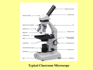
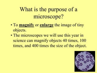
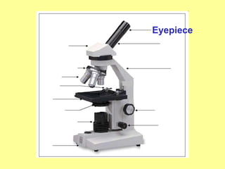
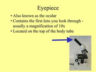
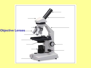
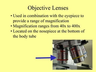
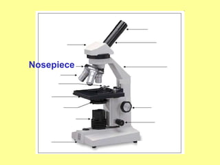
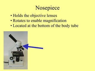
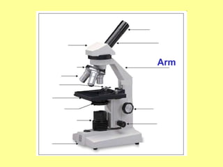
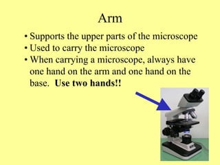
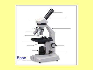
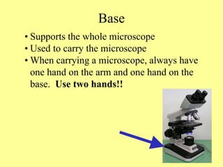
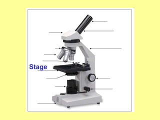
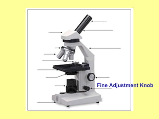
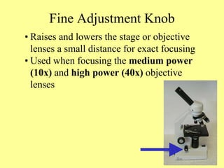
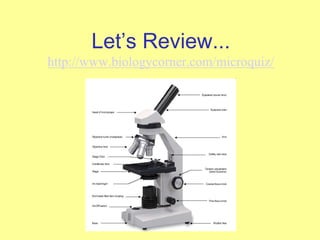
A microscope is an instrument that is used to magnify small objects. Some microscopes can even be used to observe an object at the cellular level, allowing scientists to see the shape of a cell, its nucleus, mitochondria, and other organelles. While the modern microscope has many parts, the most important pieces are its lenses. It is through the microscope’s lenses that the image of an object can be magnified and observed in detail. A simple light microscope manipulates how light enters the eye using a convex lens, where both sides of the lens are curved outwards. When light reflects off of an object being viewed under the microscope and passes through the lens, it bends towards the eye. This makes the object look bigger than it actually is.
Over the course of the microscope’s history, hile some older microscopes had only one lens, modern microscopes make use of multiple lenses to enlarge an image. There are two sets of lenses in both the compound microscope and the dissecting microscope (also called the stereo microscope). Both of these microscopes have an objective lens, which is closer to the object, and an eyepiece, which is the lens you look through. The eyepiece lens typically magnifies an object to appear ten times its actual size, while the magnification of the objective lens can vary. Compound microscopes can have up to four objective lenses of different magnifications, and the microscope can be adjusted to choose the magnification that best suits the viewer’s needs. The total magnification that a certain combination of lenses provides is determined by multiplying the magnifications of the eyepiece and the objective lens being used. For example, if both the eyepiece and the objective lens magnify an object ten times, the object would appear one hundred tThe dissecting microscope provides a lower magnification than the compound microscope, but produces a three-dimensional image. This makes the dissecting microscope good for viewing objects that are larger than a few cells but too small to see in detail with the human eye. The compound microscope is typically used for observing objects at the cellular level.Over the course of the microscope’s history, technological innovations have made the microscope easier to use and have improved the quality of the images produced. The compound microscope, which consists of at least two lenses, was invented in 1590 by Dutch spectacle-makers Zacharias and Hans Jansen. Some of the earliest microscopes were also made by a Dutchman named Antoine Van Leeuwenhoek. Leeuwenhoek’s microscopes consisted of a small glass ball set inside a metal frame. He became known for using his microscopes to observe freshwater, single-celled microorganisms that he called “animalcules.”
While some older microscopes had only one loutwards. When light reflects off of an object being viewed under the microscope and passes through the lens, it bends towards the eye. This makes the object look bigger than it actually is.. Some of
2. Starting with use of a simple lens in ancient
times, to the first compound microscope
around 1590, and up to the microscopes you
are using in 7th grade life science, the
microscope has allowed scientists to make
discoveries about the “invisible world.”
The microscope has
become one of the most
recognizable symbols of
science.
4. What is the purpose of a
microscope?
• To magnify or enlarge the image of tiny
objects.
• The microscopes we will use this year in
science can magnify objects 40 times, 100
times, and 400 times the size of the object.
5. What type of microscope will we
be using in class this year?
• In 7th grade science, we use a compound
light microscope for most of our lab
activities.
• A light microscope uses light as a source
of energy for viewing objects.
• A compound microscope contains two
lenses for magnifying a specimen at the
same time, the eyepiece (ocular) and one of
the objective lenses.
7. Eyepiece
• Also known as the ocular
• Contains the first lens you look through -
usually a magnification of 10x
• Located on the top of the body tube
9. Objective Lenses
• Used in combination with the eyepiece to
provide a range of magnification
• Magnification ranges from 40x to 400x
• Located on the nosepiece at the bottom of
the body tube
13. Arm
• Supports the upper parts of the microscope
• Used to carry the microscope
• When carrying a microscope, always have
one hand on the arm and one hand on the
base. Use two hands!!
15. Base
• Supports the whole microscope
• Used to carry the microscope
• When carrying a microscope, always have
one hand on the arm and one hand on the
base. Use two hands!!
19. Stage Clip
• Helps to hold the slide in place
• Usually one on each side of the hole (stage
opening) = 2 stage clips
• The stage opening allows light to pass from
the light source to the lenses.
21. Light Source
• Provides light necessary for viewing the
specimen
• Usually either a mirror or illuminator
• Sends light through the stage opening to
the diaphragm
23. Diaphragm
• Wheel or lever located below the stage
opening
• Regulates the amount of light that can enter
the lenses
• May need to be adjusted based on the
thickness of the specimen being studied
27. Fine Adjustment Knob
• Raises and lowers the stage or objective
lenses a small distance for exact focusing
• Used when focusing the medium power
(10x) and high power (40x) objective
lenses