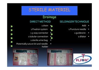
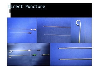
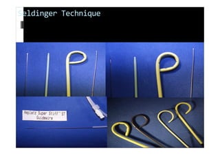
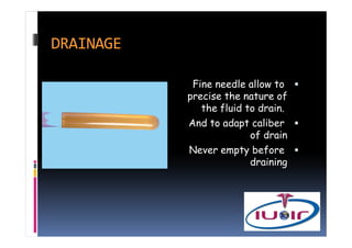
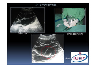
1. Biopsy results have a positive diagnosis rate between 70-100%. Performance is least for lymphoma biopsies.
2. Abdominal drainage procedures can be performed via direct puncture or the Seldinger technique using sterile materials including drains, fixation systems, and drainage bags.
3. After drainage procedures, the drain output is checked and patients are monitored for improvement before conducting clamping tests if drainage decreases and removing the drain. Follow up involves emptying and flushing the drain with saline.
The Indian Dental Academy is the Leader in continuing dental education , training dentists in all aspects of dentistry and
offering a wide range of dental certified courses in different formats.
The Indian Dental Academy is the Leader in continuing dental education , training dentists in all aspects of dentistry and
offering a wide range of dental certified courses in different formats.
This presentation provides sufficient material for anyone who wants is interested in interventional radiology. Here we will discuss the available facilities, mechanisms and equipments.
In my opinion this presentation will prove a footstep in interventional radiology
1. Breast Ductography:Evaluation and Surgical Planning
2.INTRODUCTION: Breast ductography, sometimes referred to as galactography, is an imaging modality used to assess lesions resulting in discharge from the breasts. It offers helpful information for surgical approach and planning and aids in accurately finding the lump within breast tissue.
3. ANATOMY
4. Indications:If a single duct has a discharge, breast ductography is recommended.
Contraindications: When there are two or more discharging ducts, breast ductography is not recommended since the cause is either physiological or systemic.
5. Surgery: It helps locate the mass within breast tissue and provides useful information for surgical planning.
Excision of lesions: Surgery for breast ductography involves excising the majority of lesions found in ducts, such as intraductal papillomas.
Ultrasound-Guided Percutaneous Biopsy: For smaller lesions, ultrasound-guided percutaneous biopsy can be used.
Duct Excision with Dye: The duct is excised after it is filled with dye.
6. Procedure : A blunt-tipped sialogram needle is used in breast ductography procedure to perform the ductogram.
Once the aberrant duct has been located and cannulated, 1-2 mL of contrast agent is administered.
Images taken from standard mammography are obtained.
7. The tip of the cannula is inserted into the duct opening once the duct causing the discharge has been located.
The cannula is then carefully straightened. The cannula typically falls in the duct to the hub in patients. There is no force applied. Cannulation shouldn't cause any pain for the patient.
8. Before injecting contrast, watch the tubing for a short while after cannulation.
Since the duct is cannulated and the tubing is now part of a closed system, it is occasionally possible to view the contents of the duct refluxing into the tubing. This indicates that the duct producing the discharge has been cannulated if it is observed.
9. When contrast is injected into the cannula, duct contents occasionally form a droplet (arrow) around the cannula if they do not reflux into the tubing. Duct contents are displaced by the contrast. Observing this verifies that the duct causing the discharge has been cannulated.
10. Following the injection of 0.2 to 0.4 mL of contrast, the cannula is secured to the nipple with two pieces of paper tape. Leaving the cannula in situ allows for additional contrast injections when duct evaluation is required. Additionally, the cannula lessens the quantity of contrast that compression forces out of the duct.
11. Intraductal Abnormalities
Filling defects. Fusiform or tubular dilatation of ducts
Abrupt ductal cut off.
Causes of Abnormalities
The intraductal abnormalities observed on mammography can have both pathological and artifactual causes.
12. Complications: Duct Perforation, Pain or Burning Sensation, Infection.
13. It matters more how a patient perceives the discharge than what kind of discharge it is.
The International Union of Interventional Radiologists (IUOIR) is a non-profit association of interventional radiologists all over the world
The IUOIR offers:
1- Training of Radiologists in situ and follow up the training to build independent IR unit.
2- One to two months observership in Europe and USA.
3-Treatment of patients in your country as well as receiving of patients in IUOIR' Centers in Europe.
4- May offer one year fellowship.
Flu Vaccine Alert in Bangalore Karnatakaaddon Scans
As flu season approaches, health officials in Bangalore, Karnataka, are urging residents to get their flu vaccinations. The seasonal flu, while common, can lead to severe health complications, particularly for vulnerable populations such as young children, the elderly, and those with underlying health conditions.
Dr. Vidisha Kumari, a leading epidemiologist in Bangalore, emphasizes the importance of getting vaccinated. "The flu vaccine is our best defense against the influenza virus. It not only protects individuals but also helps prevent the spread of the virus in our communities," he says.
This year, the flu season is expected to coincide with a potential increase in other respiratory illnesses. The Karnataka Health Department has launched an awareness campaign highlighting the significance of flu vaccinations. They have set up multiple vaccination centers across Bangalore, making it convenient for residents to receive their shots.
To encourage widespread vaccination, the government is also collaborating with local schools, workplaces, and community centers to facilitate vaccination drives. Special attention is being given to ensuring that the vaccine is accessible to all, including marginalized communities who may have limited access to healthcare.
Residents are reminded that the flu vaccine is safe and effective. Common side effects are mild and may include soreness at the injection site, mild fever, or muscle aches. These side effects are generally short-lived and far less severe than the flu itself.
Healthcare providers are also stressing the importance of continuing COVID-19 precautions. Wearing masks, practicing good hand hygiene, and maintaining social distancing are still crucial, especially in crowded places.
Protect yourself and your loved ones by getting vaccinated. Together, we can help keep Bangalore healthy and safe this flu season. For more information on vaccination centers and schedules, residents can visit the Karnataka Health Department’s official website or follow their social media pages.
Stay informed, stay safe, and get your flu shot today!
TEST BANK for Operations Management, 14th Edition by William J. Stevenson, Ve...kevinkariuki227
TEST BANK for Operations Management, 14th Edition by William J. Stevenson, Verified Chapters 1 - 19, Complete Newest Version.pdf
Couples presenting to the infertility clinic- Do they really have infertility...Sujoy Dasgupta
Dr Sujoy Dasgupta presented the study on "Couples presenting to the infertility clinic- Do they really have infertility? – The unexplored stories of non-consummation" in the 13th Congress of the Asia Pacific Initiative on Reproduction (ASPIRE 2024) at Manila on 24 May, 2024.
New Directions in Targeted Therapeutic Approaches for Older Adults With Mantl...i3 Health
i3 Health is pleased to make the speaker slides from this activity available for use as a non-accredited self-study or teaching resource.
This slide deck presented by Dr. Kami Maddocks, Professor-Clinical in the Division of Hematology and
Associate Division Director for Ambulatory Operations
The Ohio State University Comprehensive Cancer Center, will provide insight into new directions in targeted therapeutic approaches for older adults with mantle cell lymphoma.
STATEMENT OF NEED
Mantle cell lymphoma (MCL) is a rare, aggressive B-cell non-Hodgkin lymphoma (NHL) accounting for 5% to 7% of all lymphomas. Its prognosis ranges from indolent disease that does not require treatment for years to very aggressive disease, which is associated with poor survival (Silkenstedt et al, 2021). Typically, MCL is diagnosed at advanced stage and in older patients who cannot tolerate intensive therapy (NCCN, 2022). Although recent advances have slightly increased remission rates, recurrence and relapse remain very common, leading to a median overall survival between 3 and 6 years (LLS, 2021). Though there are several effective options, progress is still needed towards establishing an accepted frontline approach for MCL (Castellino et al, 2022). Treatment selection and management of MCL are complicated by the heterogeneity of prognosis, advanced age and comorbidities of patients, and lack of an established standard approach for treatment, making it vital that clinicians be familiar with the latest research and advances in this area. In this activity chaired by Michael Wang, MD, Professor in the Department of Lymphoma & Myeloma at MD Anderson Cancer Center, expert faculty will discuss prognostic factors informing treatment, the promising results of recent trials in new therapeutic approaches, and the implications of treatment resistance in therapeutic selection for MCL.
Target Audience
Hematology/oncology fellows, attending faculty, and other health care professionals involved in the treatment of patients with mantle cell lymphoma (MCL).
Learning Objectives
1.) Identify clinical and biological prognostic factors that can guide treatment decision making for older adults with MCL
2.) Evaluate emerging data on targeted therapeutic approaches for treatment-naive and relapsed/refractory MCL and their applicability to older adults
3.) Assess mechanisms of resistance to targeted therapies for MCL and their implications for treatment selection
Report Back from SGO 2024: What’s the Latest in Cervical Cancer?bkling
Are you curious about what’s new in cervical cancer research or unsure what the findings mean? Join Dr. Emily Ko, a gynecologic oncologist at Penn Medicine, to learn about the latest updates from the Society of Gynecologic Oncology (SGO) 2024 Annual Meeting on Women’s Cancer. Dr. Ko will discuss what the research presented at the conference means for you and answer your questions about the new developments.
Explore natural remedies for syphilis treatment in Singapore. Discover alternative therapies, herbal remedies, and lifestyle changes that may complement conventional treatments. Learn about holistic approaches to managing syphilis symptoms and supporting overall health.
- Video recording of this lecture in English language: https://youtu.be/lK81BzxMqdo
- Video recording of this lecture in Arabic language: https://youtu.be/Ve4P0COk9OI
- Link to download the book free: https://nephrotube.blogspot.com/p/nephrotube-nephrology-books.html
- Link to NephroTube website: www.NephroTube.com
- Link to NephroTube social media accounts: https://nephrotube.blogspot.com/p/join-nephrotube-on-social-media.html
Prix Galien International 2024 Forum ProgramLevi Shapiro
June 20, 2024, Prix Galien International and Jerusalem Ethics Forum in ROME. Detailed agenda including panels:
- ADVANCES IN CARDIOLOGY: A NEW PARADIGM IS COMING
- WOMEN’S HEALTH: FERTILITY PRESERVATION
- WHAT’S NEW IN THE TREATMENT OF INFECTIOUS,
ONCOLOGICAL AND INFLAMMATORY SKIN DISEASES?
- ARTIFICIAL INTELLIGENCE AND ETHICS
- GENE THERAPY
- BEYOND BORDERS: GLOBAL INITIATIVES FOR DEMOCRATIZING LIFE SCIENCE TECHNOLOGIES AND PROMOTING ACCESS TO HEALTHCARE
- ETHICAL CHALLENGES IN LIFE SCIENCES
- Prix Galien International Awards Ceremony
6. DRAINAGE
Fine needle allow to
precise the nature of
the fluid to drain.
And to adapt caliber
of drain
Never empty before
draining
7. INTERVENTIONAL PROCEDURE
Radiologist perform disinfection with antiseptic iodinated
solution (Povidone).
Locale anesthesia (Lidocaïne 1%).
Large skin incision (caliber of drain)
US-guided puncture and drain positioning.
Technician may help for gain and depth adjustment of the US
machine, Doppler activation and good contact between probe
and skin by alcohol or betadine
Fixation of drain by radiologist (2 zones of fixation)
Dressing is done by the technician.
Drainage bag is left dependant (never under aspiration)
9. DRAINAGE
If guidwire too soft: risk of outside curve (curling)
If guidwire too rigid : risk posterior wall injury and
dissemination.
No ‘locking’ pigtail catheter in abcess except
transrectal or vaginal abcess.
Kinking of catheter in the wall
10. AFTER THE INTERVENTIONAL PROCEDURE
Verification of discharge flow in the drain.
Follow-up form & potential specimens joined.
Pt. lying on point of puncture (compression)
Patient sent back to his ward.
11. FOLLOW-UP AFTER INTERVENTIONAL PROCEDURE
Verification of discharge flow in the drain.
Clinical state improvement
Follow-up when no more discharge comes out.
Clamping Test (2-3j)
If persistance : search for fistula
12. AFTER INTERVENTIONAL PROCEDURE
Verification of discharge flow in the drain.
Follow-up: Emptying – flush with10 cc normal saline
with re-aspiration - AB IV : no flushing
Decreasing discharge
Clamping Test after follow-up US and clinical
improvement.
13. Interventional Ultrasound
PATIENT
Skin cleaning in 4 steps
- detersion with cleaning solution
- Rince with Sodium Chloride
- Dry with sterile gauze
- Disinfection with antiseptic solution
In case of wound:
Cover the probe with sterile protection
Cover the lesion with transparent sterile dressing
14. INTERVENTIONAL RADIOLOGY
CT
Advantages
anatomy•
Content•
Disadvantages
Long•
Axial only or oblique axial (limited)•
Mobility•
15. INTERVENTIONAL RADIOLOGY
US/CT
Position /Gantry Dimension•
Laser beam•
Monitor in the room•
IV (ureter, necrosis)•
Cooperation (apnea)•
Needle guide•
18. INTERVENTIONAL RADIOLOGY
US/CT
Liver
US
Rules: Pass through normal liver
Biopsy of the lesion’s wall
Needle retrieval during blocked expiration
Ambulatory (outpatient)
Prevent shoulder pain after (20%)
19. ……
Breast cancer – ovarian masses
Peritoneal carcinomatosis with -
ascites (cytology non
contributive)
origin : type de cancer? -
20. INTERVENTIONAL RADIOLOGY
US/CT
Pancreas
CT or US
Rules: Use the technique that best shows the lesion
Avois gastric puncture, otherwise 20G aspiration
always sufficient
If suspected multicystic lesion avoid colon puncture
Risks: Hemorrhage by vascular injury
Acute pancréatitis if normal pancreas is injured
Passing through normal liver