
Recommended
Recommended
More Related Content
Similar to interanal anatomy 1.pptx
Similar to interanal anatomy 1.pptx (20)
Recently uploaded
Recently uploaded (20)
interanal anatomy 1.pptx
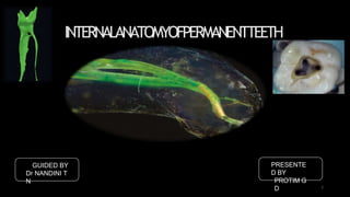
- 1. INTERNALANATOMYOFPERMANENTTEETH GUIDED BY Dr NANDINI T N PRESENTE D BY PROTIM G D 1
- 2. CONTENTS: • INTRODUCTION • PULP CHAMBERANATOMY • APICAL CONSIDERATIONS • CANAL CURVATURESAND TYPES • C SHAPED CANALS • INDIVIDUAL TOOTH ANATOMYANDVARIATIONS • COMMONANOMALIES • CONCLUSION • REFERENCES 2
- 3. INTRODUCTI ON: ROOT CANAL PULP CHAMB ER Versiani MA, Basrani B, Sousa-Neto MD, editors. The root canal anatomy in permanent dentition. Springer; 2018 Jun 18. Ingle JI, Bakland LK, Baumgartner JC. Ingle's endodontics/John I. Ingle, Leif K. Bakland, J. Craig Baumgartner. Hamilton, Ont.:BC Decker,; 2008. Hargreaves KM, Berman LH. Cohen's pathways of the pulp expert consult. Elsevier Health Sciences; 2015 Oct 2. 3 ROO T CANA LS
- 4. INTRODUCTI ON: Latera l canal s Apical foramin a Accessor y canals Apical ramification s Furcatio n canals Canal orifice s Versiani MA, Basrani B, Sousa-Neto MD, editors. The root canal anatomy in permanent dentition. Springer; 2018 Jun 18. Ingle JI, Bakland LK, Baumgartner JC. Ingle's endodontics/John I. Ingle, Leif K. Bakland, J. Craig Baumgartner. Hamilton, Ont.:BC Decker,; 2008. Hargreaves KM, Berman LH. Cohen's pathways of the pulp expert consult. Elsevier Health Sciences; 2015 Oct 2. 4
- 5. INTRODUCTI ON: Versiani MA, Basrani B, Sousa-Neto MD, editors. The root canal anatomy in permanent dentition. Springer; 2018 Jun 18. Ingle JI, Bakland LK, Baumgartner JC. Ingle's endodontics/John I. Ingle, Leif K. Bakland, J. Craig Baumgartner. Hamilton, Ont.:BC Decker,; 2008. Hargreaves KM, Berman LH. Cohen's pathways of the pulp expert consult. Elsevier Health Sciences; 2015 Oct 2. 5
- 6. PULP CHAMBER ANATOMY: 6 Pulp chamber roof Cusps, mamelons, incisal edges Projections and prominences Pulp horns Versiani MA, Basrani B, Sousa-Neto MD, editors. The root canal anatomy in permanent dentition. Springer; 2018 Jun 18. Ingle JI, Bakland LK, Baumgartner JC. Ingle's endodontics/John I. Ingle, Leif K. Bakland, J. Craig Baumgartner. Hamilton, Ont.:BC Decker,; 2008. Hargreaves KM, Berman LH. Cohen's pathways of the pulp expert consult. Elsevier Health Sciences; 2015 Oct 2.
- 7. 7 PULP CHAMBER ANATOMY: Acc to Krasner and Rankow, Versiani MA, Basrani B, Sousa-Neto MD, editors. The root canal anatomy in permanent dentition. Springer; 2018 Jun 18. Ingle JI, Bakland LK, Baumgartner JC. Ingle's endodontics/John I. Ingle, Leif K. Bakland, J. Craig Baumgartner. Hamilton, Ont.:BC Decker,; 2008. Hargreaves KM, Berman LH. Cohen's pathways of the pulp expert consult. Elsevier Health Sciences; 2015 Oct 2.
- 8. 8 PULP CHAMBER ANATOMY: Magnificatio n Ultrasound tips Illuminatio n Additional aids! Versiani MA, Basrani B, Sousa-Neto MD, editors. The root canal anatomyin permanent dentition. Springer; 2018 Jun18.
- 9. 9 PULP CHAMBER ANATOMY: Versiani MA, Basrani B, Sousa-Neto MD, editors. The root canal anatomy in permanent dentition. Springer; 2018 Jun 18.
- 10. 10 CLASSIFICATI ON: Acc to Weine et al, Radiograph ic Sectionin g Division of main root canal from pulp chamber to root apex Weine FS. Endodontic therapy. CV Mosby; 2003 Sep 1.
- 11. 11 CLASSIFICATI ON: Acc to Vertucci et al, 200 maxillary 2nd premolars Dye staining Ingle JI, Bakland LK, Baumgartner JC. Ingle's endodontics/John I. Ingle, Leif K. Bakland, J. Craig Baumgartner. Hamilton, Ont.: BCDecker,; Hargreaves KM, Berman LH. Cohen's pathways of the pulp expert consult. Elsevier Health Sciences; 2008.
- 12. 12 CLASSIFICATI ON: Acc to Gulabivala et al, Burmese population Hargreaves KM, Berman LH. Cohen's pathways of the pulp expert consult. Elsevier Health Sciences; Ingle JI, Bakland LK, Baumgartner JC. Ingle's endodontics/John I. Ingle, Leif K. Bakland, J. Craig Baumgartner. Hamilton, Ont.:BC Decker,; 2008.
- 13. CLASSIFICATI ON: Acc to Sert and Beyarli, 1400 male and female Turkish origin 14 additional morpholog y Male mandibular central Female mandibular central Female mandibular central Female mandibular central Male mandibula r canine 1-3-1- 2 1-2-4- 2 1-3-4- 1 1-2-3-2-1- 3 1-2-4- 31 Sert S, Bayirli GS. Evaluation of the root canal configurations of the mandibular and maxillary permanent teeth by gender in the Turkishpo1p3ulation. Journal of endodontics. 2004 Jun 1;30(6):391-8.
- 14. CLASSIFICATI ON: Acc to Sert and Beyarli, 1400 male and female Turkish origin 14 additional morpholog y Female mandibular central Female mandibular lateral Female mandibular central Female mandibular lateral Female mandibular central 2-3- 2 1-2-3- 2 1-2-3-2- 1 1-2-3- 1 2-1/2- 1 Sert S, Bayirli GS. Evaluation of the root canal configurations of the mandibular and maxillary permanent teeth by gender in the Turkishpo1p4ulation. Journal of endodontics. 2004 Jun 1;30(6):391-8.
- 15. 15 CLASSIFICATI ON: Acc to Sert and Beyarli, 1400 male and female Turkish origin 14 additional morpholog y Male maxillary 1st molar Female maxillary 2nd molar Female maxillary 2nd molar (single root) Male maxillary first molar 3-2- 1 2-3-1-3-1- 4 2-3-2-1- 2 3-2- 1 Sert S, Bayirli GS. Evaluation of the root canal configurations of the mandibular and maxillary permanent teeth by gender in the Turkishpopulation. Journal of endodontics. 2004 Jun 1;30(6):391-8.
- 17. CLASSIFICATI ON: Need for new classification??? 17 Ahmed HM, Versiani MA, De‐Deus G, Dummer PM. A new system for classifying root and root canal morphology.International endodontic journal. 2017Aug;50(8):761-70.
- 18. CLASSIFICATI ON: Ahmed HM, Versiani MA, De‐Deus G, Dummer PM. A new system for classifying root and root canal morphology. International endodontic journal. 2017Aug;50(8):761-70. Versiani MA, Basrani B, Sousa-Neto MD, editors. The root canal anatomy in permanent dentition. Springer; 2018 Jun 18. 18
- 19. CLASSIFICATI ON: Ahmed HM, Versiani MA, De‐Deus G, Dummer PM. A new system for classifying root and root canal morphology. International endodontic journal. 2017Aug;50(8):761-70. Versiani MA, Basrani B, Sousa-Neto MD, editors. The root canal anatomy in permanent dentition. Springer; 2018 Jun 18. 19
- 20. CLASSIFICATI ON: Ahmed HM, Versiani MA, De‐Deus G, Dummer PM. A new system for classifying root and root canal morphology. International endodontic journal. 2017Aug;50(8):761-70. Versiani MA, Basrani B, Sousa-Neto MD, editors. The root canal anatomy in permanent dentition. Springer; 2018 Jun 18. 20
- 21. 21 ISTHMU S: Root canal Lateral canals (isthmuse s, accessory canals) Main canal Hargreaves KM, Berman LH. Cohen's pathways of the pulp expert consult. Elsevier Health Sciences; Ingle JI, Bakland LK, Baumgartner JC. Ingle's endodontics/John I. Ingle, Leif K. Bakland, J. Craig Baumgartner. Hamilton, Ont.: BCDecker,; 2008.
- 22. ISTHMU S: An isthmus also known as transverse anastomosis or a narrow ribbon shaped communication between two root canals that may contain vital tissue, necrotic pulps, biofilms or residual filling material.- Cohen 11th edition Type of tooth Root level Patient age Hsu YY, Kim S. The resected root surface. The issue of canal isthmuses. Dental Clinics of NorthAmerica. 1997 Jul;41(3):529-40. 22 Hargreaves KM, Berman LH. Cohen's pathways of the pulp expert consult. Elsevier Health Sciences;
- 23. ISTHMU S: Classificatio n Acc to Hsu and Kim, Hsu YY, Kim S. The resected root surface. The issue of canal isthmuses. Dental Clinics of NorthAmerica. 1997 Jul;41(3):529-40. 23 Hargreaves KM, Berman LH. Cohen's pathways of the pulp expert consult. Elsevier Health Sciences;
- 24. ISTHMU S: Micro-CT images Versiani MA, Basrani B, Sousa-Neto MD, editors. The root canal anatomy in permanent dentition. Springer; 2018 Jun 18. 24
- 25. 25 ISTHMU S: Prevalence rates 15% in anterior teeth Maxillary premolars 16% apical third 52% middle third More prevalent in MB root of maxillary molars 30-50% junction of middle and apical third Mesial roots of mandibular molars 80% junction of middle and apical third More in distal root Versiani MA, Basrani B, Sousa-Neto MD, editors. The root canal anatomy in permanent dentition. Springer; 2018 Jun 18. Ingle JI, Bakland LK, Baumgartner JC. Ingle's endodontics/John I. Ingle, Leif K. Bakland, J. Craig Baumgartner. Hamilton, Ont.:BC Decker,; 2008. Hargreaves KM, Berman LH. Cohen's pathways of the pulp expert consult. Elsevier Health Sciences; 2015 Oct 2.
- 26. 26 ACCESSORY CANALS: Accessory canals are minute canals that extend in a horizontal, vertical and lateral direction from pulp space towards the periodontium. Apic al third 74% Middl e third 11% Coron al third 15% Versiani MA, Basrani B, Sousa-Neto MD, editors. The root canal anatomy in permanent dentition. Springer; 2018 Jun 18. Ingle JI, Bakland LK, Baumgartner JC. Ingle's endodontics/John I. Ingle, Leif K. Bakland, J. Craig Baumgartner. Hamilton, Ont.:BC Decker,; 2008. Hargreaves KM, Berman LH. Cohen's pathways of the pulp expert consult. Elsevier Health Sciences; 2015 Oct 2.
- 27. 27 ACCESSORY CANALS: Entrapment of periodontal vessels Calcificatio n Hertwigs epithelial root sheath Passage of irritants Pulp to periodontium Accessory canal located in the coronal and middle third of root- Lateral canal Accessory canal located in the bifurcation and trifurcation of multirooted teeth- Furcation canals Versiani MA, Basrani B, Sousa-Neto MD, editors. The root canal anatomy in permanent dentition. Springer; 2018 Jun 18. Ingle JI, Bakland LK, Baumgartner JC. Ingle's endodontics/John I. Ingle, Leif K. Bakland, J. Craig Baumgartner. Hamilton, Ont.:BC Decker,; 2008. Hargreaves KM, Berman LH. Cohen's pathways of the pulp expert consult. Elsevier Health Sciences; 2015 Oct 2.
- 28. 28 ACCESSORY CANALS: Acc to Vertucci and Williams, Mandibular first molars Ingle JI, Bakland LK, Baumgartner JC. Ingle's endodontics/John I. Ingle, Leif K. Bakland, J. Craig Baumgartner. Hamilton, Ont.: BCDecker,; 2008. Hargreaves KM, Berman LH. Cohen's pathways of the pulp expert consult. Elsevier Health Sciences;
- 29. 29 ACCESSORY CANALS: Versiani MA, Basrani B, Sousa-Neto MD, editors. The root canal anatomy in permanent dentition. Springer; 2018 Jun 18.
- 30. ACCESSORY CANALS: Versiani MA, Basrani B, Sousa-Neto MD, editors. The root canal anatomy in permanent dentition. Springer; 2018 Jun 18. 30
- 31. ACCESSORY CANALS: Micro-CT Mandibular premolar Versiani MA, Basrani B, Sousa-Neto MD, editors. The root canal anatomy in permanent dentition. Springer; 2018 Jun 18. 31
- 32. ACCESSORY CANALS: Identification??? ?? Localized thickening of PDL Lesion lateral surface of the root CBC T Acc to Weine, lateral lesions radiographically can be classified as: Type I Type II Type III Versiani MA, Basrani B, Sousa-Neto MD, editors. The root canal anatomy in permanent dentition. Springer; 2018 Jun 18. Ingle JI, Bakland LK, Baumgartner JC. Ingle's endodontics/John I. Ingle, Leif K. Bakland, J. Craig Baumgartner. Hamilton, Ont.:BC Decker,; 2008. Hargreaves KM, Berman LH. Cohen's pathways of the pulp expert consult. Elsevier Health Sciences; 2015 Oct 2. 32
- 33. ACCESSORY CANALS: Versiani MA, Basrani B, Sousa-Neto MD, editors. The root canal anatomy in permanent dentition. Springer; 2018 Jun 18. Ingle JI, Bakland LK, Baumgartner JC. Ingle's endodontics/John I. Ingle, Leif K. Bakland, J. Craig Baumgartner. Hamilton, Ont.:BC Decker,; 2008. Hargreaves KM, Berman LH. Cohen's pathways of the pulp expert consult. Elsevier Health Sciences; 2015 Oct 2. 33
- 34. CANAL CURVATURE: Pruett’ s metho d Schneider’ s method Weine’s method Versiani MA, Basrani B, Sousa-Neto MD, editors. The root canal anatomy in permanent dentition. Springer; 2018 Jun 18. 34
- 35. 35 CANAL CURVATURE: Schneider ’s method Line drawn parallel to the long axis of canal Second line drawn from apical foramen to intersect with the first line at a point where the canal began to leave the long axis Measured in degrees Straight (<5 degrees) Moderate 10-20 degree Severe 25-70 degree Schneider SW. A comparison of canal preparations in straight and curved root canals. Oral surgery, Oral medicine, Oral pathology. 1971 Aug 1;32(2):271-5.
- 36. 36 Weine’ s metho d CANAL CURVATURE: Weine FS. Endodontic therapy. CV Mosby; 2003 Sep 1. Straight line drawn from canal orifice to the point of curvature Second line drawn from the apex of apical curvature to the first line Angle measured at the point of intersection
- 37. 37 CANAL CURVATURE: Pruett’ s metho d Angle of curvature Radius of curvature Length of radius of circle measured in millimetres Abruptness/severity Smaller r.o.c More abrupt a.o.c Effect on instrument fatigue Pruett JP, Clement DJ, Carnes Jr DL. Cyclic fatigue testing of nickel-titanium endodontic instruments. Journal of endodontics. 1997 Feb 1;23(2):77-85.
- 38. 38 CANAL CURVATURE: Coronal and apical part of rc curvature Schneid er Heigh t radiu s Canal access angle Distanc e Most accurate First study to use CAA for calculation of root canal curvature Acc to Shiva Sadeghi Sadeghi S, Poryousef V.A novel approach in assessment of root canal curvature. Iranian endodontic journal.2009;4(4):131.
- 39. 39 APICAL CONSIDERATIONS: The apical anatomy of root canal is complex and variable which includes ramifications and lateral canals that are frequently present. Landmark studies, Dummer, 1984 Wu, 2000 Kuttler, 1955 Stein, 1990 DUMMER PM, McGINN JH, REES DG. The position and topography of the apical canal constriction and apical foramen. International Endodontic Journal. 1984 Oct;17(4):192-8. Kuttler Y. Microscopic investigation of root apexes. The Journal of the American Dental Association. 1955 May 1;50(5):544-52. Wu MK, Wesselink PR, Walton RE. Apical terminus location of root canal treatment procedures. Oral Surgery, Oral Medicine, Oral Pathology, Oral Radiology, and Endodontology. 2000 Jan 1;89(1):99-103.
- 40. 40 APICAL CONSIDERATIONS: Apical delta Intricate system of spaces within the root canal that allows free passage of blood vessels and nerves from periapical compartment to the pulp tissue. Versiani MA, Basrani B, Sousa-Neto MD, editors. The root canal anatomy in permanent dentition. Springer; 2018 Jun 18. Ingle JI, Bakland LK, Baumgartner JC. Ingle's endodontics/John I. Ingle, Leif K. Bakland, J. Craig Baumgartner. Hamilton,Ont.: BC Decker,; 2008.
- 41. 41 APICAL CONSIDERATIONS: Ricucci and Siqueira, 75% prevalence of apical deltas Vertucci, 73.5% in apical third, 11% in middle third and 15% in coronal third De Deus, apical deltas seen more common in posterior teeth and greater incidence in apical region Literature review DUMMER PM, McGINN JH, REES DG. The position and topography of the apical canal constriction and apical foramen. International Endodontic Journal. 1984 Oct;17(4):192-8. Stein TJ.Anatomy of root apex and histological changes with age. Oral Surg Oral Med Oral Pathol. 1990;69:266-70. • Kuttler Y. Microscopic investigation of root apexes. The Journal of the American Dental Association. 1955 May 1;50(5):544-52.
- 42. 42 APICAL CONSIDERATIONS: Acc to Dummer, Mean apex to foramen distance 0.38mm 0.36mm Only anteriors Similar to Chapman(1969) DUMMER PM, McGINN JH, REES DG. The position and topography of the apical canal constriction and apical foramen. International Endodontic Journal. 1984 Oct;17(4):192-8.
- 43. 43 APICAL CONSIDERATIONS: Acc to Kuttler, The narrowest part of the canal in all tooth types is an approx. 0.59mm from the foramen. 0.5mm in young teeth 0.75mm in adults Acc to Chapman, Vast majority of constrictions were at 0.5-1mm from apex DUMMER PM, McGINN JH, REES DG. The position and topography of the apical canal constriction and apical foramen. International Endodontic Journal. 1984 Oct;17(4):192-8. Stein TJ.Anatomy of root apex and histological changes with age. Oral Surg Oral Med Oral Pathol. 1990;69:266-70.
- 44. 44 APICAL CONSIDERATIONS: Apical constriction is the narrowest part of the canal Limit for instrumentation and obturation Mean distance between major and minor apical diameter 0.5mm young 0.67mm older en. Internat DUMMER PM, McGINN JH, REES DG. The position and topography of the apical canal constriction andapic Endodontic Journal. 1984 Oct;17(4):192-8. Stein TJ.Anatomy of root apex and histological changes with age. Oral Surg Oral Med Oral Pathol. 1990;69:266-70.
- 45. APICAL CONSIDERATIONS: Size of apical foramina Ingle JI, Bakland LK, Baumgartner JC. Ingle's endodontics/John I. Ingle, Leif K. Bakland, J. Craig Baumgartner. Hamilton, Ont.: BC Decker,;2008 45
- 46. APICAL LIMIT OF INSTRUMENTATION: Controversial? ?? Ricucci D. Apical limit of root canal instrumentation and obturation, part 1. Literature review. International Endodontic Journal. 1998 Nov;31(6):384- 93. DUMMER PM, McGINN JH, REES DG. The position and topography of the apical canal constriction and apical foramen. InternationalEndodontic Journal. 1984 Oct;17(4):192-8. Wu MK, Wesselink PR, Walton RE. Apical terminus location of root canal treatment procedures. Oral Surgery, Oral Medicine, Oral Pathology, Oral Radiology, and Endodontology. 2000 Jan 1;89(1):99-103. The apical constriction is advocated generally to be the ideal and most practical point of termination of endodontic procedures and results in optimal healing conditions. Necrotic pulp??? Vital pulp??? 46
- 47. 47 APICAL LIMIT OF INSTRUMENTATION: Controversial? ?? Ricucci D, Russo J, Rutberg M, Burleson JA, Spångberg LS. A prospective cohort study of endodontic treatments of 1,369 root canals: results after 5 years. Oral Surgery, Oral Medicine, Oral Pathology, Oral Radiology, and Endodontology. 2011 Dec 1;112(6):825-42. Wu MK, Wesselink PR, Walton RE. Apical terminus location of root canal treatment procedures. Oral Surgery, Oral Medicine, Oral Pathology, Oral Radiology, and Endodontology. 2000 Jan 1;89(1):99- Vital pulp??? 2-3 mm short of the apex Prevents extrusion into the periradicular spaces Promotes healing Apical 3mm-pulp stump Necrotic pulp??? At or within 2mm of radiographic apex Apical level of bacterial infection Clinical identification difficult As close to canal terminus as possible
- 48. C SHAPED CANALS: Failure of hertwig’s epithelial root sheath to fuse on the lingual or buccal surface. Cooke and Cox, 1979 Versiani MA, Basrani B, Sousa-Neto MD, editors. The root canal anatomy in permanent dentition. Springer; 2018 Jun 18. 48
- 49. C SHAPED CANALS: Ribbon shaped Acc to Melton in 1991, Type I Type II Type III Versiani MA, Basrani B, Sousa-Neto MD, editors. The root canal anatomy in permanent dentition. Springer; 2018 Jun 18. 49
- 50. C SHAPED CANALS: Fan et al Micro-CT sections Type I Type II Type III Type IV Type V Versiani MA, Basrani B, Sousa-Neto MD, editors. The root canal anatomy in permanent dentition. Springer; 2018 Jun 18. 50
- 51. C SHAPED CANALS: Radiographic basis by Fan, Mesial and distal canal merged and exited as one (I) Mesial and distal canal exited as 2 separate (II) One canal superimposed, other continuous to apex (III) Versiani MA, Basrani B, Sousa-Neto MD, editors. The root canal anatomy in permanent dentition. Springer; 2018 Jun 18. 51
- 52. C SHAPED CANALS: Micro-CT images 52 Versiani MA, Basrani B, Sousa-Neto MD, editors. The root canal anatomy in permanent dentition. Springer; 2018 Jun 18.
- 53. C SHAPED CANALS: Micro-CT images Versiani MA, Basrani B, Sousa-Neto MD, editors. The root canal anatomy in permanent dentition. Springer; 2018 Jun 18. 53
- 54. C SHAPED CANALS: Gao et al, Based on 3- D imaging Versiani MA, Basrani B, Sousa-Neto MD, editors. The root canal anatomy in permanent dentition. Springer; 2018 Jun 18. 54
- 55. C SHAPED CANALS: Versiani MA, Basrani B, Sousa-Neto MD, editors. The root canal anatomy in permanent dentition. Springer; 2018 Jun 18. 55
- 56. C SHAPED CANALS: Versiani MA, Basrani B, Sousa-Neto MD, editors. The root canal anatomy in permanent dentition. Springer; 2018 Jun 18. 56
- 57. C SHAPED CANALS: Versiani MA, Basrani B, Sousa-Neto MD, editors. The root canal anatomy in permanent dentition. Springer; 2018 Jun 18. 57
- 58. C SHAPED CANALS: Min et al, Morphology of pulp chamber floor 3mm above the orifice Versiani MA, Basrani B, Sousa-Neto MD, editors. The root canal anatomy in permanent dentition. Springer; 2018 Jun 18. 58
- 59. C SHAPED CANALS: Descending canal morphology 2mm below orifice 1/3rd between orifice and apex Midpoint between orifice and apex( MB, ML) (MB ML and distal) 3mm from the apex Versiani MA, Basrani B, Sousa-Neto MD, editors. The root canal anatomy in permanent dentition. Springer; 2018 Jun 18. 59
- 60. 60 C SHAPED CANALS: Prevalenc e Highest prevalence in mandibular 2nd molars- 2.7% to 44.9% Mandibular premolars Asian population Chinese and mongoloid Decker,; 2008. Hargreaves KM, Berman LH. Cohen's pathways of the pulp expert consult. Elsevier Health Sciences; Cooke HG, Cox FL. C-shaped canal configurations in mandibular molars. The Journal of theAmerican DentalAssociation. 1979 Nov 1;99(5):836- 9. Versiani MA, Basrani B, Sousa-Neto MD, editors. The root canal anatomy in permanent dentition. Springer; 2018 Jun 18. Ingle JI, Bakland LK, Baumgartner JC. Ingle's endodontics/John I. Ingle, Leif K. Bakland, J. Craig Baumgartner. Hamilton, Ont.: BC
- 61. 61 INDIVIDUAL TOOTH ANATOMY: Maxillary central incisor Single root canal Wider mesiodistally Oval in adults Diameter of foramina CEJ triangular in younger 3 pulp horns 0.4mm major 0.2mm minor 12% exhibited accessory foramina Versiani MA, Basrani B, Sousa-Neto MD, editors. The root canal anatomy in permanent dentition. Springer; 2018 Jun 18. Ingle JI, Bakland LK, Baumgartner JC. Ingle's endodontics/John I. Ingle, Leif K. Bakland, J. Craig Baumgartner. Hamilton, Ont.: BC Decker,;2008
- 62. 62 INDIVIDUAL TOOTH ANATOMY: Variation s Shovel shaped incisor crowns common in Asian population Acc to Pecora and Cruz Filho Radicular grooves seen in 0.9% of population Fusion Gemination Dens invaginatus Hargreaves KM, Berman LH. Cohen's pathways of the pulp expert consult. Elsevier Health Sciences; 2015 Oct 2.
- 63. INDIVIDUAL TOOTH ANATOMY: Case report One main canal 2 distinct canals in apical third Only case report with type V morphology Calvert G. Maxillary central incisor with type V canal morphology: Case report and literature review. Journal of endodontics. 2014 Oct 1;40(10):1684-7. 63
- 64. INDIVIDUAL TOOTH ANATOMY: Maxillary lateral incisor Mostly single rooted 2 rooted documented Fusion / Gemination Macrodont crown Developmental radicular groove Versiani MA, Basrani B, Sousa-Neto MD, editors. The root canal anatomy in permanent dentition. Springer; 2018 Jun 18. 64 Ingle JI, Bakland LK, Baumgartner JC. Ingle's endodontics/John I. Ingle, Leif K. Bakland, J. Craig Baumgartner. Hamilton, Ont.: BC Decker,;2008
- 65. INDIVIDUAL TOOTH ANATOMY: Maxillary lateral incisor Diameter of foramina 0.2mm accessory 0.4mm major Major apical foramen to anatomic apex 0.3mm 2 pulp horns Versiani MA, Basrani B, Sousa-Neto MD, editors. The root canal anatomy in permanent dentition. Springer; 2018 Jun 18. 65 Ingle JI, Bakland LK, Baumgartner JC. Ingle's endodontics/John I. Ingle, Leif K. Bakland, J. Craig Baumgartner. Hamilton, Ont.: BC Decker,;2008
- 66. INDIVIDUAL TOOTH ANATOMY: Variation s Fusio n Accessory root Geminatio n Dens invaginatus Versiani MA, Basrani B, Sousa-Neto MD, editors. The root canal anatomy in permanent dentition. Springer; 2018 Jun 18. 66 Ingle JI, Bakland LK, Baumgartner JC. Ingle's endodontics/John I. Ingle, Leif K. Bakland, J. Craig Baumgartner. Hamilton, Ont.: BC Decker,;2008
- 67. INDIVIDUAL TOOTH ANATOMY: Maxillary canine Wider labiolingually No pulp horn Single canal 2 canal (3.5%) Join in apical third Exit single foramen Versiani MA, Basrani B, Sousa-Neto MD, editors. The root canal anatomy in permanent dentition. Springer; 2018 Jun 18. 67 Ingle JI, Bakland LK, Baumgartner JC. Ingle's endodontics/John I. Ingle, Leif K. Bakland, J. Craig Baumgartner. Hamilton, Ont.: BC Decker,;2008
- 68. INDIVIDUAL TOOTH ANATOMY: Maxillary canine Diameter of foramina 0.2mm accessory 0.5mm major Major apical foramen to anatomic apex 0.3mm Accessory foramina 12% Versiani MA, Basrani B, Sousa-Neto MD, editors. The root canal anatomy in permanent dentition. Springer; 2018 Jun 18. 68 Ingle JI, Bakland LK, Baumgartner JC. Ingle's endodontics/John I. Ingle, Leif K. Bakland, J. Craig Baumgartner. Hamilton, Ont.: BC Decker,;2008
- 69. 69 INDIVIDUAL TOOTH ANATOMY: Variation s Dens invaginatus Tubercle/talon cusp Lingual surface of crown Dens invaginatus 2 roots Ingle JI, Bakland LK, Baumgartner JC. Ingle's endodontics/John I. Ingle, Leif K. Bakland, J. Craig Baumgartner. Hamilton, Ont.: BC Decker,;2008 Hargreaves KM, Berman LH. Cohen's pathways of the pulp expert consult. Elsevier Health Sciences; 2015 Oct 2.
- 70. 70 INDIVIDUAL TOOTH ANATOMY: Maxillary first premolar Ethnicity Asian higher incidence one canal Mostly 2 root canals Palatal aspect of buccal groove Developmental depression Versiani MA, Basrani B, Sousa-Neto MD, editors. The root canal anatomy in permanent dentition. Springer; 2018 Jun 18. Ingle JI, Bakland LK, Baumgartner JC. Ingle's endodontics/John I. Ingle, Leif K. Bakland, J. Craig Baumgartner. Hamilton, Ont.: BC Decker,;2008
- 71. 71 INDIVIDUAL TOOTH ANATOMY: Maxillary first premolar Palatal orifice slightly larger 2 pulp horns Buccal larger Pulp chamber wider buccolingually 2 canals Buccal, palatal 3 canals MB DB Palatal Longitudinal sulcus Buccal surface of buccal root 2 canals in buccal root Acc to Mattuell a et al, Green D. Stereomicroscopic study of 700 root apices of maxillary and mandibular posterior teeth. Oral Surgery, Oral Medicine, Oral Pathology and Oral Radiology. 1960 Jun 1;13(6):728-33.
- 72. 72 INDIVIDUAL TOOTH ANATOMY: Micro- CT Hargreaves KM, Berman LH. Cohen's pathways of the pulp expert consult. Elsevier Health Sciences; 2015 Oct 2.
- 73. 73 INDIVIDUAL TOOTH ANATOMY: Maxillary 2nd premolar Wider buccolingually 1, 2 or 3 roots and canals 2 pulp horns Buccal larger Versiani MA, Basrani B, Sousa-Neto MD, editors. The root canal anatomy in permanent dentition. Springer; 2018 Jun 18. Ingle JI, Bakland LK, Baumgartner JC. Ingle's endodontics/John I. Ingle, Leif K. Bakland, J. Craig Baumgartner. Hamilton, Ont.: BC Decker,;2008
- 74. 74 INDIVIDUAL TOOTH ANATOMY: variation s Taurodontis m Distal root concavity Dens invaginatus 3 roots with 3 canals Often bilateral Green D. Stereomicroscopic study of 700 root apices of maxillary and mandibular posterior teeth. Oral Surgery, Oral Medicine, Oral Pathology and Oral Radiology. 1960 Jun 1;13(6):728-33. Ingle JI, Bakland LK, Baumgartner JC. Ingle's endodontics/John I. Ingle, Leif K. Bakland, J. Craig Baumgartner. Hamilton, Ont.:BC Decker,; 2008
- 75. 75 INDIVIDUAL TOOTH ANATOMY: Micro- CT Hargreaves KM, Berman LH. Cohen's pathways of the pulp expert consult. Elsevier Health Sciences; 2015 Oct 2.
- 76. 76 INDIVIDUAL TOOTH ANATOMY: Maxillary 1st molar Palatal wider mesiodistally 1 canal MB broad buccolingually 2 canals 3 roots ( MB DB P) Versiani MA, Basrani B, Sousa-Neto MD, editors. The root canal anatomy in permanent dentition. Springer; 2018 Jun 18. Ingle JI, Bakland LK, Baumgartner JC. Ingle's endodontics/John I. Ingle, Leif K. Bakland, J. Craig Baumgartner. Hamilton, Ont.: BC Decker,;2008
- 77. 77 INDIVIDUAL TOOTH ANATOMY: Maxillary 1st molar Palatal root buccal curvature Apical third Depression distal surface of MB root 3 roots most common 95% 2 roots rare 3.8% Fusion (6.2%) Single canal with single apical foramen- predominant DB and P Fusio n Green D. Stereomicroscopic study of 700 root apices of maxillary and mandibular posterior teeth. Oral Surgery, Oral Medicine, Oral Pathology and Oral Radiology. 1960 Jun 1;13(6):728-33.
- 78. 78 INDIVIDUAL TOOTH ANATOMY: Maxillary 1st molar Hargreaves KM, Berman LH. Cohen's pathways of the pulp expert consult. Elsevier Health Sciences; 2015 Oct 2.
- 79. INDIVIDUAL TOOTH ANATOMY: variation s C shaped canals Taurodontism Root fusion Ingle JI, Bakland LK, Baumgartner JC. Ingle's endodontics/John I. Ingle, Leif K. Bakland, J. Craig Baumgartner. Hamilton, Ont.: BC Decker,;2008 79
- 80. 80 INDIVIDUAL TOOTH ANATOMY: Case reports Barbizam JV, Ribeiro RG, Tanomaru Filho M. Unusual anatomy of permanent maxillary molars. Journal of endodontics. 2004 Sep1;30(9):668- 71.
- 81. INDIVIDUAL TOOTH ANATOMY: Case reports Kottoor J, Velmurugan N, Sudha R, Hemamalathi S. Maxillary first molar with seven root canals diagnosed with cone-beam computedtomography scanning: a case report. Journal of endodontics. 2010 May1;36(5):915-21. 81
- 82. INDIVIDUAL TOOTH ANATOMY: Mesiobuccal 2 Greater incidence in males The orifice of MB2 is located either mesial to or in the subpulpal groove within 3.5mm palatally and 2mm Mesially from MB1 Prevalenc e 69.6% Geographic location 82 Martins JN, Marques D, Silva EJ, Caramês J, MataA, Versiani MA. Second mesiobuccal root canal in maxillary molars—A systematic review and meta-analysis of prevalence studies using cone beam computed tomography.Archives of oral biology. 2020 May 1;113:104589.
- 83. 83 INDIVIDUAL TOOTH ANATOMY: Maxillary 2nd molars MB broader b/l Flutes on mesial and distal Closer High incidence of fusion Mostly 3 roots Palatal broad mesiodistally 1 canal in each root Versiani MA, Basrani B, Sousa-Neto MD, editors. The root canal anatomy in permanent dentition. Springer; 2018 Jun 18. Ingle JI, Bakland LK, Baumgartner JC. Ingle's endodontics/John I. Ingle, Leif K. Bakland, J.Craig
- 84. 84 INDIVIDUAL TOOTH ANATOMY: Variation s 4 rooted 2 palatal 1 canal each 41 palatal, 1 MB 2 DB Single rooted Single canal Taurodontis m MB2 31.1%- 46.9% Green D. Stereomicroscopic study of 700 root apices of maxillary and mandibular posterior teeth. Oral Surgery, Oral Medicine, Oral Pathology and Oral Radiology. 1960 Jun 1;13(6):728-33. Ingle JI, Bakland LK, Baumgartner JC. Ingle's endodontics/John I. Ingle, Leif K. Bakland, J. Craig Baumgartner. Hamilton, Ont.:BC Decker,; 2008
- 85. INDIVIDUAL TOOTH ANATOMY: Mandibular centrals Ovoid/ hourglass shaped Single canal Root broad labiolingually Developmental depression Mesial and distal 2 canals 26% Single apical foramen 2 pulp horns Larger buccal Rounded lingual Versiani MA, Basrani B, Sousa-Neto MD, editors. The root canal anatomy in permanent dentition. Springer; 2018 Jun 18. 85 Ingle JI, Bakland LK, Baumgartner JC. Ingle's endodontics/John I. Ingle, Leif K. Bakland, J. Craig Baumgartner. Hamilton, Ont.: BC Decker,;2008
- 86. INDIVIDUAL TOOTH ANATOMY: Mandibular centrals Diameter of foramen Accessory foramen 0.2mm Major foramen 0.3mm AF- AA 0.2M M Talon cusp Labial/lingu al Dens invaginatus Fusion Geminatio n 2 canals 2 separate foramina Versiani MA, Basrani B, Sousa-Neto MD, editors. The root canal anatomy in permanent dentition. Springer; 2018 Jun 18. 86 Ingle JI, Bakland LK, Baumgartner JC. Ingle's endodontics/John I. Ingle, Leif K. Bakland, J. Craig Baumgartner. Hamilton, Ont.: BC Decker,;2008
- 87. 87 INDIVIDUAL TOOTH ANATOMY: Micro- CT Hargreaves KM, Berman LH. Cohen's pathways of the pulp expert consult. Elsevier Health Sciences; 2015 Oct 2.
- 88. 88 INDIVIDUAL TOOTH ANATOMY: Mandibular laterals Canal round/ribbo n shaped Depressions present on mesial and distal root surface Single rooted Pulp chamber wider labiolingually 2 canals 28.1% Versiani MA, Basrani B, Sousa-Neto MD, editors. The root canal anatomy in permanent dentition. Springer; 2018 Jun 18. Ingle JI, Bakland LK, Baumgartner JC. Ingle's endodontics/John I. Ingle, Leif K. Bakland, J. Craig Baumgartner. Hamilton, Ont.: BC Decker,;2008
- 89. 89 INDIVIDUAL TOOTH ANATOMY: variation s Dens invaginatus Fusion Gemination Bifid roots Hargreaves KM, Berman LH. Cohen's pathways of the pulp expert consult. Elsevier Health Sciences; 2015 Oct 2.
- 90. INDIVIDUAL TOOTH ANATOMY: Mandibular canines 2 roots 1.7%- 6.2% Development al depressions Mesial n distal Single root Wider labiolingually Versiani MA, Basrani B, Sousa-Neto MD, editors. The root canal anatomy in permanent dentition. Springer; 2018 Jun 18. 90 Ingle JI, Bakland LK, Baumgartner JC. Ingle's endodontics/John I. Ingle, Leif K. Bakland, J. Craig Baumgartner. Hamilton, Ont.: BC Decker,;2008
- 91. INDIVIDUAL TOOTH ANATOMY: Mandibular canines Single canal Single AF Diameter of foramen Accessory foramen 0.2mm Major foramen 0.3mm AF-AA 0.35m m Fusio n Dens invaginatus 2 canals with single AF 2 roots 2 canals Versiani MA, Basrani B, Sousa-Neto MD, editors. The root canal anatomy in permanent dentition. Springer; 2018 Jun 18. 91 Ingle JI, Bakland LK, Baumgartner JC. Ingle's endodontics/John I. Ingle, Leif K. Bakland, J. Craig Baumgartner. Hamilton, Ont.: BC Decker,;2008
- 92. INDIVIDUAL TOOTH ANATOMY: Versiani MA, Basrani B, Sousa-Neto MD, editors. The root canal anatomy in permanent dentition. Springer; 2018 Jun 18. 92 Ingle JI, Bakland LK, Baumgartner JC. Ingle's endodontics/John I. Ingle, Leif K. Bakland, J. Craig Baumgartner. Hamilton, Ont.: BC Decker,;2008
- 93. INDIVIDUAL TOOTH ANATOMY: Mandibular 1st premolars Ethnic origin 3 rooted rare Tome’s root Single rooted 2 rooted common 2 pulp horns Large buccal Small lingual Versiani MA, Basrani B, Sousa-Neto MD, editors. The root canal anatomy in permanent dentition. Springer; 2018 Jun 18. 93 Ingle JI, Bakland LK, Baumgartner JC. Ingle's endodontics/John I. Ingle, Leif K. Bakland, J. Craig Baumgartner. Hamilton, Ont.: BC Decker,;2008
- 94. INDIVIDUAL TOOTH ANATOMY: Mandibular 1st premolars 27.8% incidence More than one canal- Kartal and Yanikoglu Enigma to endodontists!! Ingle JI, Bakland LK, Baumgartner JC. Ingle's endodontics/John I. Ingle, Leif K. Bakland, J. Craig Baumgartner. Hamilton, Ont.: BC Decker,;2008 94
- 95. INDIVIDUAL TOOTH ANATOMY: Variation s Ingle JI, Bakland LK, Baumgartner JC. Ingle's endodontics/John I. Ingle, Leif K. Bakland, J. Craig Baumgartner. Hamilton, Ont.: BC Decker,;2008 95
- 96. 96 INDIVIDUAL TOOTH ANATOMY: Variation s Dens invaginatus Most common Deep mesial radicular invagination(13- 27%) Multiple apical foramina (15%-20%) More prone to bifurcated canals(23-30%) Type I Most prevalent Kottoor J, Albuquerque D, Velmurugan N, Kuruvilla J. Root anatomy and root canal configuration of human permanent mandibular premolars: a systematic review. Anatomy research international.2013;2013.
- 97. 97 INDIVIDUAL TOOTH ANATOMY: Mandibular 2nd premolars 2 rooted rare (0.3%) Lingual pulp horn larger Developmental depression Distal surface Single rooted Versiani MA, Basrani B, Sousa-Neto MD, editors. The root canal anatomy in permanent dentition. Springer; 2018 Jun 18. Ingle JI, Bakland LK, Baumgartner JC. Ingle's endodontics/John I. Ingle, Leif K. Bakland, J. Craig Baumgartner. Hamilton, Ont.: BC Decker,;2008
- 98. INDIVIDUAL TOOTH ANATOMY: Mandibular 2nd premolars Single AF 91.6% 2 or more canals 8.9% Single canal 91.1% Green D. Stereomicroscopic study of 700 root apices of maxillary and mandibular posterior teeth. Oral Surgery, Oral Medicine, Oral Pathology and Oral Radiology. 1960 Jun 1;13(6):728-33. Ingle JI, Bakland LK, Baumgartner JC. Ingle's endodontics/John I. Ingle, Leif K. Bakland, J. Craig Baumgartner. Hamilton, Ont.: BC Decker,;2008 98
- 99. INDIVIDUAL TOOTH ANATOMY: Variation s Ingle JI, Bakland LK, Baumgartner JC. Ingle's endodontics/John I. Ingle, Leif K. Bakland, J. Craig Baumgartner. Hamilton, Ont.: BC Decker,;2008 99
- 100. 10 0 INDIVIDUAL TOOTH ANATOMY: Mandibular 1st molar Roots broader buccolinguall y 4 pulp horns Occasionally 3 2 roots Versiani MA, Basrani B, Sousa-Neto MD, editors. The root canal anatomy in permanent dentition. Springer; 2018 Jun 18. Ingle JI, Bakland LK, Baumgartner JC. Ingle's endodontics/John I. Ingle, Leif K. Bakland, J. Craig Baumgartner. Hamilton, Ont.: BC Decker,;2008
- 101. 10 1 INDIVIDUAL TOOTH ANATOMY: Mandibular 1st molar Mesia l Dista l MM 1%- 15% Orifices Developmental groove Distal DB and DL Mid distal rare MB and ML MM occasionally 2 distal roots DL root smaller Curves buccally Mesial root curves distally towards apex Green D. Stereomicroscopic study of 700 root apices of maxillary and mandibular posterior teeth.Oral Surgery, Oral Medicine, Oral Pathology and Oral Radiology. 1960 Jun 1;13(6):728-33. Hargreaves KM, Berman LH. Cohen's pathways of the pulp expert consult. Elsevier Health Sciences; 2015 Oct 2.
- 103. INDIVIDUAL TOOTH ANATOMY: Mandibular 1st molar Usually 1 canal Radix paramolaris is an extra root located mesiobuccally Radix entomolaris is a Supernumery root located distolingually 103 Ingle JI, Bakland LK, Baumgartner JC. Ingle's endodontics/John I. Ingle, Leif K. Bakland, J. Craig Baumgartner. Hamilton, Ont.: BC Decker,;2008
- 104. 10 4 INDIVIDUAL TOOTH ANATOMY: No of roots related to ethnicity Type II and IV most common mesial root Distal root Type I foll by type II and IV MM incidence of 2.6% Root canal isthmuses 55% mesial root 20% distal root Mandibular 1st molar de Pablo ÓV, Estevez R, Sánchez MP, Heilborn C, Cohenca N. Root anatomy and canal configuration of the permanent mandibular first molar:a systematic review. Journal of endodontics. 2010 Dec 1;36(12):1919-31.
- 105. INDIVIDUAL TOOTH ANATOMY: Case report Independen t Confluen t Fi n Petridis XM, Dechouniotis GP, Kondylidou V, Georgopoulou MK. Middle mesial canal in mandibular molars: review and clinical case reports. ENDO (Lond Engl). 2012 Jun 1;6(2):143-52. 105
- 106. INDIVIDUAL TOOTH ANATOMY: Case report Petridis XM, Dechouniotis GP, Kondylidou V, Georgopoulou MK. Middle mesial canal in mandibular molars: review and clinical case reports. ENDO (Lond Engl). 2012 Jun 1;6(2):143-52. 106
- 107. INDIVIDUAL TOOTH ANATOMY: Mandibular 2nd molar Single root fusion 21.8% Root concavities Mesial and distal Broader buccolingually 2 roots Close together Roots sweep distally Apices close Versiani MA, Basrani B, Sousa-Neto MD, editors. The root canal anatomy in permanent dentition. Springer; 2018 Jun 18. 107 Ingle JI, Bakland LK, Baumgartner JC. Ingle's endodontics/John I. Ingle, Leif K. Bakland, J. Craig Baumgartner. Hamilton, Ont.: BC Decker,;2008
- 108. INDIVIDUAL TOOTH ANATOMY: Mandibular 2nd molar Root concavities Mesial and distal 1 canal in mesial 14% 2 mesial canals 1 distal Distolingual root incidence 2.2% Highest incidence of c shaped canals Versiani MA, Basrani B, Sousa-Neto MD, editors. The root canal anatomy in permanent dentition. Springer; 2018 Jun 18. 108 Ingle JI, Bakland LK, Baumgartner JC. Ingle's endodontics/John I. Ingle, Leif K. Bakland, J. Craig Baumgartner. Hamilton, Ont.: BC Decker,;2008 May have a common foramen or separate Cooke HG, Cox FL. C-shaped canal configurations in mandibular molars. The Journal of theAmerican DentalAssociation. 1979 Nov 1;99(5):836- 9.
- 109. INDIVIDUAL TOOTH ANATOMY: Variation s Supernumery roots Fused/single root Taurodontis m Versiani MA, Basrani B, Sousa-Neto MD, editors. The root canal anatomy in permanent dentition. Springer; 2018 Jun 18. 109 Ingle JI, Bakland LK, Baumgartner JC. Ingle's endodontics/John I. Ingle, Leif K. Bakland, J. Craig Baumgartner. Hamilton, Ont.: BC Decker,;2008
- 110. INDIVIDUAL TOOTH ANATOMY: Variation s 110 Ingle JI, Bakland LK, Baumgartner JC. Ingle's endodontics/John I. Ingle, Leif K. Bakland, J. Craig Baumgartner. Hamilton, Ont.: BC Decker,;2008
- 111. PULP STONES: Tru e Fals e Pulp stones are compact degenerative masses of calcified tissue Based on morphology Degenerating cells which mineralize Dentine lined by odontoblasts Goga R, Chandler NP, Oginni AO. Pulp stones: a review. International Endodontic Journal. 2008Jun;41(6):457-68. Hargreaves KM, Goodis HE, Tay FR, editors. Seltzer and Bender's dental pulp. Chicago: Quintessence;2012. 111
- 112. PULP STONES: Fre e Embedde d Adheren t Based on location Laminate d Non laminate d Higher incidenc e in molars ( 45.2%) Dentin dysplasia Dentinogenesis imperfect Van Der Woude syndrome Goga R, Chandler NP, Oginni AO. Pulp stones: a review. International Endodontic Journal. 2008Jun;41(6):457-68. Hargreaves KM, Goodis HE, Tay FR, editors. Seltzer and Bender's dental pulp. Chicago: Quintessence;2012. 112
- 113. 113 ANOMALIE S: Rajendran R. Shafer's textbook of oral pathology. Elsevier India; 2009.
- 114. ANOMALIE S: Dens invaginatus Infolding of dental papilla during tooth development Deep infolding of enamel and dentin starting from foramen caecum Socrates in 1856 Maxillary lateral incisors Extend upto roots Rapid and aggressive proliferation of a part of IEE invading dental papilla- Rushton Classification by Oehelers in 1957, Hülsmann M. Dens invaginatus: aetiology, classification, prevalence, diagnosis, and treatment considerations. International endodontic journal. 1997 Mar;30(2):79-90. 114
- 115. ANOMALIE S: Treatmen t Composite filling No communication invagination and RC Separate apical/lateral foramen RC treatment Obturatio n Irrigation followed by ultrasonic Warm vertical Thermoplasti c techniques Efficien t Hülsmann M. Dens invaginatus: aetiology, classification, prevalence, diagnosis, and treatment considerations. International endodontic journal. 1997 Mar;30(2):79-90. 115
- 116. Case report Chen YH, Tseng CC, Harn WM. Dens invaginatus: review of formation and morphology with 2 case reports. Oral Surgery, OralMedicine,1O16ral Pathology, Oral Radiology, and Endodontology. 1998 Sep 1;86(3):347-52.
- 117. 117 ANOMALIE S: Case report Bharti R, Chandra A, Tikku AP, Wadhwani KK. “Taurodontism” an endodontic challenge: a case report. Journal of oral science. 2009;51(3):471-4. Tooth no 16 Canals short and deeply placed orifices Apical third of file Lateral condensation Warm vertical
- 118. EXTERNAL ANATOMICAL CLUES DEPICTING EXTRA CANALS: Gutmann J. Grossman's Endodontic Practice-13 [sup] th Edition. Journal of Conservative Dentistry. 2016 Sep1;19(5):494-. 118
- 120. CLINICAL TIPS: Ruddle CJ. LOCATING CANALS. DENTISTRY TODAY. 2017 Feb. Transilluminatio n Angled radiographs Oval orifice Apically curved small file One orifice not in the centre 2nd canal More separation between orifices Less canal curvature Red line test White line test Buccal/lingual direction 2nd canal First file inserted distal canal of mandibular molar Champagne bubble test Dyes Ultrasonics Magnificatio n 120
- 121. CONCLUSI ON: 121
- 122. REFEREN CES: 122 • Versiani MA, Basrani B, Sousa-Neto MD, editors. The root canal anatomy in permanent dentition. Springer; 2018 Jun 18. • Ingle JI, Bakland LK, Baumgartner JC. Ingle's endodontics/John I. Ingle, Leif K. Bakland, J. Craig Baumgartner. Hamilton,Ont.: BC Decker,; 2008. • Hargreaves KM, Berman LH. Cohen's pathways of the pulp expert consult. Elsevier Health Sciences; 2015 Oct 2. • Weine FS. Endodontic therapy. CV Mosby; 2003 Sep 1. • Sert S, Bayirli GS. Evaluation of the root canal configurations of the mandibular and maxillary permanent teeth by gender inthe Turkish population. Journal of endodontics. 2004 Jun1;30(6):391-8. • Ahmed HM, Versiani MA, De‐Deus G, Dummer PM. A new system for classifying root and root canal morphology.International endodontic journal. 2017Aug;50(8):761-70. • Schneider SW.A comparison of canal preparations in straight and curved root canals. Oral surgery, Oral medicine, Oral pathology. 1971 Aug 1;32(2):271-5. • Pruett JP, Clement DJ, Carnes Jr DL. Cyclic fatigue testing of nickel-titanium endodontic instruments. Journal of endodontics. 1997 Feb 1;23(2):77-85. • Sadeghi S, Poryousef V.A novel approach in assessment of root canal curvature. Iranian endodontic journal.2009;4(4):131. • DUMMER PM, McGINN JH, REES DG. The position and topography of the apical canal constriction and apicalforamen. International Endodontic Journal. 1984 Oct;17(4):192-8. • Stein TJ.Anatomy of root apex and histological changes with age. Oral Surg Oral Med Oral Pathol. 1990;69:266-70. • Kuttler Y. Microscopic investigation of root apexes. The Journal of the American Dental Association. 1955 May 1;50(5):544-52. • Cooke HG, Cox FL. C-shaped canal configurations in mandibular molars. The Journal of theAmerican DentalAssociation. 1979 Nov 1;99(5):836-9. • Calvert G. Maxillary central incisor with type V canal morphology: Case report and literature review. Journal of endodontics. 2014 Oct 1;40(10):1684-7. • Green D. Stereomicroscopic study of 700 root apices of maxillary and mandibular posterior teeth. Oral Surgery, Oral Medicine, Oral Pathology and Oral Radiology. 1960 Jun 1;13(6):728-33. • Barbizam JV, Ribeiro RG, Tanomaru Filho M. Unusual anatomy of permanent maxillary molars. Journal of endodontics.2004 Sep 1;30(9):668-71.
- 123. REFEREN CES: 123 • Kottoor J, Velmurugan N, Sudha R, Hemamalathi S. Maxillary first molar with seven root canals diagnosed with cone-beam computed tomography scanning: a case report. Journal of endodontics. 2010 May 1;36(5):915-21. • Martins JN, Marques D, Silva EJ, Caramês J, Mata A, Versiani MA. Second mesiobuccal root canal in maxillary molars—Asystematic review and meta-analysis of prevalence studies using cone beam computed tomography. Archives of oral biology. 2020 May1;113:104589. • Kottoor J, Albuquerque D, Velmurugan N, Kuruvilla J. Root anatomy and root canal configurationof human permanent mandibular premolars: a systematic review.Anatomy research international. 2013;2013. • de Pablo ÓV, Estevez R, Sánchez MP, Heilborn C, Cohenca N. Root anatomy and canal configuration of the permanent mandibular first molar: a systematic review. Journal of endodontics. 2010 Dec1;36(12):1919-31. • Petridis XM, Dechouniotis GP, Kondylidou V, Georgopoulou MK. Middle mesial canal in mandibularmolars: review and clinical case reports. ENDO (Lond Engl). 2012 Jun1;6(2):143-52. • Goga R, Chandler NP, Oginni AO. Pulp stones: a review. International Endodontic Journal.2008 Jun;41(6):457-68. • Hargreaves KM, Goodis HE, Tay FR, editors. Seltzer and Bender's dental pulp. Chicago: Quintessence; 2012. • Hülsmann M. Dens invaginatus: aetiology, classification, prevalence, diagnosis, and treatment considerations. International endodontic journal. 1997 Mar;30(2):79-90. • Chen YH, Tseng CC, Harn WM. Dens invaginatus: review of formation and morphology with 2 casereports. Oral Surgery, Oral Medicine, Oral Pathology, Oral Radiology, and Endodontology. 1998 Sep 1;86(3):347-52. • Bharti R, Chandra A, Tikku AP, Wadhwani KK. “Taurodontism” an endodontic challenge: a case report. Journal of oral science. 2009;51(3):471-4. • Wu MK, Wesselink PR, Walton RE. Apical terminus location of root canal treatment procedures. Oral Surgery, Oral Medicine, Oral Pathology, Oral Radiology, and Endodontology. 2000 Jan 1;89(1):99-103. • Ricucci D, Russo J, Rutberg M, Burleson JA, Spångberg LS. A prospective cohort study of endodontic treatments of 1,369 root canals: results after 5 years. Oral Surgery, Oral Medicine, OralPathology, Oral Radiology, and Endodontology. 2011 Dec 1;112(6):825-42.
- 124. THANK YOU! 124