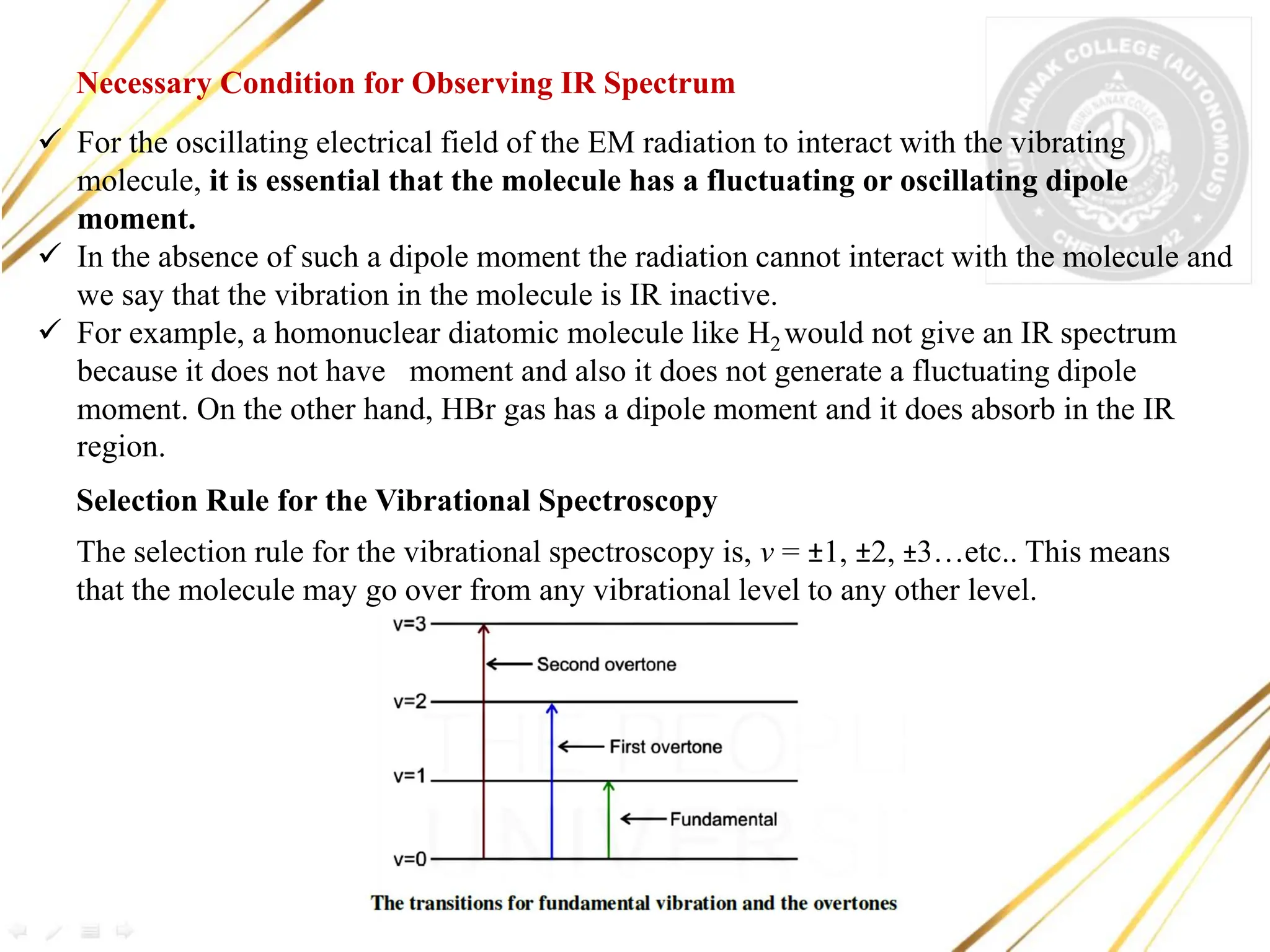
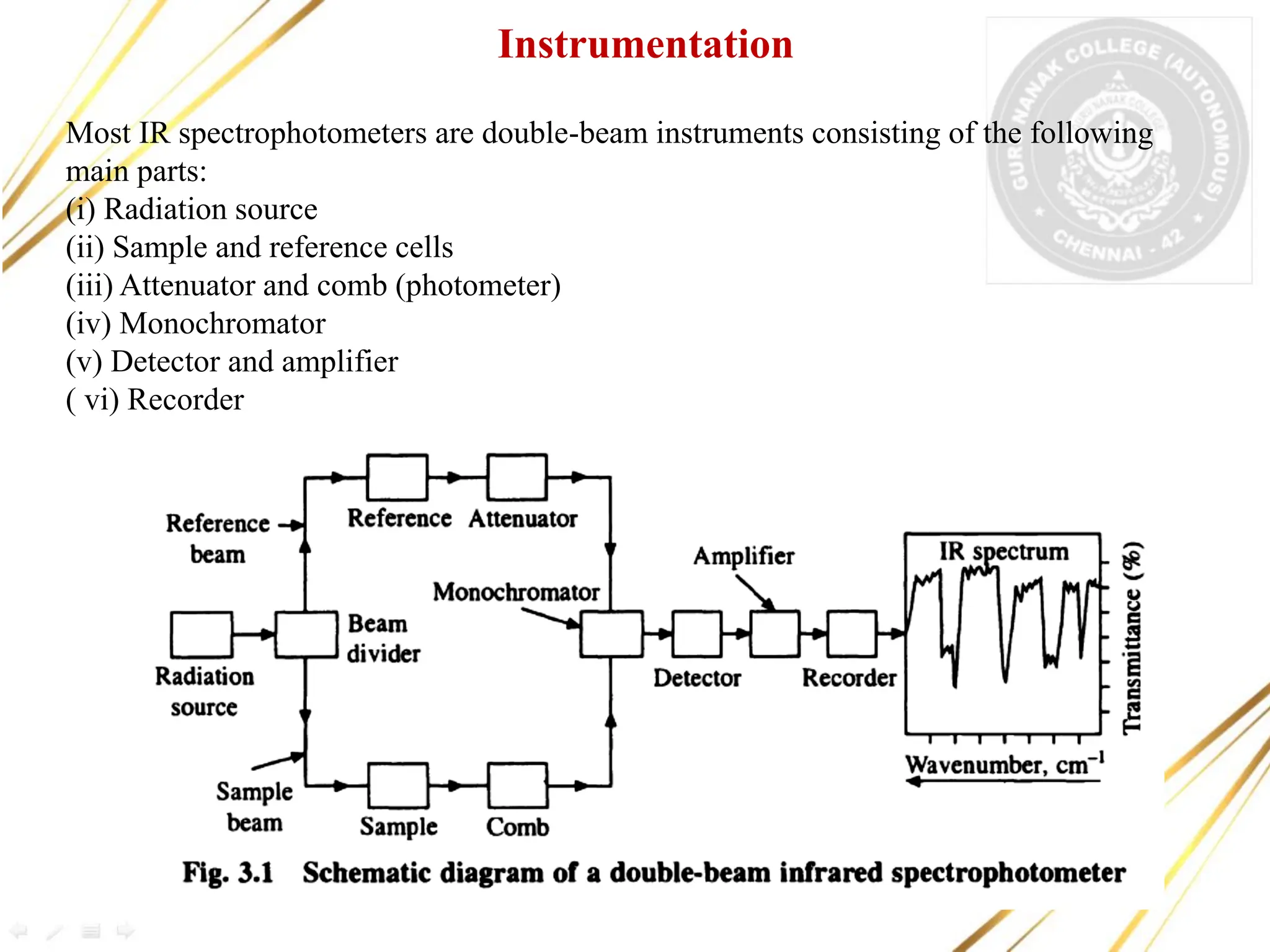
Name of the Paper : III B.Sc. Analytical Chemistry II
Semester : VI
Department :CHEMISTRY
Name of the Faculty : Dr.S.E.NOORJAHAN
Designation : ASSISTANT PROFESSOR
Topic/Title: Infrared Spectroscopy
Learning outcomes
•
• •
A student would have gained knowledge about the
following:
❖ Principle of IR spectroscopy
❖ Instrumentation of IR spectroscopy
❖ Types of vibrations
❖ Spectrum of IR
❖ Fingerprint region
❖ Applications of IR
❖ Spectra of simple molecules
Introduction
▪ Infrared spectroscopy deals with the recording of the absorption of radiations in
the infrared region of the electromagnetic spectrum.
▪ The position of a given infrared absorption is expressed in terms of wavelength in
terms of wavenumber v (cm-1) since it is directly proportional to energy.
▪ The infrared region (4000-600cm-1)practical use to organic chemists.
▪ The absorption of infrared radiation by a molecule occurs due to quantized
vibrational and rotational energy changes when it is subjected to infrared irradiation.
Thus, IR spectra are often called vibrational-rotational spectra.
▪ Unlike UV spectra which have relatively few absorption bands, IR spectra have a
large number of absorption bands and therefore provide plenty of structural
information about a molecule.
▪ Different bands observed in an IR spectrum correspond to various functional groups
and bonds present in the molecule.
▪ Thus, IR spectroscopy is most widely used for the detection of functional groups
▪ and identification of organic compounds.
✓ For the oscillating electrical field of the EM radiation to interact with the vibrating
molecule, it is essential that the molecule has a fluctuating or oscillating dipole
moment.
✓ In the absence of such a dipole moment the radiation cannot interact with the molecule and
we say that the vibration in the molecule is IR inactive.
✓ For example, a homonuclear diatomic molecule like H2 would not give an IR spectrum
because it does not have moment and also it does not generate a fluctuating dipole
moment. On the other hand, HBr gas has a dipole moment and it does absorb in the IR
region.
Necessary Condition for Observing IR Spectrum
Selection Rule for the Vibrational Spectroscopy
The selection rule for the vibrational spectroscopy is, v = ±1, ±2, ±3…etc.. This means
that the molecule may go over from any vibrational level to any other level.
Vibrational Modes or Degrees of Freedom
For a linear molecule containing N atoms, the total vibrational modes are (3N-5) and
for a nonlinear molecule of the same size, the total number of vibrational modes is
(3N-6).
Total degrees of freedom (3N) = Translational + Rotational + Vibrational degrees
of freedom
Where N – number of atoms in a molecule
Thus, the linear CO2 molecule will have 3(3) -5 = 4 vibrational modes whereas, water a
non linear triatomic molecule would have only 3(3)-6 = 3 vibrational modes.
Normal Modes of Vibrations
A molecule can vibrate only in certain modes, known as normal modes.
The two types (modes) of fundamental molecular vibrations known are: (a) stretching
and (b) bending vibrations (deformations).
(i) Stretching Vibrations
In stretching vibrations, the distance between two atoms increases or decreases, but
the atoms remain in the same bond axis. Stretching vibrations are of two types:
(a) Symmetrical stretching. In this mode of vibration, the movement of atoms
with respect to the common (or central) atom is simultaneously in the same
direction along the same bond axis.
(b) Asymmetrical Stretching. In this vibration, one atom approaches the common
atom while the other departs from it.
(ii) Bending Vibrations (Deformations)
In such vibrations, the positions of the atoms change with respect to their original bond
axes (bond angle changes). Bending vibrations are of four types:
(a) Scissoring. In this mode of vibration, the movement of atoms is in the opposite
direction with change in their bond axes as well as in the bond angle they form with the
central atom.
(b) Rocking. In this vibration, the movement of atoms takes place in the same direction
with change in their bond axes. Scissoring and rocking are in-plane bendings.
(c) Wagging. In this vibration, two atoms simultaneously move above and below
the plane with respect to the common atom.
(d) Twisting. In this mode of vibration, one of the atom moves up and the other
moves down the plane with respect to the common atom.
Bending vibrations (deformations) of a CH2 group (+ and- signs indicate
movements perpendicular to the plane of the paper)
Bending vibrations of a band require lesser energy than its stretching vibrations.
Thus, the absorption bands resulting from bending vibrations appear at lower
wavenumbers those that resulting from stretching vibrations of the same band
The carbon dioxide molecule is linear and has four fundamental vibrations (3 x 3)- 5 = 4.
Thus, four theoretical fundamental bands are expected but actually it shows only two. The
symmetrical Stretching vibration in carbon dioxide is IR inactive because it produces no
change in the dipole moment of the molecule. The two bending vibrations are equivalent
and absorb at the same wavenumber (667.3 cm-1)Thus, the IR spectrum of carbon dioxide
shows only two fundamental absorption bands, one at 2350 cm-1 due to asymmetrical
stretching vibration, and the other at 667.3 cm-1 due to the two bending vibrations.
FUNDAMENTAL VIBRATIONS OF CARBON DIOXIDE
Fundamental vibrations of the linear carbon dioxide molecule ( + and - signs
indicate movements perpendicular to the plane of paper)
Calculation of Vibrational Frequencies
The stretching absorption frequency of a bond, can be calculated by Hooke's law
where
m1 +m2 atoms linked to the particular bond, the force constant of the bond in dynes/cm
c the velocity of light (2.998 x 1010 cm/sec) and v the stretching vibrational frequency of
the bond in cm-1 (wavenumber).
The force constant of a bond is related to its bond strength. The value of force constant is
approximately 5 X 10⁵ dynes/cm for single bonds and approximately twice and thrice of
this value for double and triple bonds, respectively.
is the reduced mass, m 1 and m 2 are the masses (g) of the
The frequency of the C-H Stretching vibration can be calculated as follows:
The atomic mass of carbon is 12 and that of hydrogen is 1. Thus,
Most IR spectrophotometers are double-beam instruments consisting of the following
main parts:
(i) Radiation source
(ii) Sample and reference cells
(iii) Attenuator and comb (photometer)
(iv) Monochromator
(v) Detector and amplifier
( vi) Recorder
Instrumentation
Sources of IR Radiation
The best sources available and commonly employed in the IR spectrophotometers are
• Nernst or Glower filament lamp
• Globar
• Ni-Cr (Nichrome) wire
• Carbon dioxide LASER
Nernst Glower: It is a cylindrical rod made up of a mixture of zirconium, yttrium and
erbium oxides (diameter of 12 mm and length of about 20 mm). It is electrically
connected through platinum wire and heated by the passage of an electrical current to a
temperature of about 1200 to 2200 K.
Globar: It is a silicon carbide rod (diameter of 5 mm and length of about 50 mm) which
is electrically heated to about 1500 K. The electrical contacts have a risk of arcing
therefore water cooling of the contacts is used. The spectral output is comparable with the
Nernst glower, except at short wavelengths (less than 5 μm) where its output becomes
larger.
Incandescent nichrome wire: It is a tightly wound coil of nichrome (Ni-Cr) wire,
electrically heated to 1100 K. It produces a lower intensity of radiation than the Nernst or
Globar sources, but has a longer working life.
Carbon dioxide lasers: The tuneable carbon dioxide lasers produce closely spaced
discrete lines in a limited range of IR radiation (900 to 1100 cm1). Though the tuneable
laser has limited range but it provides a much larger intensity of all the lines in the range.
The laser can be tuned to any of these lines and can be used for the quantitative
determination of different species like, ammonia, benzene and nitrogen dioxide etc.
(ii) Sample and Reference Cells
Reference and sample beams pass through the reference cell and sample cell, respectively.
Glass and quartz cannot be used as windows of cells and optical prisms, etc. because they
absorb strongly in most of the IR region. Thus, alkali metal halides such as NaCl, NaBr,
KCl and KBr are most commonly used as these are transparent to most of the IR region.
(iii) Attenuator and Comb (Photometer)
The reference beam passes through the attenuator and the sample beam through the comb.
Then the two beams can be alternately reflected out of the optical system and to the
entrance slit of the monochromator with the help of several mirrors. Thus, the photometer
combines the reference and sample beams into a single beam of alternating segments. The
comb allows balancing of the two beams.
(iv) Monochromator
The combined beam passes through the prism or grating of the monochromator which
disperses the beam into various frequencies. Since the prism or grating rotates slowly, it
sends individual frequency bands to the detector, thus allowing a scan of frequency bands.
Gratings that give better resolutions than prisms consist of a series of parallel and straight
thin lines on a smooth reflecting surface; the spacing between lines is of the order of few
angstrom (Ä) depending on the desired wavelength range.
(v) Detector and Amplifier
The detector is a thermocouple which measures radiant energy by means of its heating
effect that produces current. Due to difference in the intensity of the two beams falling on
the detector, an alternating current starts flowing from the detector to the amplifier where
it is amplified and relayed to the recorder.
(vi) Recorder
It records IR spectra as a plot of wavelengths λ or wavenumbers of absorbed
radiations against the intensity of absorption in terms of transmittance T or Absorbance A.
Sample Handling
Infrared spectra of compounds may be recorded in the vapour phase, as pure liquids, in
solution and in the solid state. The sample should be dry because water absorbs near -3710
cm-1 and near -1630 cm-1•
(i) In Vapour Phase
The vapour or gas is introduced into a special cell which is usually about 10 cm long and the
walls of its both the ends are normally made of NaCl which is transparent to IR radiation.
The vapour phase technique is limited because of the too low vapour pressure of most
organic compounds to produce a useful absorption spectrum.
(ii) As a Liquid Film
A drop of neat liquid is placed between two flat plates of NaCl to give a thin film. Thick
samples of neat liquids usually absorb too strongly to give satisfactory spectrum. This is the
simplest of all sampling techniques.
(iii) In solution
Usually, a 1-5% solution of the compound is introduced into a special cell of 0.1-1 mm
thickness and made of NaCl. The most commonly used solvents in IR spectroscopy are
carbon disulphide, carbon tetrachloride and chloroform. The selected solvent should be inert
to the sample.
(iv) In Solid State
(a) As a mull or paste: About 2-5 mg of a solid is finely ground in a mortar with one or
two drops of the mulling agent, nujol (a high-boiling petroleum oil).
(b) As a pressed disc: A solid about 0.5-1 mg is intimately mixed with about 100 mg of
dry and powdered KBr. The mixture is pressed with special dies, under a pressure of
10,000-15,000 pounds per square inch, to give a transparent disc (pellet). The use of KBr
eliminates the problem of bands due to the mulling agent and gives better spectra.
(c) As deposited films: The spectra of solids can also be recorded by depositing a glassy
thin film of the compound on an alkali metal halide (NaCI or KBr) disc. The deposition
takes place by putting a drop of the solution of the sample (in a volatile solvent) on the
disc and then evaporating the solvent. Resins, plastics and waxy materials give satisfactory
spectra by this technique.
A representative IR spectrum
The Functional Group and Fingerprint Regions
An infrared spectrum can be divided into two areas. The area on the left (4000-1400 cm-1)
is where most of the functional groups show absorption bands. This is called the
functional group region.
❖ The area on the right (1400-600 cm-1) is called the fingerprint region because it is
Characteristic of the compound as a whole, just as a fingerprint is characteristic of an
individual.
❖ Even if two different molecules have the same functional groups, their IR spectra
will not be identical because the functional groups are not in exactly the same
environment in both compounds.
❖ This difference is reflected in the patterns of the absorption bands in the fingerprint
region.
❖ For example, 2-pentanol and 3-pentanol have the same functional groups, so they show
similar absorption bands in the functional group region. Their fingerprint regions are
different, however, because the compounds are different. Thus, a compound can be
positively identified by comparing its fingerprint region with the fingerprint region of
the spectrum of a known sample of the compound.
The IR spectra of (a) 2-pentanol and (b) 3-pentanol. The functional group
regions (4000–1400 cm-1) are very similar because the two compounds
have the same functional groups. The fingerprint region (1400–600 cm-1)
is unique for each compound.
Applications of Infrared Spectroscopy
(i) Detection of Functional groups
All functional groups absorb in a definite frequency region and hence the presence or
absence of a band in a definite frequency region tells the presence or absence of a
particular functional group in the compound. For example, the presence of a ν C=O band in
the region 1720-1740 cm-1 along with another band (usually two bands) in the region
2700-2900 cm-1 shows the presence of an aldehydic carbonyl group in the compound.
(ii) Confirmation of the ldentity of Compounds
The identity (and thus the purity) of a compound is often established by comparing its IR
spectrum with that of an authentic sample. If the IR spectra, recorded under same
conditions, are superimposable, they represent the same compound. The large number of
bands, especially in the fingerprint region, are most useful for identification. This
technique is more reliable than taking mixed m.p. or comparing other physical properties,
particularly when only small quantities of a substance are available.
(iii) Estimation of the Purity of Sampies
IR spectra of impure sample are usually blurred and have many bands which cannot be
interpreted, whereas a pure compound gives a clear IR spectrum. For example, a sample
of an alcohol containing a ketone as an impurity gives poor IR spectrum which shows
additional absorption bands due to the carbonyl group. The percentages of individual
components in a mixture can be estimated by intensity measurements of specific
absorption bands. Thus, the purity of a sample may be estimated by inspection of its IR
spectrum and comparison with a reference spectrum.
(iv) Study of Hydrogen Bonding
IR spectroscopy is useful in detecting hydrogen bonding, in estimating the strength of
hydrogen bonds and in distinguishing intermolecular and intramolecular hydrogen
bondings.
(v) Calculation of Force Constants
The frequencies of IR absorptions are commonly used to calculate the force constants of
various bonds
(vi) Determination of Orientations in Aromatic Compounds
Absorptions in the region 675-900 cm-1 due to out-of-plane bending vibrations indicate
the relative positions of substituents on the benzene ring. The position of absorption
bands in this region depends on the number of adjacent hydrogen atoms on the ring. The
o-disubstituted benzenes show a strong band in the region 735-770 cm-1 but no band in
the region 690-710 cm-1. The m-disubstituted benzenes show two strong bands in the
regions 680-725 and 750-810 cm-1, whereas p-disubstituted benzenes exhibit only one
band in the region 800-860 cm-1
(vii) Study of the Progress of Reactions
In most of the cases, the progress of an organic reaction can be followed by IR
spectroscopy. This is done by examining the IR spectra of portions of the reaction
mixture withdrawn at certain time intervals.
Infrared spectroscopy - IR...............

Infrared spectroscopy - IR...............

  • 1.
    Name of thePaper : III B.Sc. Analytical Chemistry II Semester : VI Department :CHEMISTRY Name of the Faculty : Dr.S.E.NOORJAHAN Designation : ASSISTANT PROFESSOR Topic/Title: Infrared Spectroscopy
  • 2.
    Learning outcomes • • • Astudent would have gained knowledge about the following: ❖ Principle of IR spectroscopy ❖ Instrumentation of IR spectroscopy ❖ Types of vibrations ❖ Spectrum of IR ❖ Fingerprint region ❖ Applications of IR ❖ Spectra of simple molecules
  • 3.
    Introduction ▪ Infrared spectroscopydeals with the recording of the absorption of radiations in the infrared region of the electromagnetic spectrum. ▪ The position of a given infrared absorption is expressed in terms of wavelength in terms of wavenumber v (cm-1) since it is directly proportional to energy. ▪ The infrared region (4000-600cm-1)practical use to organic chemists. ▪ The absorption of infrared radiation by a molecule occurs due to quantized vibrational and rotational energy changes when it is subjected to infrared irradiation. Thus, IR spectra are often called vibrational-rotational spectra. ▪ Unlike UV spectra which have relatively few absorption bands, IR spectra have a large number of absorption bands and therefore provide plenty of structural information about a molecule. ▪ Different bands observed in an IR spectrum correspond to various functional groups and bonds present in the molecule. ▪ Thus, IR spectroscopy is most widely used for the detection of functional groups ▪ and identification of organic compounds.
  • 4.
    ✓ For theoscillating electrical field of the EM radiation to interact with the vibrating molecule, it is essential that the molecule has a fluctuating or oscillating dipole moment. ✓ In the absence of such a dipole moment the radiation cannot interact with the molecule and we say that the vibration in the molecule is IR inactive. ✓ For example, a homonuclear diatomic molecule like H2 would not give an IR spectrum because it does not have moment and also it does not generate a fluctuating dipole moment. On the other hand, HBr gas has a dipole moment and it does absorb in the IR region. Necessary Condition for Observing IR Spectrum Selection Rule for the Vibrational Spectroscopy The selection rule for the vibrational spectroscopy is, v = ±1, ±2, ±3…etc.. This means that the molecule may go over from any vibrational level to any other level.
  • 5.
    Vibrational Modes orDegrees of Freedom For a linear molecule containing N atoms, the total vibrational modes are (3N-5) and for a nonlinear molecule of the same size, the total number of vibrational modes is (3N-6). Total degrees of freedom (3N) = Translational + Rotational + Vibrational degrees of freedom Where N – number of atoms in a molecule Thus, the linear CO2 molecule will have 3(3) -5 = 4 vibrational modes whereas, water a non linear triatomic molecule would have only 3(3)-6 = 3 vibrational modes. Normal Modes of Vibrations A molecule can vibrate only in certain modes, known as normal modes. The two types (modes) of fundamental molecular vibrations known are: (a) stretching and (b) bending vibrations (deformations). (i) Stretching Vibrations In stretching vibrations, the distance between two atoms increases or decreases, but the atoms remain in the same bond axis. Stretching vibrations are of two types: (a) Symmetrical stretching. In this mode of vibration, the movement of atoms with respect to the common (or central) atom is simultaneously in the same direction along the same bond axis. (b) Asymmetrical Stretching. In this vibration, one atom approaches the common atom while the other departs from it.
  • 6.
    (ii) Bending Vibrations(Deformations) In such vibrations, the positions of the atoms change with respect to their original bond axes (bond angle changes). Bending vibrations are of four types: (a) Scissoring. In this mode of vibration, the movement of atoms is in the opposite direction with change in their bond axes as well as in the bond angle they form with the central atom. (b) Rocking. In this vibration, the movement of atoms takes place in the same direction with change in their bond axes. Scissoring and rocking are in-plane bendings. (c) Wagging. In this vibration, two atoms simultaneously move above and below the plane with respect to the common atom. (d) Twisting. In this mode of vibration, one of the atom moves up and the other moves down the plane with respect to the common atom.
  • 7.
    Bending vibrations (deformations)of a CH2 group (+ and- signs indicate movements perpendicular to the plane of the paper) Bending vibrations of a band require lesser energy than its stretching vibrations. Thus, the absorption bands resulting from bending vibrations appear at lower wavenumbers those that resulting from stretching vibrations of the same band
  • 8.
    The carbon dioxidemolecule is linear and has four fundamental vibrations (3 x 3)- 5 = 4. Thus, four theoretical fundamental bands are expected but actually it shows only two. The symmetrical Stretching vibration in carbon dioxide is IR inactive because it produces no change in the dipole moment of the molecule. The two bending vibrations are equivalent and absorb at the same wavenumber (667.3 cm-1)Thus, the IR spectrum of carbon dioxide shows only two fundamental absorption bands, one at 2350 cm-1 due to asymmetrical stretching vibration, and the other at 667.3 cm-1 due to the two bending vibrations. FUNDAMENTAL VIBRATIONS OF CARBON DIOXIDE Fundamental vibrations of the linear carbon dioxide molecule ( + and - signs indicate movements perpendicular to the plane of paper)
  • 9.
    Calculation of VibrationalFrequencies The stretching absorption frequency of a bond, can be calculated by Hooke's law where m1 +m2 atoms linked to the particular bond, the force constant of the bond in dynes/cm c the velocity of light (2.998 x 1010 cm/sec) and v the stretching vibrational frequency of the bond in cm-1 (wavenumber). The force constant of a bond is related to its bond strength. The value of force constant is approximately 5 X 10⁵ dynes/cm for single bonds and approximately twice and thrice of this value for double and triple bonds, respectively. is the reduced mass, m 1 and m 2 are the masses (g) of the The frequency of the C-H Stretching vibration can be calculated as follows: The atomic mass of carbon is 12 and that of hydrogen is 1. Thus,
  • 10.
    Most IR spectrophotometersare double-beam instruments consisting of the following main parts: (i) Radiation source (ii) Sample and reference cells (iii) Attenuator and comb (photometer) (iv) Monochromator (v) Detector and amplifier ( vi) Recorder Instrumentation
  • 11.
    Sources of IRRadiation The best sources available and commonly employed in the IR spectrophotometers are • Nernst or Glower filament lamp • Globar • Ni-Cr (Nichrome) wire • Carbon dioxide LASER Nernst Glower: It is a cylindrical rod made up of a mixture of zirconium, yttrium and erbium oxides (diameter of 12 mm and length of about 20 mm). It is electrically connected through platinum wire and heated by the passage of an electrical current to a temperature of about 1200 to 2200 K. Globar: It is a silicon carbide rod (diameter of 5 mm and length of about 50 mm) which is electrically heated to about 1500 K. The electrical contacts have a risk of arcing therefore water cooling of the contacts is used. The spectral output is comparable with the Nernst glower, except at short wavelengths (less than 5 μm) where its output becomes larger. Incandescent nichrome wire: It is a tightly wound coil of nichrome (Ni-Cr) wire, electrically heated to 1100 K. It produces a lower intensity of radiation than the Nernst or Globar sources, but has a longer working life. Carbon dioxide lasers: The tuneable carbon dioxide lasers produce closely spaced discrete lines in a limited range of IR radiation (900 to 1100 cm1). Though the tuneable laser has limited range but it provides a much larger intensity of all the lines in the range. The laser can be tuned to any of these lines and can be used for the quantitative determination of different species like, ammonia, benzene and nitrogen dioxide etc.
  • 12.
    (ii) Sample andReference Cells Reference and sample beams pass through the reference cell and sample cell, respectively. Glass and quartz cannot be used as windows of cells and optical prisms, etc. because they absorb strongly in most of the IR region. Thus, alkali metal halides such as NaCl, NaBr, KCl and KBr are most commonly used as these are transparent to most of the IR region. (iii) Attenuator and Comb (Photometer) The reference beam passes through the attenuator and the sample beam through the comb. Then the two beams can be alternately reflected out of the optical system and to the entrance slit of the monochromator with the help of several mirrors. Thus, the photometer combines the reference and sample beams into a single beam of alternating segments. The comb allows balancing of the two beams. (iv) Monochromator The combined beam passes through the prism or grating of the monochromator which disperses the beam into various frequencies. Since the prism or grating rotates slowly, it sends individual frequency bands to the detector, thus allowing a scan of frequency bands. Gratings that give better resolutions than prisms consist of a series of parallel and straight thin lines on a smooth reflecting surface; the spacing between lines is of the order of few angstrom (Ä) depending on the desired wavelength range. (v) Detector and Amplifier The detector is a thermocouple which measures radiant energy by means of its heating effect that produces current. Due to difference in the intensity of the two beams falling on the detector, an alternating current starts flowing from the detector to the amplifier where it is amplified and relayed to the recorder.
  • 13.
    (vi) Recorder It recordsIR spectra as a plot of wavelengths λ or wavenumbers of absorbed radiations against the intensity of absorption in terms of transmittance T or Absorbance A. Sample Handling Infrared spectra of compounds may be recorded in the vapour phase, as pure liquids, in solution and in the solid state. The sample should be dry because water absorbs near -3710 cm-1 and near -1630 cm-1• (i) In Vapour Phase The vapour or gas is introduced into a special cell which is usually about 10 cm long and the walls of its both the ends are normally made of NaCl which is transparent to IR radiation. The vapour phase technique is limited because of the too low vapour pressure of most organic compounds to produce a useful absorption spectrum. (ii) As a Liquid Film A drop of neat liquid is placed between two flat plates of NaCl to give a thin film. Thick samples of neat liquids usually absorb too strongly to give satisfactory spectrum. This is the simplest of all sampling techniques. (iii) In solution Usually, a 1-5% solution of the compound is introduced into a special cell of 0.1-1 mm thickness and made of NaCl. The most commonly used solvents in IR spectroscopy are carbon disulphide, carbon tetrachloride and chloroform. The selected solvent should be inert to the sample.
  • 14.
    (iv) In SolidState (a) As a mull or paste: About 2-5 mg of a solid is finely ground in a mortar with one or two drops of the mulling agent, nujol (a high-boiling petroleum oil). (b) As a pressed disc: A solid about 0.5-1 mg is intimately mixed with about 100 mg of dry and powdered KBr. The mixture is pressed with special dies, under a pressure of 10,000-15,000 pounds per square inch, to give a transparent disc (pellet). The use of KBr eliminates the problem of bands due to the mulling agent and gives better spectra. (c) As deposited films: The spectra of solids can also be recorded by depositing a glassy thin film of the compound on an alkali metal halide (NaCI or KBr) disc. The deposition takes place by putting a drop of the solution of the sample (in a volatile solvent) on the disc and then evaporating the solvent. Resins, plastics and waxy materials give satisfactory spectra by this technique. A representative IR spectrum
  • 15.
    The Functional Groupand Fingerprint Regions An infrared spectrum can be divided into two areas. The area on the left (4000-1400 cm-1) is where most of the functional groups show absorption bands. This is called the functional group region. ❖ The area on the right (1400-600 cm-1) is called the fingerprint region because it is Characteristic of the compound as a whole, just as a fingerprint is characteristic of an individual. ❖ Even if two different molecules have the same functional groups, their IR spectra will not be identical because the functional groups are not in exactly the same environment in both compounds. ❖ This difference is reflected in the patterns of the absorption bands in the fingerprint region. ❖ For example, 2-pentanol and 3-pentanol have the same functional groups, so they show similar absorption bands in the functional group region. Their fingerprint regions are different, however, because the compounds are different. Thus, a compound can be positively identified by comparing its fingerprint region with the fingerprint region of the spectrum of a known sample of the compound.
  • 16.
    The IR spectraof (a) 2-pentanol and (b) 3-pentanol. The functional group regions (4000–1400 cm-1) are very similar because the two compounds have the same functional groups. The fingerprint region (1400–600 cm-1) is unique for each compound.
  • 18.
    Applications of InfraredSpectroscopy (i) Detection of Functional groups All functional groups absorb in a definite frequency region and hence the presence or absence of a band in a definite frequency region tells the presence or absence of a particular functional group in the compound. For example, the presence of a ν C=O band in the region 1720-1740 cm-1 along with another band (usually two bands) in the region 2700-2900 cm-1 shows the presence of an aldehydic carbonyl group in the compound. (ii) Confirmation of the ldentity of Compounds The identity (and thus the purity) of a compound is often established by comparing its IR spectrum with that of an authentic sample. If the IR spectra, recorded under same conditions, are superimposable, they represent the same compound. The large number of bands, especially in the fingerprint region, are most useful for identification. This technique is more reliable than taking mixed m.p. or comparing other physical properties, particularly when only small quantities of a substance are available. (iii) Estimation of the Purity of Sampies IR spectra of impure sample are usually blurred and have many bands which cannot be interpreted, whereas a pure compound gives a clear IR spectrum. For example, a sample of an alcohol containing a ketone as an impurity gives poor IR spectrum which shows additional absorption bands due to the carbonyl group. The percentages of individual components in a mixture can be estimated by intensity measurements of specific absorption bands. Thus, the purity of a sample may be estimated by inspection of its IR spectrum and comparison with a reference spectrum.
  • 19.
    (iv) Study ofHydrogen Bonding IR spectroscopy is useful in detecting hydrogen bonding, in estimating the strength of hydrogen bonds and in distinguishing intermolecular and intramolecular hydrogen bondings. (v) Calculation of Force Constants The frequencies of IR absorptions are commonly used to calculate the force constants of various bonds (vi) Determination of Orientations in Aromatic Compounds Absorptions in the region 675-900 cm-1 due to out-of-plane bending vibrations indicate the relative positions of substituents on the benzene ring. The position of absorption bands in this region depends on the number of adjacent hydrogen atoms on the ring. The o-disubstituted benzenes show a strong band in the region 735-770 cm-1 but no band in the region 690-710 cm-1. The m-disubstituted benzenes show two strong bands in the regions 680-725 and 750-810 cm-1, whereas p-disubstituted benzenes exhibit only one band in the region 800-860 cm-1 (vii) Study of the Progress of Reactions In most of the cases, the progress of an organic reaction can be followed by IR spectroscopy. This is done by examining the IR spectra of portions of the reaction mixture withdrawn at certain time intervals.