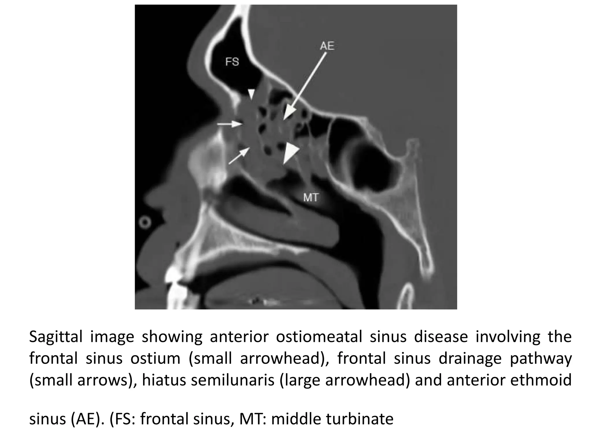
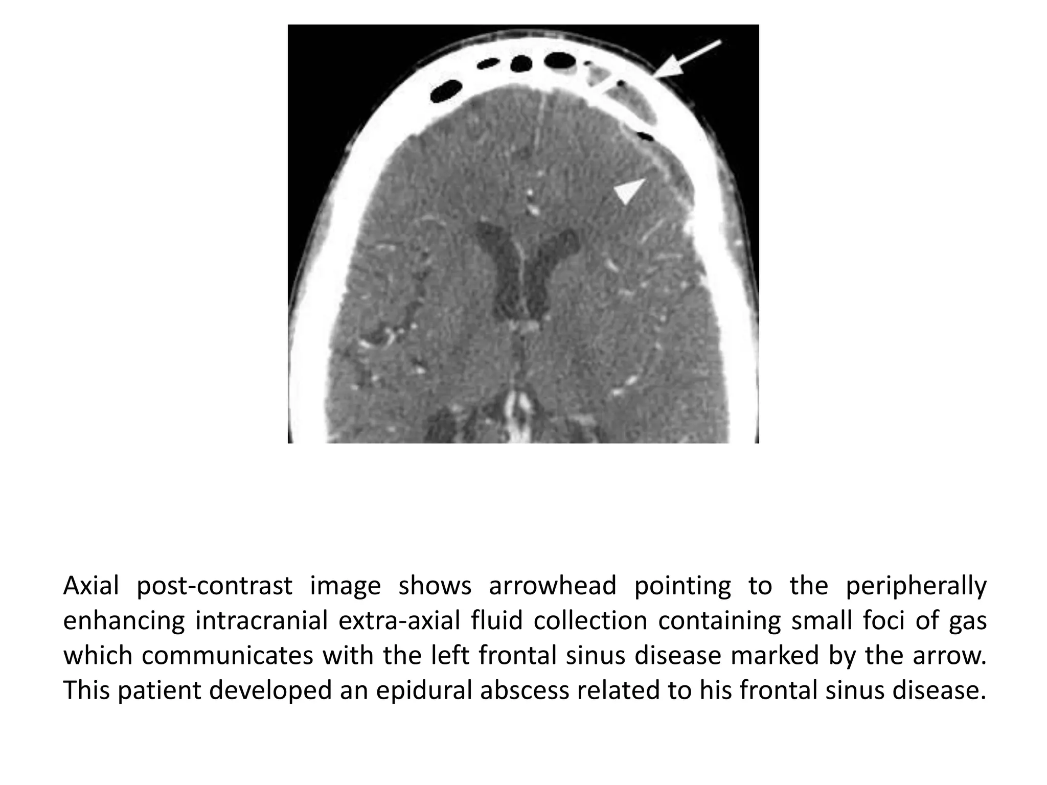
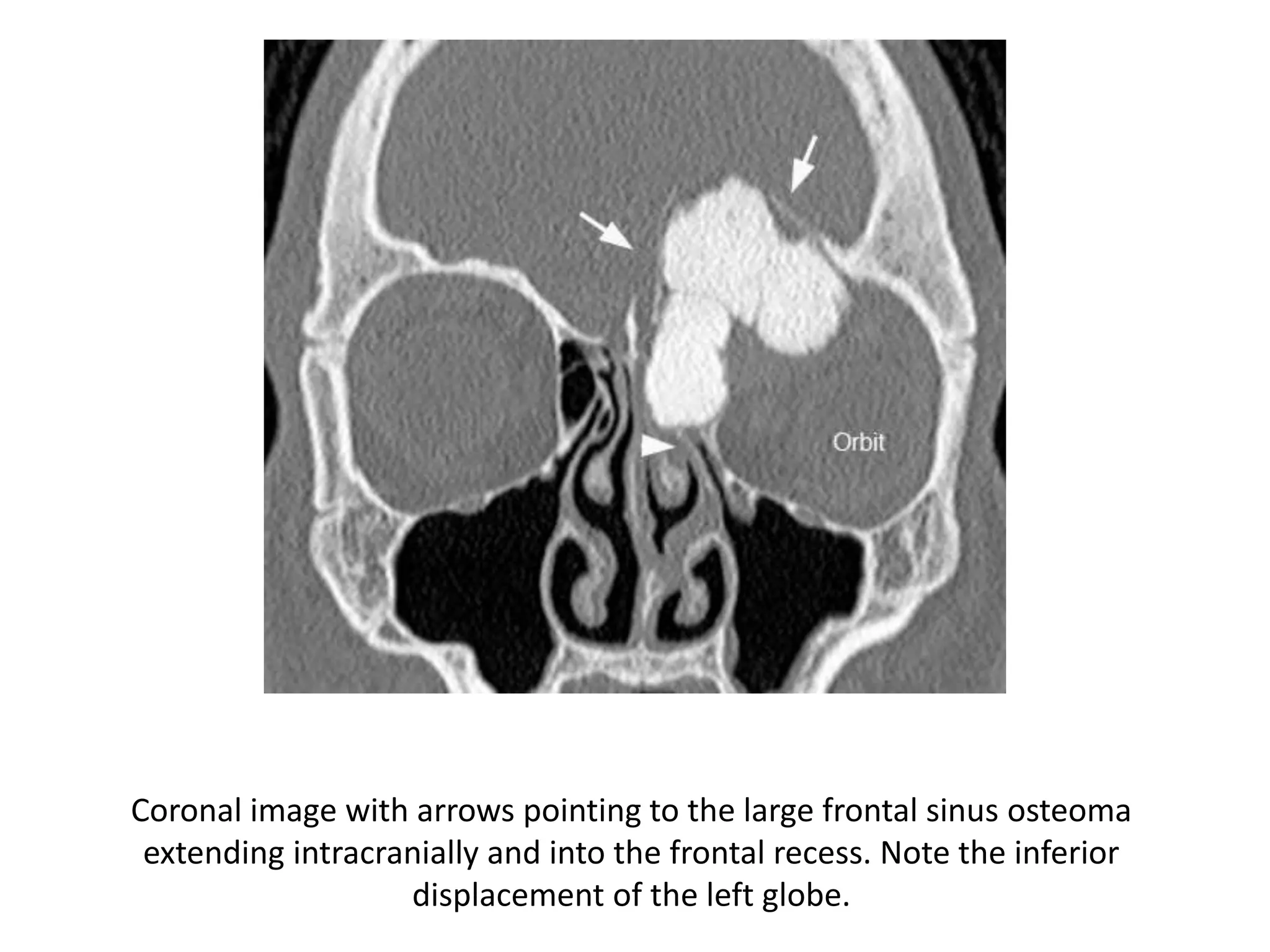
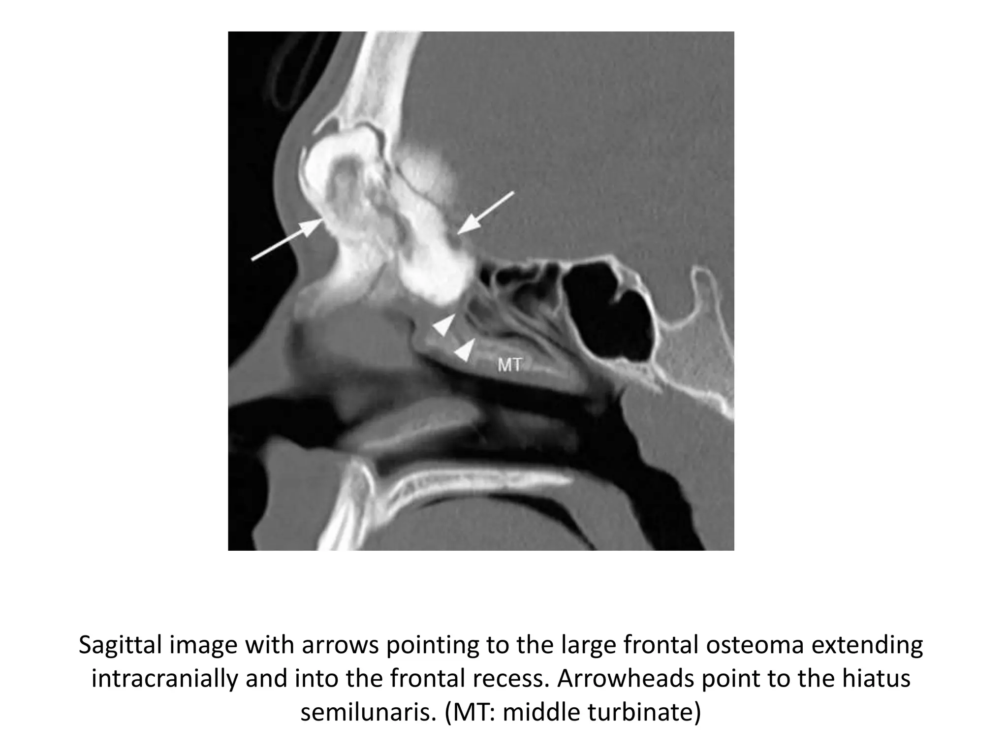
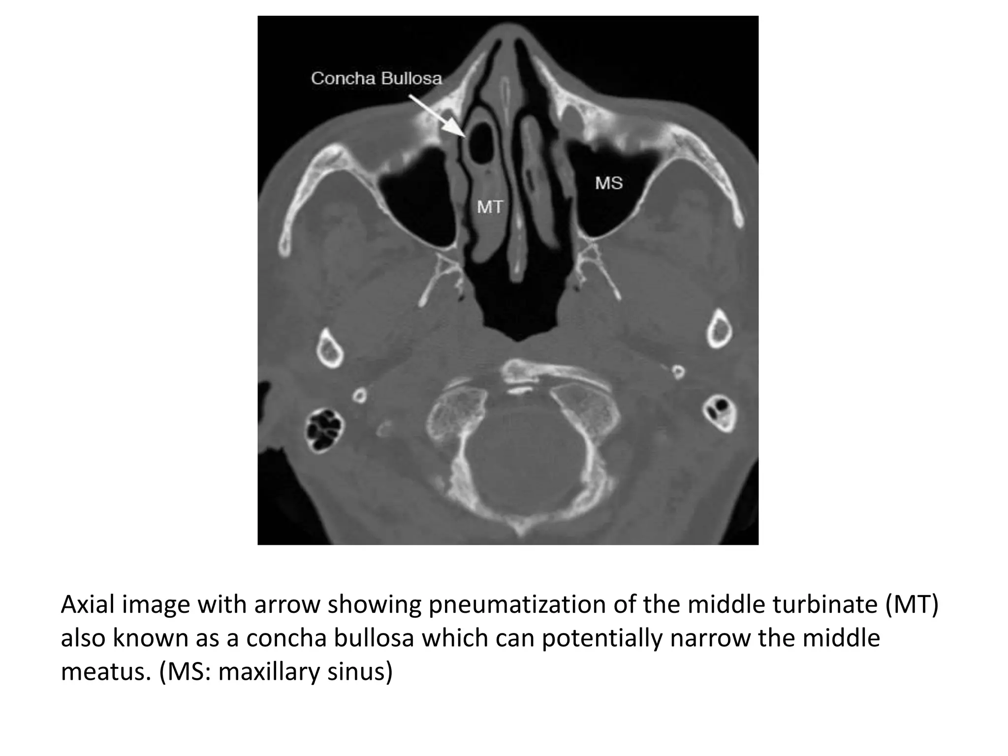
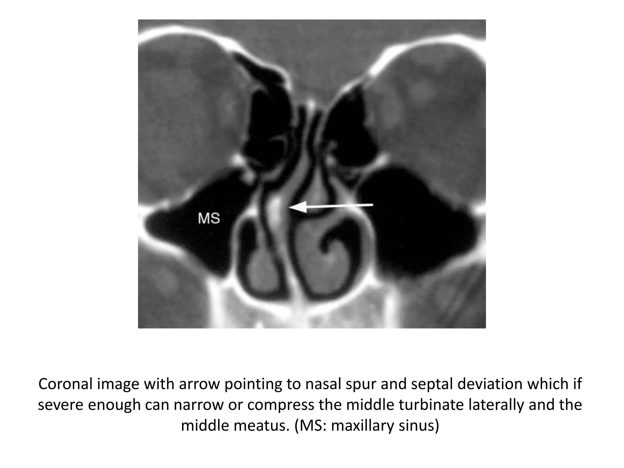
This document describes the anatomy and pathologies of the paranasal sinuses seen on CT imaging. It discusses the normal anatomy of the frontal and maxillary sinuses including common anatomic variants. Common inflammatory diseases are described such as acute sinusitis and its sequelae including mucus retention cysts, mucoceles, and intracranial extension. Other pathologies mentioned include osteomas.
Anatomy of lateral wall of nose with relevanceMalarvizhi R
June 2014, a ppt for DLO and MS ENT postgraduate students lecture by Prof Dr.G.Gananathan MS DLO FICS, then HOD & Prof of MMC, on endoscopic and ct relevence to lateral wall of nose and paranasal sinus.
Anatomy of lateral wall of nose with relevanceMalarvizhi R
June 2014, a ppt for DLO and MS ENT postgraduate students lecture by Prof Dr.G.Gananathan MS DLO FICS, then HOD & Prof of MMC, on endoscopic and ct relevence to lateral wall of nose and paranasal sinus.
Lateral skull base anatomy and applied science by Dr, bomkar bamBomkar Bam
the lateral skull base is complex anatomy that is usually students finds difficult to understand. here concise literature is made to understand the skull base more easily.
Ct anatomy of paranasal sinuses( PNS) pk.pdf pptDr pradeep Kumar
This presentation includes cross sectional anatomy like axial,saggital and coronal images of paranasal sinuses and most important variation of paranasal sinus.This help alot. Must read topic for radiology resident. Thanks
surgical anatomy of nose is a humble attempt to make the anatomy of nose simpler and easy for medical students and fellow physicians. at the end of the presentation the students will be able to identify all the structures.
263778731218 Abortion Clinic /Pills In Harare ,sisternakatoto
263778731218 Abortion Clinic /Pills In Harare ,ABORTION WOMEN’S CLINIC +27730423979 IN women clinic we believe that every woman should be able to make choices in her pregnancy. Our job is to provide compassionate care, safety,affordable and confidential services. That’s why we have won the trust from all generations of women all over the world. we use non surgical method(Abortion pills) to terminate…Dr.LISA +27730423979women Clinic is committed to providing the highest quality of obstetrical and gynecological care to women of all ages. Our dedicated staff aim to treat each patient and her health concerns with compassion and respect.Our dedicated group ABORTION WOMEN’S CLINIC +27730423979 IN women clinic we believe that every woman should be able to make choices in her pregnancy. Our job is to provide compassionate care, safety,affordable and confidential services. That’s why we have won the trust from all generations of women all over the world. we use non surgical method(Abortion pills) to terminate…Dr.LISA +27730423979women Clinic is committed to providing the highest quality of obstetrical and gynecological care to women of all ages. Our dedicated staff aim to treat each patient and her health concerns with compassion and respect.Our dedicated group of receptionists, nurses, and physicians have worked together as a teamof receptionists, nurses, and physicians have worked together as a team wwww.lisywomensclinic.co.za/
Knee anatomy and clinical tests 2024.pdfvimalpl1234
This includes all relevant anatomy and clinical tests compiled from standard textbooks, Campbell,netter etc..It is comprehensive and best suited for orthopaedicians and orthopaedic residents.
Ozempic: Preoperative Management of Patients on GLP-1 Receptor Agonists Saeid Safari
Preoperative Management of Patients on GLP-1 Receptor Agonists like Ozempic and Semiglutide
ASA GUIDELINE
NYSORA Guideline
2 Case Reports of Gastric Ultrasound
NVBDCP.pptx Nation vector borne disease control programSapna Thakur
NVBDCP was launched in 2003-2004 . Vector-Borne Disease: Disease that results from an infection transmitted to humans and other animals by blood-feeding arthropods, such as mosquitoes, ticks, and fleas. Examples of vector-borne diseases include Dengue fever, West Nile Virus, Lyme disease, and malaria.
Pulmonary Thromboembolism - etilogy, types, medical- Surgical and nursing man...VarunMahajani
Disruption of blood supply to lung alveoli due to blockage of one or more pulmonary blood vessels is called as Pulmonary thromboembolism. In this presentation we will discuss its causes, types and its management in depth.
New Drug Discovery and Development .....NEHA GUPTA
The "New Drug Discovery and Development" process involves the identification, design, testing, and manufacturing of novel pharmaceutical compounds with the aim of introducing new and improved treatments for various medical conditions. This comprehensive endeavor encompasses various stages, including target identification, preclinical studies, clinical trials, regulatory approval, and post-market surveillance. It involves multidisciplinary collaboration among scientists, researchers, clinicians, regulatory experts, and pharmaceutical companies to bring innovative therapies to market and address unmet medical needs.
Title: Sense of Taste
Presenter: Dr. Faiza, Assistant Professor of Physiology
Qualifications:
MBBS (Best Graduate, AIMC Lahore)
FCPS Physiology
ICMT, CHPE, DHPE (STMU)
MPH (GC University, Faisalabad)
MBA (Virtual University of Pakistan)
Learning Objectives:
Describe the structure and function of taste buds.
Describe the relationship between the taste threshold and taste index of common substances.
Explain the chemical basis and signal transduction of taste perception for each type of primary taste sensation.
Recognize different abnormalities of taste perception and their causes.
Key Topics:
Significance of Taste Sensation:
Differentiation between pleasant and harmful food
Influence on behavior
Selection of food based on metabolic needs
Receptors of Taste:
Taste buds on the tongue
Influence of sense of smell, texture of food, and pain stimulation (e.g., by pepper)
Primary and Secondary Taste Sensations:
Primary taste sensations: Sweet, Sour, Salty, Bitter, Umami
Chemical basis and signal transduction mechanisms for each taste
Taste Threshold and Index:
Taste threshold values for Sweet (sucrose), Salty (NaCl), Sour (HCl), and Bitter (Quinine)
Taste index relationship: Inversely proportional to taste threshold
Taste Blindness:
Inability to taste certain substances, particularly thiourea compounds
Example: Phenylthiocarbamide
Structure and Function of Taste Buds:
Composition: Epithelial cells, Sustentacular/Supporting cells, Taste cells, Basal cells
Features: Taste pores, Taste hairs/microvilli, and Taste nerve fibers
Location of Taste Buds:
Found in papillae of the tongue (Fungiform, Circumvallate, Foliate)
Also present on the palate, tonsillar pillars, epiglottis, and proximal esophagus
Mechanism of Taste Stimulation:
Interaction of taste substances with receptors on microvilli
Signal transduction pathways for Umami, Sweet, Bitter, Sour, and Salty tastes
Taste Sensitivity and Adaptation:
Decrease in sensitivity with age
Rapid adaptation of taste sensation
Role of Saliva in Taste:
Dissolution of tastants to reach receptors
Washing away the stimulus
Taste Preferences and Aversions:
Mechanisms behind taste preference and aversion
Influence of receptors and neural pathways
Impact of Sensory Nerve Damage:
Degeneration of taste buds if the sensory nerve fiber is cut
Abnormalities of Taste Detection:
Conditions: Ageusia, Hypogeusia, Dysgeusia (parageusia)
Causes: Nerve damage, neurological disorders, infections, poor oral hygiene, adverse drug effects, deficiencies, aging, tobacco use, altered neurotransmitter levels
Neurotransmitters and Taste Threshold:
Effects of serotonin (5-HT) and norepinephrine (NE) on taste sensitivity
Supertasters:
25% of the population with heightened sensitivity to taste, especially bitterness
Increased number of fungiform papillae
Report Back from SGO 2024: What’s the Latest in Cervical Cancer?bkling
Are you curious about what’s new in cervical cancer research or unsure what the findings mean? Join Dr. Emily Ko, a gynecologic oncologist at Penn Medicine, to learn about the latest updates from the Society of Gynecologic Oncology (SGO) 2024 Annual Meeting on Women’s Cancer. Dr. Ko will discuss what the research presented at the conference means for you and answer your questions about the new developments.
These simplified slides by Dr. Sidra Arshad present an overview of the non-respiratory functions of the respiratory tract.
Learning objectives:
1. Enlist the non-respiratory functions of the respiratory tract
2. Briefly explain how these functions are carried out
3. Discuss the significance of dead space
4. Differentiate between minute ventilation and alveolar ventilation
5. Describe the cough and sneeze reflexes
Study Resources:
1. Chapter 39, Guyton and Hall Textbook of Medical Physiology, 14th edition
2. Chapter 34, Ganong’s Review of Medical Physiology, 26th edition
3. Chapter 17, Human Physiology by Lauralee Sherwood, 9th edition
4. Non-respiratory functions of the lungs https://academic.oup.com/bjaed/article/13/3/98/278874
Tom Selleck Health: A Comprehensive Look at the Iconic Actor’s Wellness Journeygreendigital
Tom Selleck, an enduring figure in Hollywood. has captivated audiences for decades with his rugged charm, iconic moustache. and memorable roles in television and film. From his breakout role as Thomas Magnum in Magnum P.I. to his current portrayal of Frank Reagan in Blue Bloods. Selleck's career has spanned over 50 years. But beyond his professional achievements. fans have often been curious about Tom Selleck Health. especially as he has aged in the public eye.
Follow us on: Pinterest
Introduction
Many have been interested in Tom Selleck health. not only because of his enduring presence on screen but also because of the challenges. and lifestyle choices he has faced and made over the years. This article delves into the various aspects of Tom Selleck health. exploring his fitness regimen, diet, mental health. and the challenges he has encountered as he ages. We'll look at how he maintains his well-being. the health issues he has faced, and his approach to ageing .
Early Life and Career
Childhood and Athletic Beginnings
Tom Selleck was born on January 29, 1945, in Detroit, Michigan, and grew up in Sherman Oaks, California. From an early age, he was involved in sports, particularly basketball. which played a significant role in his physical development. His athletic pursuits continued into college. where he attended the University of Southern California (USC) on a basketball scholarship. This early involvement in sports laid a strong foundation for his physical health and disciplined lifestyle.
Transition to Acting
Selleck's transition from an athlete to an actor came with its physical demands. His first significant role in "Magnum P.I." required him to perform various stunts and maintain a fit appearance. This role, which he played from 1980 to 1988. necessitated a rigorous fitness routine to meet the show's demands. setting the stage for his long-term commitment to health and wellness.
Fitness Regimen
Workout Routine
Tom Selleck health and fitness regimen has evolved. adapting to his changing roles and age. During his "Magnum, P.I." days. Selleck's workouts were intense and focused on building and maintaining muscle mass. His routine included weightlifting, cardiovascular exercises. and specific training for the stunts he performed on the show.
Selleck adjusted his fitness routine as he aged to suit his body's needs. Today, his workouts focus on maintaining flexibility, strength, and cardiovascular health. He incorporates low-impact exercises such as swimming, walking, and light weightlifting. This balanced approach helps him stay fit without putting undue strain on his joints and muscles.
Importance of Flexibility and Mobility
In recent years, Selleck has emphasized the importance of flexibility and mobility in his fitness regimen. Understanding the natural decline in muscle mass and joint flexibility with age. he includes stretching and yoga in his routine. These practices help prevent injuries, improve posture, and maintain mobilit
Flu Vaccine Alert in Bangalore Karnatakaaddon Scans
As flu season approaches, health officials in Bangalore, Karnataka, are urging residents to get their flu vaccinations. The seasonal flu, while common, can lead to severe health complications, particularly for vulnerable populations such as young children, the elderly, and those with underlying health conditions.
Dr. Vidisha Kumari, a leading epidemiologist in Bangalore, emphasizes the importance of getting vaccinated. "The flu vaccine is our best defense against the influenza virus. It not only protects individuals but also helps prevent the spread of the virus in our communities," he says.
This year, the flu season is expected to coincide with a potential increase in other respiratory illnesses. The Karnataka Health Department has launched an awareness campaign highlighting the significance of flu vaccinations. They have set up multiple vaccination centers across Bangalore, making it convenient for residents to receive their shots.
To encourage widespread vaccination, the government is also collaborating with local schools, workplaces, and community centers to facilitate vaccination drives. Special attention is being given to ensuring that the vaccine is accessible to all, including marginalized communities who may have limited access to healthcare.
Residents are reminded that the flu vaccine is safe and effective. Common side effects are mild and may include soreness at the injection site, mild fever, or muscle aches. These side effects are generally short-lived and far less severe than the flu itself.
Healthcare providers are also stressing the importance of continuing COVID-19 precautions. Wearing masks, practicing good hand hygiene, and maintaining social distancing are still crucial, especially in crowded places.
Protect yourself and your loved ones by getting vaccinated. Together, we can help keep Bangalore healthy and safe this flu season. For more information on vaccination centers and schedules, residents can visit the Karnataka Health Department’s official website or follow their social media pages.
Stay informed, stay safe, and get your flu shot today!
9. Routine study
Coronal cuts
3-5mm sections
from the posterior wall of the sphenoid to anterior
wall of the frontal sinus
Cuts parallel to hard palate
No contrast
Axial cuts
3-5mm sections from the hard palate to end of the
frontal sinus
Full study
Axial sections Coronal sections Brain
10. CT Sinus Anatomy
Frontal Sinus: Normal Anatomy & Variants
• The frontal sinuses can have variable drainage depending on the anatomy
of the frontal sinus drainage pathway (FSDP).
• The frontal sinuses have a superior and inferior compartment of the FSDP.
• The frontal sinus ostium drains into the superior compartment which then
communicates directly with the inferior compartment.
• The inferior compartment is a narrow space that either is formed by the
ethmoid infundibulum or middle meatus depending on the anterior
attachment of the uncinate process.
• If the anterior uncinate process attaches superiorly to the skull base, then
the inferior compartment of the FSDP is the ethmoid infundibulum which
then communicates with the middle meatus via the hiatus semilunaris.
• If the anterior uncinate process attaches to the lamina papyracea, then the
inferior compartment of the FSDP is the middle meatus.
17. Coronal image with arrows demonstrating the inferior compartment of FSDP
draining to ethmoid infundibulum because anterior process of uncinate
attaches to skull base. (FS: frontal sinus, EB: ethmoid bulla, U: uncinate, MT:
middle turbinate)
18. Coronal image with arrows demonstrating the inferior compartment of the
FSDP includes the middle meatus because the anterior uncinate process
attaches to the lamina papyracea. (FS: frontal sinus, LP: lamina papyracea)
19. Axial image with arrows demonstrating hypoplastic frontal sinuses (FS).
20. Coronal image with arrows demonstrating overly pneumatized frontal
sinuses. Also note enlarged ethmoid bulla air cells (arrowheads).
21. Coronal image with arrow pointing to pneumatized crista galli. Pneumatized
crista galli may communicate with the frontal recess and can potentially
obstruct the frontal sinus ostium. Incidentally noted is a tripod fracture
involving the left maxillary sinus
22. Frontal Sinus: Inflammatory Sinus Disease
and Sequela
• Frontal sinus inflammatory disease can occur in isolation due
to involvement of the ostium and frontal recess or as part of
the anterior ostiomeatal complex (OMC) pattern which
involves the frontal sinus drainage pathway, anterior
ethmoid and maxillary sinuses.
• Inflammatory frontal sinus disease can result in mucus
retention cysts, mucoceles or intracranial extension.
• Occasionally a benign osteoma can be present, and if large
enough can obstruct the ostium or extend intracranially.
Osteomas most commonly occur in the frontal sinuses.
23. Coronal image with arrow pointing to isolated right frontal sinus (FS) dis
24. Coronal image illustrating sinus disease involving the anterior ethmoid sinus
(arrows) and maxillary sinus (MS). Pattern of sinus disease indicates
involvement of the anterior ostiomeatal unit. (MT: middle turbinate)
25. Coronal image demonstrating more diffuse anterior ostiomeatal sinus disease.
Small arrow showing involvement of frontal recess and anterior ethmoid region,
long arrow pointing to maxillary sinus ostium and infundibulum and arrowheads
showing involvement of the hiatus semilunaris. Right maxillary sinus (MS) is
hypoplastic
27. Sagitttal image demonstrating both anterior and posterior ostiomeatal sinus disease. There is
mucosal thickening involving the FSDP, AE and sphenoid sinus ostium and sphenoethmoidal
recess. (FS: frontal sinus, FSDP: frontal sinus drainage pathway, U: uncinate, HS: hiatus
semilunaris, AE: anterior ethmoid, MT: middle turbinate, SpS: sphenoid sinus, double
arrowheads: sphenoid sinus ostium and sphenoethmoidal recess)
28. Axial image of the frontal sinuses (FS) with arrow pointing to a mucus
retention cyst along the non-dependant right frontal sinus air cell.
29. Coronal image of the frontal sinuses with large arrow pointing to mucus
retention cyst
30. Coronal image with small arrow pointing to superior orbital roof and large
arrow pointing to expansile frontal sinus mucocele extending into the orbit
causing inferolateral displacement of the left globe. (CG: crista galli)
31. Sagittal image with arrow demonstrating large frontal sinus mucocele with
orbital extension causing inferior displacement of the globe.
32. Axial image with arrow pointing to the expansile left frontal mucocele that
has encroached into the orbit and is displacing the globe.
33. Axial post-contrast image shows arrowhead pointing to the peripherally
enhancing intracranial extra-axial fluid collection containing small foci of gas
which communicates with the left frontal sinus disease marked by the arrow.
This patient developed an epidural abscess related to his frontal sinus disease.
34. Coronal post-contrast image with arrows demonstrating large epidural
abscess with both intracranial and orbital extension in a different patient.
Also note the extensive unilateral sinus disease.
35. Axial image with arrows pointing to large left frontal sinus osteoma extending
intracranially and obstructing the contralateral right frontal sinus (FS).
36. Coronal image with arrows pointing to the large frontal sinus osteoma
extending intracranially and into the frontal recess. Note the inferior
displacement of the left globe.
37. Sagittal image with arrows pointing to the large frontal osteoma extending
intracranially and into the frontal recess. Arrowheads point to the hiatus
semilunaris. (MT: middle turbinate)
38. Maxillary Sinus: Normal Anatomy &
Variations
• The maxillary sinuses usually develop symmetrically.
• The maxillary sinus ostium drains into the
infundibulum which joins the hiatus semilunaris and
drains into the middle meatus.
• The anterior ostiomeatal unit (OMU) is comprised of
the frontal sinus ostium, frontal sinus drainage
pathway (FSDP), maxillary sinus ostium, infundibulum,
and middle meatus.
• These important structures connect the frontal,
anterior ethmoid and maxillary sinuses
39. Maxillary sinuses
• Ostium drains into ethmoid infundibulum to
hiatus semilunare to middle meatus
• Uncinate process=the medial wall of the
ethmoid infundibulum
• The lateral wall of the ethmoid infundibulum =
Orbital floor
• Ethmoid infundibulum connects the max.sinus
to middle meatus via hiatus semilunare
40. Axial image of the maxillary sinuses at the level of the nasal septum
marked by arrowhead. (MS: maxillary sinus, NLD: nasolacrimal duct,
IT: inferior turbinate)
41. Coronal image with arrow pointing to maxillary sinus ostium (MO) with (..)
illustrating the infundibulum joining the hiatus semilunaris (*). (MS: maxillary
sinus, MT: middle turbinate)
42. Sagittal image showing hiatus semilunaris (*) with uncinate process and
ethmoid bulla (EB) superiorly. (MT: middle turbinate, FS: frontal sinus, FSDP:
frontal sinus drainage pathyway)
43. Axial images with arrows showing hypoplastic maxillary sinuses. The nasal
septum is also absent. A septum can be absent due to either congenital or
acquired (surgery, cocaine abuse or Wegener's granulomatosis) disorders.
44. Axial image with arrow showing pneumatization of the middle turbinate (MT)
also known as a concha bullosa which can potentially narrow the middle
meatus. (MS: maxillary sinus)
45. Coronal image with arrowhead showing concha bullosa of middle turbinate.
Concha bullosa can potentially obstruct the maxillary sinus drainage pathway
by narrowing the infundibulum and middle meatus
46. Coronal image with arrow pointing to paradoxical curvature of the middle
turbinate (MT) with convexity of the bone directed laterally. Paradoxical
curvature can potentially narrow or obstruct the infundibulum or middle
meatus.
47. Coronal image with arrow pointing to nasal spur and septal deviation which if
severe enough can narrow or compress the middle turbinate laterally and the
middle meatus. (MS: maxillary sinus)
48. Coronal image with arrows illustrating thin bony septum in the
maxillary sinuses (MS).
49. Maxillary Sinus: Inflammatory Sinus Disease and
Sequela
• Maxillary sinus mucociliary drainage flows through the sinus ostium
into the infundibulum which joins the hiatus semilunaris and drains
into the middle meatus.
• The middle meatus is also the final drainage for the frontal and
anterior ethmoid sinuses.
• The anterior ostiomeatal unit comprises the frontal sinus ostium,
frontal recess, maxillary sinus ostium infundibulum and middle
meatus. Therefore, a relatively common pattern of inflammatory sinus
disease involves the anterior ostiomeatal unit.
• Acute sinus disease may be associated with air-fluid levels which if
present commonly occur in the maxillary sinuses. However, it is
important to remember that many patients with acute sinusitis will not
have air-fluid levels.
• Acute sinusitis can also have a "bubbly or foamy" appearance. Rarely
acute sinus disease can be aggressive with bony erosion. Mucus
retention cysts are commonly seen and less commonly polyps and
50. Axial image showing mucosal thicknening and an air-fluid level in
the maxillary sinus (MS).
51. Coronal image with (*) showing obstruction of the infundibulum and on left
side involvement of the hiatus semilunaris. Small arrows also demonstrate
sinus disease of the anterior ethmoid air cells and larger arrows point to
bilateral maxillary sinus mucosal thickening. Pattern of sinus disease involves
the anterior ostiomeatal unit.
52. Coronal image demonstrating more extensive pattern of anterior ostiomeatal
unit sinus disease with short arrow pointing to frontal recess, long arrow
pointing to maxillary sinus ostium and infundibulum region with arrowheads
marking the area of the hiatus semilunaris and middle meatus. Right maxillary
sinus (MS) is hypoplastic.
53. Coronal image with small arrows illustrating involvement of the maxillary
sinus ostium and infundibulum and large arrow pointing to maxillary sinus
mucosal thickening. (U: uncinate, EB: ethmoid bulla)
54. Sagittal image with small arrowhead pointing to frontal sinus ostium, short
arrows showing frontal sinus drainage involvement and large arrowhead
showing involvement of hitus semilunaris again demonstrating anterior
ostiomeatal pattern of disease. There is also involvement of the anterior
ethmoid sinus (AE). (FS: frontal sinus, MT: middle turbinate)
55. Axial image with arrow pointing to air-fluid level in maxillary
sinus in acute sinusitis. Note the slightly bubbly appearing fluid.
56. Axial image demonstrating additional case of acute sinusitis
with arrows pointing to air-fluid levels in the ethmoid and
sphenoid sinuses.
57. Coronal image with large arrow pointing to "foamy" maxillary
sinus disease.
58. Axial image demonstrating air-fluid level in left maxillary sinus
with arrow pointing to bony erosion of the posterior maxillary
sinus wall in a case of acute sinusitis.
59. Axial image in soft tissue window with arrow pointing to
posterior extension of sinus disease through the posterior
maxillary sinus wall into the retroantral fat pad.
60. Coronal image with arrow pointing to right maxillary sinus mucus retention
cyst (MRC). Notice how sinus air partially surrounds the MRC in contrast to a
mucocele which completely fills and expands the sinus.
62. Coronal image with arrow pointing to maxillary sinus polyp.
Often on imaging a polyp and mucus retention cyst cannot be
differentiated, but is usually of little clinical consequence.
63. Axial image demonstrates an antrochoanal polyp that completely fills the
maxillary sinus with arrow pointing to widened infundibular region with
extension into the middle meatus and nasal cavity.
64.
65. allergic fungal sinusitis complicated by compression of right optic nerve. Painless
decreased vision had been present in the right eye for 2 months. Coronal (A–C)
and axial (D) CT images show high-attenuation opacification of left maxillary, left
ethmoidal, and bilateral sphenoidal sinuses with bone expansion and thinning.
Compression of right optic nerve (straight arrow, B and D) is caused by
expanded right anterior clinoid process (asterisk, B and D). Bone dehiscence is
present at left lamina papyracea (curved arrow, A and D) and around left optic
nerve (arrowhead, B and D), and internal carotid arteries (arrows, C). These
structures are at risk of injury during functional endoscopic sinus surgery.
66.
67. Ethmoid Sinus: Normal Anatomy &
Variants
• The ethmoid sinus can have a variable number
of air cells. Additionally, the ethmoid sinuses
are divided into groups of cells by bony basal
lamellae.
• The most important one is the basal lamellae
of the middle turbinate which separates the
ethmoid into anterior and posterior groups
with different drainage patterns.
68. Ethmoid complex
Ethmoid bones lies between the orbits.
• Horizontal plate ; (cribriform plate), perforated for
transmission of olfactory nerve
• Vertical plate; extends above the horizontal plate
intracranially as the cresta Galli , the gyrus rectus+
olfactory bulb rests upon the olfactory fossa on either
side of the CG
• Ethmoid sinuses: is separated
from the orbit by lamina papyrecea
69.
70. Axial view shows small arrows demonstrating bony canal for anterior and
posterior ethmoidal arteries (CG: crista galli, AC: anterior clinoid process, OC:
optic canal, SP: sphenoid sinus).
71. Anatomic variations are common in general population and
1. Concha bullosa (CB): is defined as aeration in the middle turbinate. It may be
unilateral or bilateral.
• A CB in the MT may enlarge to obstruct the middle meatus or the
infundibulum.
2. Nasal septal deviation: it is asymmetrical bowing of the nasal septum. Bony
spurs are often associated with septal deviation. Septal deviations are usually
congenital but may be post-traumatic finding in some
3. Paradoxical middle turbinate: here the convexity of the turbinate facing laterally
instead of medial direction and is a bilateral finding. This will lead to the
stenosis of the middle meatus and it depends upon the degree of paradoxical
curve.
4. Variation in the uncinate process: the course of the free edge of UP varies as
follows:
• - It may be attached to the base of skull superiorly.
• - It can curve medially towards the nasal septum with the free edge
• - Atelactatic UP: the free edge of the UP can adhere to the orbital
5. The Haller cells: are ethmoid air cells that extend along the medial roof of the
maxillary sinus. It may cause narrowing of the infundibulum when they are
large.
72. 6. The Onodi cells: are the lateral and posterior extensions of the posterior
ethmoid air cells. These cells may surround the optic nerve tract and put the
nerve at risk during the surgery.
7. The Giant BE: are the largest of the ethmoidal cells. The BE may enlarge to
narrow or obstruct the middle meatus and cause infection.
8. Medial deviation or dehiscence of lamina papyracea: It may be congenital
finding or a result of prior facial trauma. The intraorbital contents are at risk
during surgery.
9. The aerated crista galli: when aeration of the crista galli occur, these cells may
communicate with the frontal recess, obstruction of this ostium can lead to
the chronic sinusitis and mucocele formation .
10. Asymmetry of ethmoid roof height: there is a higher incidence of intracranial
penetration during the FESS when this anatomic variation occurs. The
intracranial penetration is more likely to occur on the side where the position
of the roof is lower .
11- Aggar Nasi Cells : The most anterior ethmoid air cell.
73. Axial image showing normal ethmoid sinus anatomy. Arrows point to the
lateral attachment of the basal lamellae to lamina papyracea separating
anterior and posterior ethmoid sinuses (AE: anterior ethmoid, PE: posterior
ethmoid, NS: nasal septum, SpS: sphenoid sinus)
74. Sagittal image with arrowhead demonstrating anterior ethmoid drainage to
hiatus semilunaris and middle meatus. Arrow showing posterior ethmoid
drainage to sphenoethmoidal recess and superior meatus. (AG: agger nasi
cell, AE: anterior ethmoid, PE: posterior ethmoid, MT: middle turbinate)
75. Coronal image with arrowhead showing lateral attachment of basal lamellae
to lamina papyracea marked by the arrow. (CG: crista galli, *: cribriform plate,
FE: fovea ethmoidalis, MT: middle turbinate, IT: inferior turbinate)
76. Sagittal image with arrow showing vertical attachment of basal lamellae to
anterior skull base separating the anterior ethmoid (AE) and posterior
ethmoid (PE) sinuses. (FS: frontal sinus, AG: agger nasi cell, SpS: sphenoid
sinus, MT: middle turbinate)
77. Axial image demonstrating an Agger Nasi air cell which is the most anterior
ethmoid air cell. (AE: anterior ethmoid, PE: posterior ethmoid, SpS: sphenoid
sinus)
78. Coronal image with arrowhead showing Agger Nasi air cell. (MS:
maxillary sinus, MT: middle turbinate, IT: inferior turbinate)
80. Coronal image shows ethmoid bulla air cells superior to uncinate
processes. The (*) highlight the hiatus semilunaris. Ethmoid bulla
air cells are part of the anterior ethmoid sinuses and make up the
superior border of the hiatus semilunaris. (EB:ethmoid bulla, U:
uncinate process, MT: middle turbinate)
81. Coronal image with arrows pointing to enlarged ethmoid bulla
encroaching on the OMU. Ethmoid bulla air cells can
demonstrate variable pneumatization.
82. Axial image with arrow pointing to an infraorbital ethmoid air cell (Haller cell).
If present, a Haller cell can cause narrowing of the infundibulum and maxillary
sinus ostuim potentially causing obstruction. (MS: maxillary sinus, NLD:
nasolacrimal duct)
83. Coronal image with arrowhead pointing to infraorbital ethmoid air cell (
Haller cell) which is narrowing the maxillary sinus ostium and infundibulum.
(MT: middle turbinate, MS: maxillary sinus)
84. Sagittal image with arrowhead pointing to infraorbital ethmoid
air cell (Haller cell). (FS: frontal sinus, MS: maxillary sinus)
85. Ethmoid Sinus: Inflammatory Sinus
Disease and Sequela
• Ethmoid inflammatory sinus disease can involve either the
anterior or posterior ethmoid sinuses which have separate
drainage pathways. Recall that the basal lamellae of the middle
turbinate anatomically separates the ethmoid sinuses into the
anterior ethmoid which drains into middle meatus and posterior
ethmoid which drains into the sphenoethmoidal recess and
superior meatus. As a result of the dual drainage pathways
ethmoid sinus disease can also be a part of a spectrum of
inflammatory sinus disease related to inflammatory disease of the
ostiomeatal unit, infundibulum and spheoethmoidal recess.
Ethmoid sinus disease can less commonly result in mucoceles, but
due to the thin lamina papyracea and valveless ethmoidal veins
can occasionally result in orbital extension of disease and
cavernous sinus thrombosis.
86. Axial image with arrowheads pointing to anterior ethmoid sinus disease. Long
arrows point to clear posterior ethmoid air cells. Short arrows point to clear
sphenoid sinus with (*) marking the sphenoid sinus ostium
87. Sagittal image with arrows pointing to posterior ethmoid sinus disease and
arrowheads showing involvement of the ostium and sphenoethmidal recess.
(SpS: sphenoid sinus, FS: frontal sinus, MT: middle turbinate)
88. Axial image with arrowheads pointing to both anterior and
posterior ethmoid sinus disease.
89. Coronal image with arrowhead pointing to ethmoid infundibulum sinus
disease. In this patient the arrows are pointing to the uncinate process which
connects to the skull base so the inferior compartment of the frontal sinus
drainage pathway (FSDP) is the ethmoid infundibulum. (FS: frontal sinus, MT:
middle turbinate)
90. Sagittal image demonstrates more extensive involvement of the paranasal
sinuses including frontal sinus (FS) and frontal sinus drainage pathway (large
arow), both anterior ethmoid (AE) and posterior ethmoid sinuses (PE) marked
by arrows, and sphenoid sinus (SpS) with arrowhead pointing to sphenoid
sinus ostium and sphenoethmoidal recess.
92. Axial post-contrast image with arrowheads pointing to orbital extension of
ethmoid sinus disease marked by arrow.
93. Axial post-contrast image demonstrates ethmoid sinus disease (large arrow)
with orbital extension (arrowheads) resulting in cavernous sinus thrombosis
(small arrow) and proptosis of left globe. Heterogeneous low density material
within the enlarged cavernous sinus represents thrombus.
94. Sagittal post-contrast image again demonstrating the cavernous sinus
thrombosis with clot (arrow) and orbital extension of disease marked by the
arrowheads.
95. Coronal post-contrast image with arrow demonstrating the enlarged
cavernous sinus due to sinus thrombosis with clot (arrowhead).
96. Axial image with arrow pointing to benign osteoma arising from the anterior
ethmoid sinuses (AE) with adjacent orbital extension resulting in proptosis.
Osteomas can result in narrowing and obstruction of the infundibulum and
middle meatus. They are most common in the frontal sinuses. (SpS: sphenoid
sinus)
97. Coronal image with arrow pointing to the ethmoid osteoma extending into
orbit and arrowhead showing involvement of the fovea ethmoidalis.(CG: crista
galli)
98. Sphenoid Sinus: Normal Anatomy
& Variants
• The sphenoid sinuses are highly variable in their
configuration.
• Pneumatization can extend into the greater sphenoid
wing resulting in lateral recesses.
• Additionally, pneumatization can also involve the
posterior orbital wall, pterygoid processes, and lesser
sphenoid wing.
• Important neighboring structures include the foramen
rotundum, vidian canal, optic canal and internal
carotid artery. The sphenoid sinus drains via the
ostium into the sphenoethmoidal recess.
99. Axial image shows sphenoid sinus (SpS) and the sphenoethmoidal recess
marked by the (*). (AE: anterior ethmoid, PE: posterior ethmoid, CC: carotid
canal, NS: nasal septu
100. Coronal image of the sphenoid sinus (SpS) and neighboring
structures. (FR: foramen rotundum, VC: vidian canal, OC: optic
canal, AC: anterior clinoid, PtP: pterygoid plate
101. Sagittal image showing the sphenoid sinus (SpS) with sinus
ostium (*) and arrow demonstrating the sphenoethmoidal
recess (SER). (PE: posterior ethmois sinus)
102. Axial image with large arrows pointing to pneumatized lateral
recesses of the sphenoid sinus (SpS). (FO: foramen ovale, FS:
foramen spinosum)
103. Coronal image showing pneumatized lateral recesses of sphenoid
sinus (SpS) and foramen rotundum (FR) bulging into the sinus.
Arrows point to optic canals superior to sphenoid sinus and
medial to anterior clinoid processes
104. Axial image with arrows showing bilateral onodi air cells. Onodi air cells
represent contigous extension of the posterior ethmoid air cells into the
sphenoid sinus and are closely associated with the optic nerve. Due to their
close association with the optic nerve the nerve can be at increased risk of
injury during sinus surgery.
105. Coronal image showing bilateral onodi air cells with pneumatized
anterior clinoid processes (AC) and arrows pointing to the optic
canals.
106. Axial image with arrows pointing to pneumatized
pterygoid plates (PP).
107. Axial image with arrow pointing to hypoplastic sphenoid sinus. (BO:
basi-occiput, ES: ethmoid sinuses)
108. Sphenoid Sinus: Inflammatory Sinus
Disease and Sequela
• Sphenoid sinuses drain via their ostium to the
sphenoethmoidal recess and nasopharynx.
Sphenoethmoidal recess inflammatory sinus
disease pattern is usually due to either
obstruction of the sinus in isolation or in
conjunction with the posterior ethmoidal air
cells.
• Mucoceles are not common in the sphenoid
sinus but can occur and rarely sphenoid sinus
disease can result in cavernous sinus
thrombosis or intracranial extension
109. Coronal image of the sphenoid sinuses with arrow pointing to
isolated left sphenoid sinus disease. Again notice the close
anatomic proximity to the optic canal (OC) and foramen
rotundum (FR). (AC: anterior clinoid process)
110. Sagittal image showing sphenoid sinus disease (SpS) with arrow
showing obstructed sinus ostium and arrowhead pointing to
sphenoethmoidal recess.
111. Axial image with large arrow showing left sphenoid sinus (SpS)
disease with ostruction of the ostium (arrowhead). (EB: ethmoid
bulla)
112. Axial image shows arrows pointing to a large expansile mass in
the sphenoid sinus (SpS) extending into the posterior ethmoid
sinus (PE) which was due to a large sphenoid sinus mucocele.
(AE: anterior ethmoid sinus)
113. Sagittal image demonstrating large expansile sphenoid sinus
(SpS) mucocele extending into the posterior ethmoid sinus (PE)
and is being displaced anteriorly. (NP: nasopharynx)
114. Axial post-contrast image demonstrating sphenoid sinus disease
(SpS) with cavernous sinus thrombosis (CS) and orbital
involvement. There is also ethmoid sinus disease.
116. Figure 1. Sinonasal polyposis. (A) Coronal sinus CT scan depicts an
extensive soft-tissue abnormality filling nasal cavity and sinuses.
Discrete polyps are noted extending into the middle meatus
(arrow). (B) This section is further posterior than in A, in the same
patient. Note preservation of normal ethmoid septae, despite the
significant soft-tissue abnormality.