Identification and Confirmation of Ten
Common Seized Drugs Utilizing a
Single Quadrupole Mass
Spectrometer
Shimadzu Scientific Instruments, Columbia, MD
Introduction
The use of illicit drugs continues to be an epidemic across the United States. Currently,
most laboratories require the use of a GC or GCMS system and a cumbersome
derivatization step to analyze seized drugs.
By utilizing a LCMS method, faster detection rates and a larger drug panel could be
screened simultaneously. The need for a derivatization step could also be avoided.
Instrumentation
A Shimadzu LCMS-2020 single quadrupole mass spectrometer coupled with an
integrated LC-2040C 3D UHPLC system was employed for this evaluation. The LC-
2040C 3D was equipped with a PDA detector for simultaneous analysis of analyte
absorbance and compound ionization.
Method Development
Ten common seized and illicit drugs were analyzed using a single quadrupole mass
spectrometer, LCMS-2020, with electrospray ionization (ESI) in positive mode. A
mixture of all 10 analytes was prepared using certified reference materials purchased
from Cayman Chemical. Each standard was dissolved in LCMS grade water and
diluted to 10ug/mL for initial testing.
A 1uL injection volume was used with Shimadzu’s NexLeaf CBX Potency II column
(100mm x 3.0mm, 1.8um) for complete baseline separation of all 10 analytes. Gradient
elution was required with mobile phase A being water and Mobile phase B being
Methanol both with 5mmol ammonium formate and 0.1% formic acid (Figure 1).
Time %B
0 20
7.5 95
10 95
10.01 20
15 20Figure 1: LC Gradient Parameters
The overall runtime was 15 minutes and utilized selected ion monitoring (SIM) for
quantitation and scanning with in-source collision-induced dissociation (in-source CID)
for identification and secondary confirmation. The scan event used a scan range of 50
to 500 m/z with a scan speed of 5000 u/sec. LCMS method parameters and the final
SIM channels used can be found in Table 1.
Calibration curves were prepared with neat standards ranging from 0.1ug/mL to
1ug/mL. The corresponding SIM channel for each analyte was used for external
calibration.
Method Development
Drying Gas 10.0 L/min
Interface Temperature 350°C
DL Temperature 250°C
Heat Block Temperature 400°C
Flow rate 0.3 mL/min
Column Oven
Temperature
40°C
Sample Tray Temperature 10°C
SIM Channels (m/z)
337.2; 323.2; 286.1;
328.1; 205.1; 370.1;
304.1; 150.2; 256.2
Table 1: LCMS-2020
Method parameters
Results and Discussion
A Shimadzu LCMS-2020 was used to create a SIM method for 10 common drugs
generally run by GCMS. In addition to a SIM method demonstrating chromatographic
separation (Figure 2) a scan event was added to monitor in-source fragmentation.
Figure 2: Representative SIM Chromatogram for 10 common seized drugs at 1ug/mL
Results and Discussion
Figure 3: Scan and in-source CID spectra for Naloxone and 6-MAM at 1ug/mL. Top spectrum is a
positive scan event with 0V, bottom spectrum is a positive scan event with 60V in-source CID
Two different Q-Array voltages were applied to fragment all of the analytes of interest,
60V and 75V. Naloxone and 6-MAM have the same mass and therefore could only be
differentiated using retention time. With simultaneous in-source CID, additional
identification could be completed by comparing the fragmentation patterns produced
(Figure 3). Fragments m/z 310.05 and 268.10 are only seen with Naloxone and are not
produced for 6-MAM.
Results and Discussion
Figure 4: SIM and in-source CID spectra for Acetyl Fentanyl at 1ug/mL. A.) Spectrum from SIM event,
B.) Spectrum from scan event with 60V in-source CID on LCMS-2020, C.) Spectrum from Metlin
Database1
in-source CID was also used for secondary confirmation. Figure 4 shows a comparison
of the fragmentation pattern produced using in-source CID versus a known library
spectrum from Metlin’s database1.
105.1
188.15
Results and Discussion
All calibration curves demonstrated linearity with a range from 0.1ug/mL to 1ug/mL.
Final calibration curves were determined by plotting peak area versus concentration
with a 1/C weighting factor. A correlation coefficient, R2=0.996 or better was obtained
for all analytes. Representative chromatograms and calibration curves for each analyte
can be seen in Figure 5.
All calibration curves were run in triplicate to evaluate reproducibility and accuracy. The
final method demonstrated peak area RSDs ranging from 0.15 to 1.40% with the
average accuracies between 93 and 106% for every compound.
Results and Discussion
0 500 Conc.
0.0
2.5
5.0
Area(x1,000,000)
0 500 Conc.
0.0
2.5
5.0
Area(x1,000,000)
0 500 Conc.
0.0
2.5
Area(x1,000,000)
0 500 Conc.
0.0
2.5
5.0
Area(x1,000,000)
5.0 6.0
0.0
1.0
2.0
3.0
4.0
(x100,000)
1:370.1000(+)
3.0 4.0
0.0
0.5
1.0
1.5
(x100,000)
1:205.1000(+)
3.0 4.0
0.0
1.0
2.0
3.0
(x100,000)
1:328.1000(+)
2.0 3.0
0.0
1.0
2.0
3.0
(x100,000)
1:328.1000(+)
R
2
= 0.997
R
2
= 0.999 R
2
= 0.999
R
2
= 0.998
Naloxone 6-MAM
Levamisole Heroin
0 500 Conc.
0.0
2.5
5.0
7.5
Area(x1,000,000)
0 500 Conc.
0.0
1.0
2.0
3.0
Area(x1,000,000)
4.0 5.0
0.0
0.5
1.0
1.5
(x100,000)
1:150.2000(+)
5.0 6.0
0.0
2.5
(x100,000)
1:304.1000(+)
R
2
= 0.999 R
2
= 0.998
Cocaine Methamphetamine
Figure 5: Calibration Curves for each compound and representative
chromatograms at 250 ng/mL
Results and Discussion
0 500 Conc.
0.0
2.5
5.0
Area(x1,000,000)
0 500 Conc.
0.0
2.5
Area(x1,000,000)
0 500 Conc.
0.0
0.5
1.0
Area(x10,000,000)
0 500 Conc.
0.00
0.25
0.50
0.75
1.00
Area(x10,000,000)
6.0 7.0
0.0
2.5
5.0
(x100,000)
1:323.2000(+)
6.0 7.0
0.0
2.5
5.0
(x100,000)
1:337.2000(+)
1.0 2.0
0.0
1.0
2.0
3.0
(x100,000)
1:286.1000(+)
7.0 8.0
0.0
1.0
2.0
3.0
4.0
(x100,000)
1:256.2000(+)
R
2
= 0.999
R
2
= 0.999 R
2
= 0.999
R
2
= 0.996
Diphenyhydramine Morphine
Fentanyl Acetyl Fentanyl
Figure 5 (continued): Calibration Curves for each compound and
representative chromatograms at 250 ng/mL
Conclusion
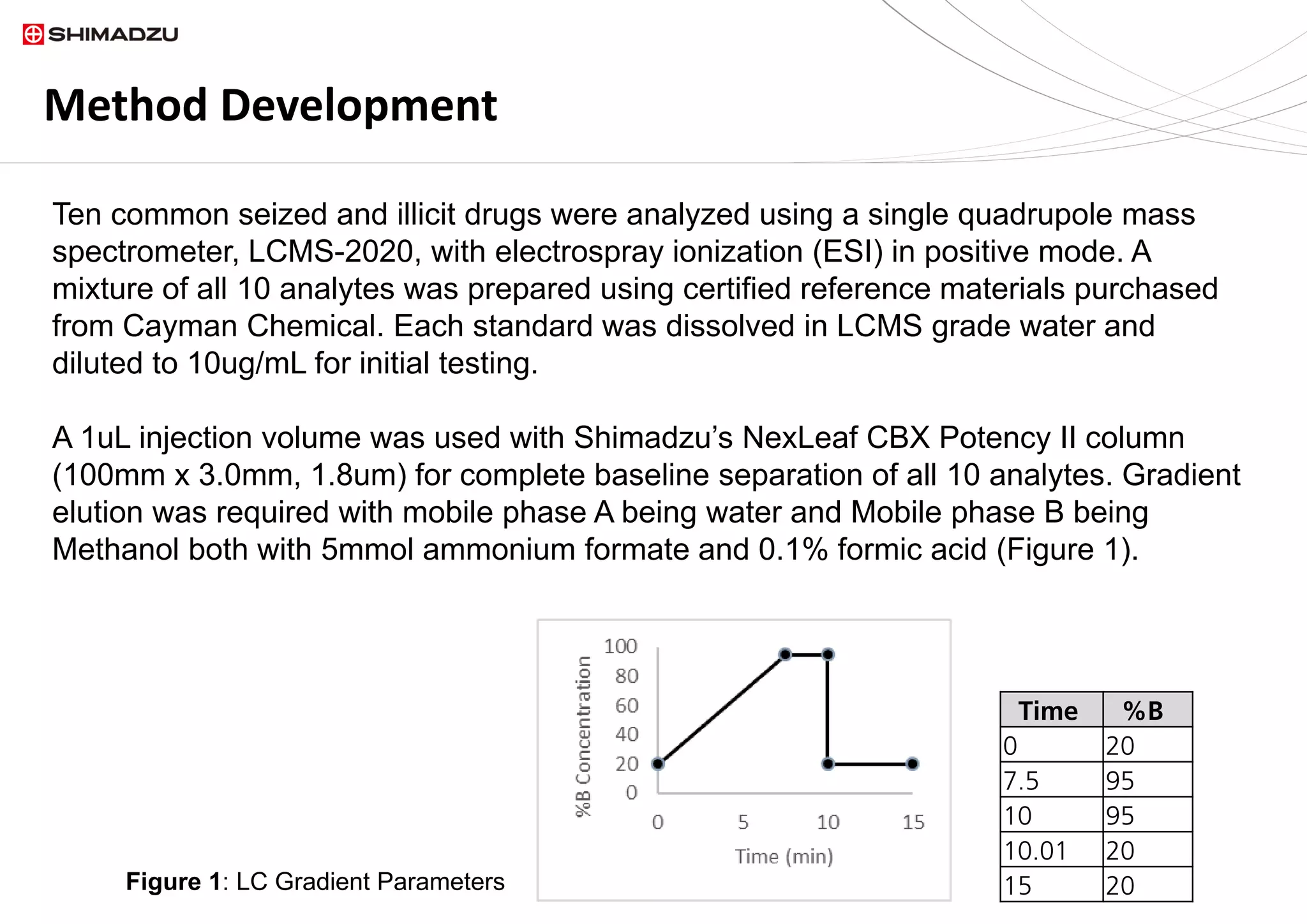
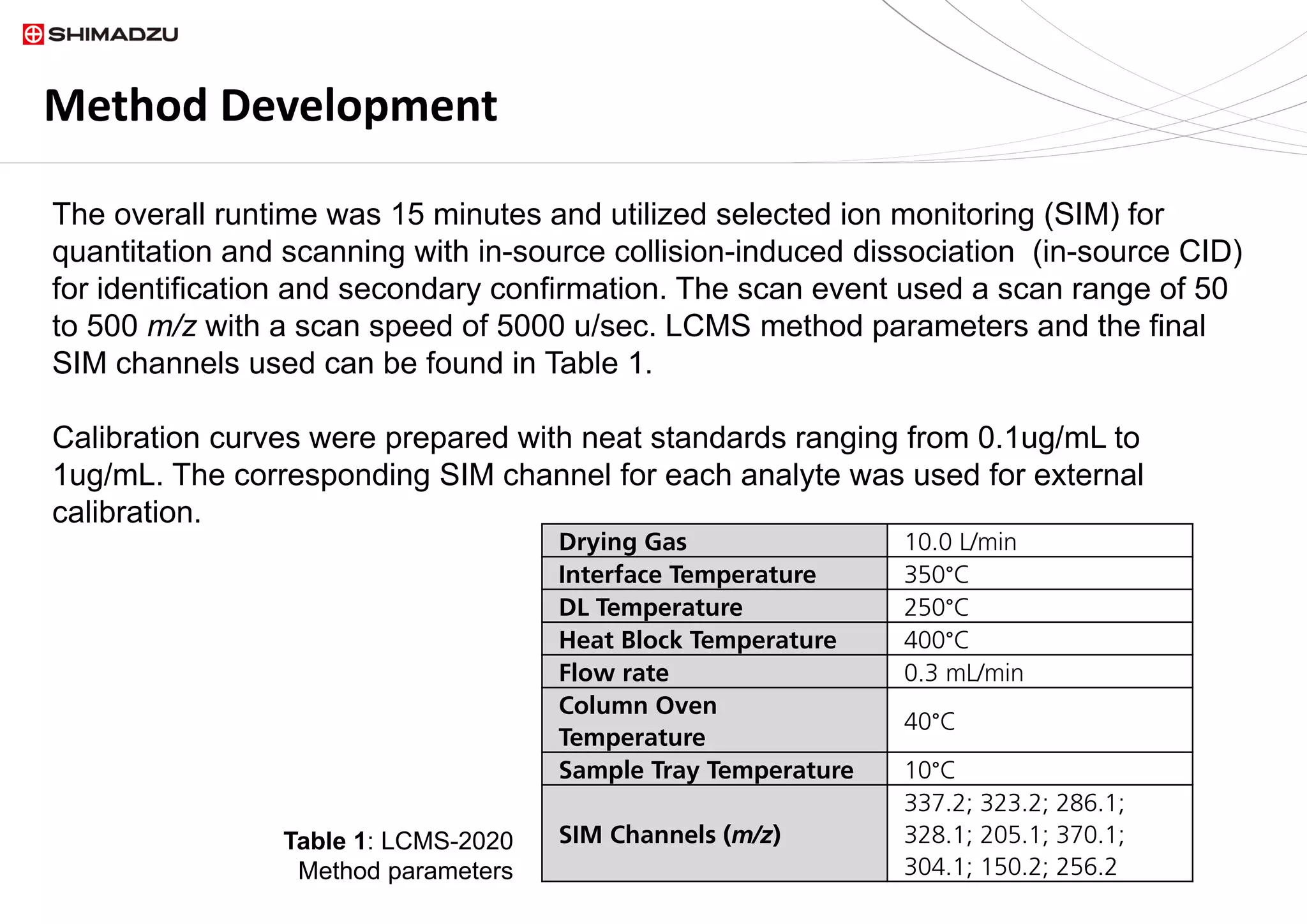
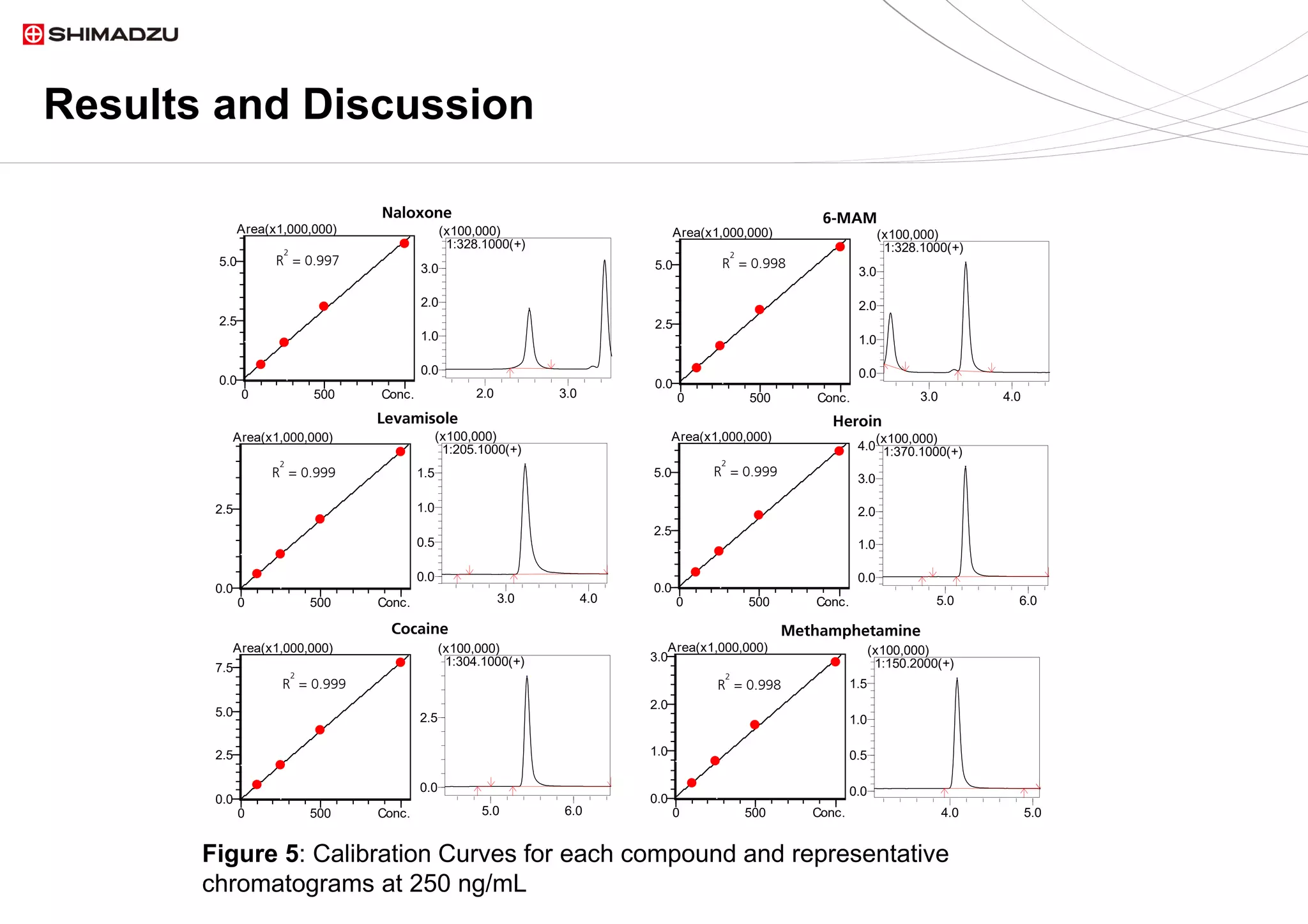
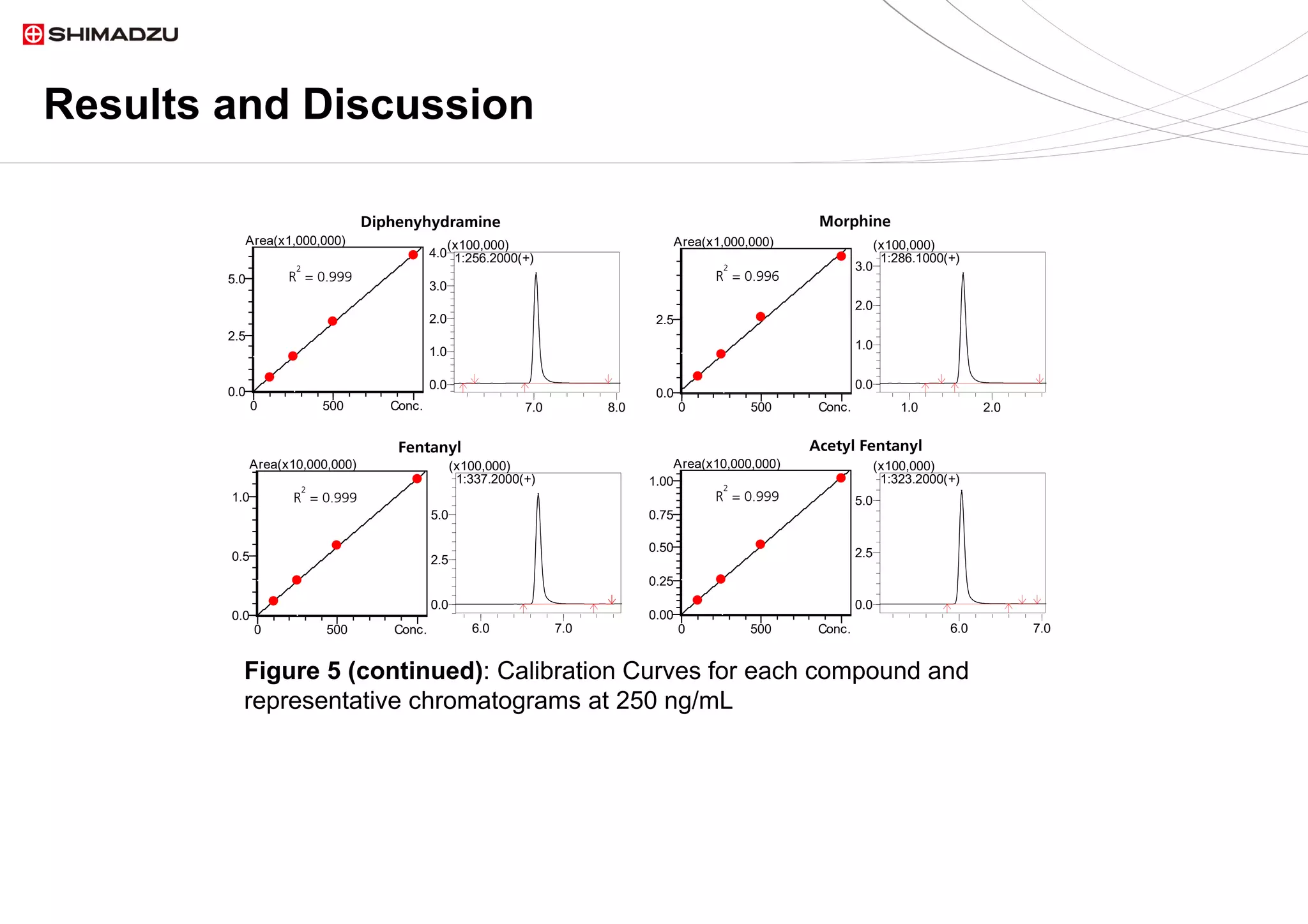
A single chromatographic method was developed for the separation and quantitation of
ten common seized drugs. The single quadrupole mass spectrometer, LCMS 2020,
demonstrated its capability for simultaneous detection and confirmation using in source
fragmentation of all analytes. Linear calibration curves were acquired for each analyte.
Further method development could be explored to increase the panel of drugs being
screened as well as determining LOQs for each analyte of interest.
Reference
1. Smith CA, O'Maille G, Want EJ, Qin C, Trauger SA, xonBrandon TR, Custodio DE, Abagyan
R, Siuzdak G. METLIN: a metabolite mass spectral database. Ther Drug Monit [Internet].
2005;27 :747-51.
Need More Info?
Thank you for viewing this presentation. Should you have any
questions or require additional information about our research,
products, or services, please visit our Web site at:
www.ssi.shimadzu.com
Follow us on Twitter @shimadzussi

Identification and Confirmation of Ten Common Seized Drugs Utilizing a Single Quadrupole Mass Spectrometer

  • 1.
    Identification and Confirmationof Ten Common Seized Drugs Utilizing a Single Quadrupole Mass Spectrometer Shimadzu Scientific Instruments, Columbia, MD
  • 2.
    Introduction The use ofillicit drugs continues to be an epidemic across the United States. Currently, most laboratories require the use of a GC or GCMS system and a cumbersome derivatization step to analyze seized drugs. By utilizing a LCMS method, faster detection rates and a larger drug panel could be screened simultaneously. The need for a derivatization step could also be avoided.
  • 3.
    Instrumentation A Shimadzu LCMS-2020single quadrupole mass spectrometer coupled with an integrated LC-2040C 3D UHPLC system was employed for this evaluation. The LC- 2040C 3D was equipped with a PDA detector for simultaneous analysis of analyte absorbance and compound ionization.
  • 4.
    Method Development Ten commonseized and illicit drugs were analyzed using a single quadrupole mass spectrometer, LCMS-2020, with electrospray ionization (ESI) in positive mode. A mixture of all 10 analytes was prepared using certified reference materials purchased from Cayman Chemical. Each standard was dissolved in LCMS grade water and diluted to 10ug/mL for initial testing. A 1uL injection volume was used with Shimadzu’s NexLeaf CBX Potency II column (100mm x 3.0mm, 1.8um) for complete baseline separation of all 10 analytes. Gradient elution was required with mobile phase A being water and Mobile phase B being Methanol both with 5mmol ammonium formate and 0.1% formic acid (Figure 1). Time %B 0 20 7.5 95 10 95 10.01 20 15 20Figure 1: LC Gradient Parameters
  • 5.
    The overall runtimewas 15 minutes and utilized selected ion monitoring (SIM) for quantitation and scanning with in-source collision-induced dissociation (in-source CID) for identification and secondary confirmation. The scan event used a scan range of 50 to 500 m/z with a scan speed of 5000 u/sec. LCMS method parameters and the final SIM channels used can be found in Table 1. Calibration curves were prepared with neat standards ranging from 0.1ug/mL to 1ug/mL. The corresponding SIM channel for each analyte was used for external calibration. Method Development Drying Gas 10.0 L/min Interface Temperature 350°C DL Temperature 250°C Heat Block Temperature 400°C Flow rate 0.3 mL/min Column Oven Temperature 40°C Sample Tray Temperature 10°C SIM Channels (m/z) 337.2; 323.2; 286.1; 328.1; 205.1; 370.1; 304.1; 150.2; 256.2 Table 1: LCMS-2020 Method parameters
  • 6.
    Results and Discussion AShimadzu LCMS-2020 was used to create a SIM method for 10 common drugs generally run by GCMS. In addition to a SIM method demonstrating chromatographic separation (Figure 2) a scan event was added to monitor in-source fragmentation. Figure 2: Representative SIM Chromatogram for 10 common seized drugs at 1ug/mL
  • 7.
    Results and Discussion Figure3: Scan and in-source CID spectra for Naloxone and 6-MAM at 1ug/mL. Top spectrum is a positive scan event with 0V, bottom spectrum is a positive scan event with 60V in-source CID Two different Q-Array voltages were applied to fragment all of the analytes of interest, 60V and 75V. Naloxone and 6-MAM have the same mass and therefore could only be differentiated using retention time. With simultaneous in-source CID, additional identification could be completed by comparing the fragmentation patterns produced (Figure 3). Fragments m/z 310.05 and 268.10 are only seen with Naloxone and are not produced for 6-MAM.
  • 8.
    Results and Discussion Figure4: SIM and in-source CID spectra for Acetyl Fentanyl at 1ug/mL. A.) Spectrum from SIM event, B.) Spectrum from scan event with 60V in-source CID on LCMS-2020, C.) Spectrum from Metlin Database1 in-source CID was also used for secondary confirmation. Figure 4 shows a comparison of the fragmentation pattern produced using in-source CID versus a known library spectrum from Metlin’s database1. 105.1 188.15
  • 9.
    Results and Discussion Allcalibration curves demonstrated linearity with a range from 0.1ug/mL to 1ug/mL. Final calibration curves were determined by plotting peak area versus concentration with a 1/C weighting factor. A correlation coefficient, R2=0.996 or better was obtained for all analytes. Representative chromatograms and calibration curves for each analyte can be seen in Figure 5. All calibration curves were run in triplicate to evaluate reproducibility and accuracy. The final method demonstrated peak area RSDs ranging from 0.15 to 1.40% with the average accuracies between 93 and 106% for every compound.
  • 10.
    Results and Discussion 0500 Conc. 0.0 2.5 5.0 Area(x1,000,000) 0 500 Conc. 0.0 2.5 5.0 Area(x1,000,000) 0 500 Conc. 0.0 2.5 Area(x1,000,000) 0 500 Conc. 0.0 2.5 5.0 Area(x1,000,000) 5.0 6.0 0.0 1.0 2.0 3.0 4.0 (x100,000) 1:370.1000(+) 3.0 4.0 0.0 0.5 1.0 1.5 (x100,000) 1:205.1000(+) 3.0 4.0 0.0 1.0 2.0 3.0 (x100,000) 1:328.1000(+) 2.0 3.0 0.0 1.0 2.0 3.0 (x100,000) 1:328.1000(+) R 2 = 0.997 R 2 = 0.999 R 2 = 0.999 R 2 = 0.998 Naloxone 6-MAM Levamisole Heroin 0 500 Conc. 0.0 2.5 5.0 7.5 Area(x1,000,000) 0 500 Conc. 0.0 1.0 2.0 3.0 Area(x1,000,000) 4.0 5.0 0.0 0.5 1.0 1.5 (x100,000) 1:150.2000(+) 5.0 6.0 0.0 2.5 (x100,000) 1:304.1000(+) R 2 = 0.999 R 2 = 0.998 Cocaine Methamphetamine Figure 5: Calibration Curves for each compound and representative chromatograms at 250 ng/mL
  • 11.
    Results and Discussion 0500 Conc. 0.0 2.5 5.0 Area(x1,000,000) 0 500 Conc. 0.0 2.5 Area(x1,000,000) 0 500 Conc. 0.0 0.5 1.0 Area(x10,000,000) 0 500 Conc. 0.00 0.25 0.50 0.75 1.00 Area(x10,000,000) 6.0 7.0 0.0 2.5 5.0 (x100,000) 1:323.2000(+) 6.0 7.0 0.0 2.5 5.0 (x100,000) 1:337.2000(+) 1.0 2.0 0.0 1.0 2.0 3.0 (x100,000) 1:286.1000(+) 7.0 8.0 0.0 1.0 2.0 3.0 4.0 (x100,000) 1:256.2000(+) R 2 = 0.999 R 2 = 0.999 R 2 = 0.999 R 2 = 0.996 Diphenyhydramine Morphine Fentanyl Acetyl Fentanyl Figure 5 (continued): Calibration Curves for each compound and representative chromatograms at 250 ng/mL
  • 12.
    Conclusion A single chromatographicmethod was developed for the separation and quantitation of ten common seized drugs. The single quadrupole mass spectrometer, LCMS 2020, demonstrated its capability for simultaneous detection and confirmation using in source fragmentation of all analytes. Linear calibration curves were acquired for each analyte. Further method development could be explored to increase the panel of drugs being screened as well as determining LOQs for each analyte of interest. Reference 1. Smith CA, O'Maille G, Want EJ, Qin C, Trauger SA, xonBrandon TR, Custodio DE, Abagyan R, Siuzdak G. METLIN: a metabolite mass spectral database. Ther Drug Monit [Internet]. 2005;27 :747-51.
  • 13.
    Need More Info? Thankyou for viewing this presentation. Should you have any questions or require additional information about our research, products, or services, please visit our Web site at: www.ssi.shimadzu.com Follow us on Twitter @shimadzussi