Downloaded 28 times
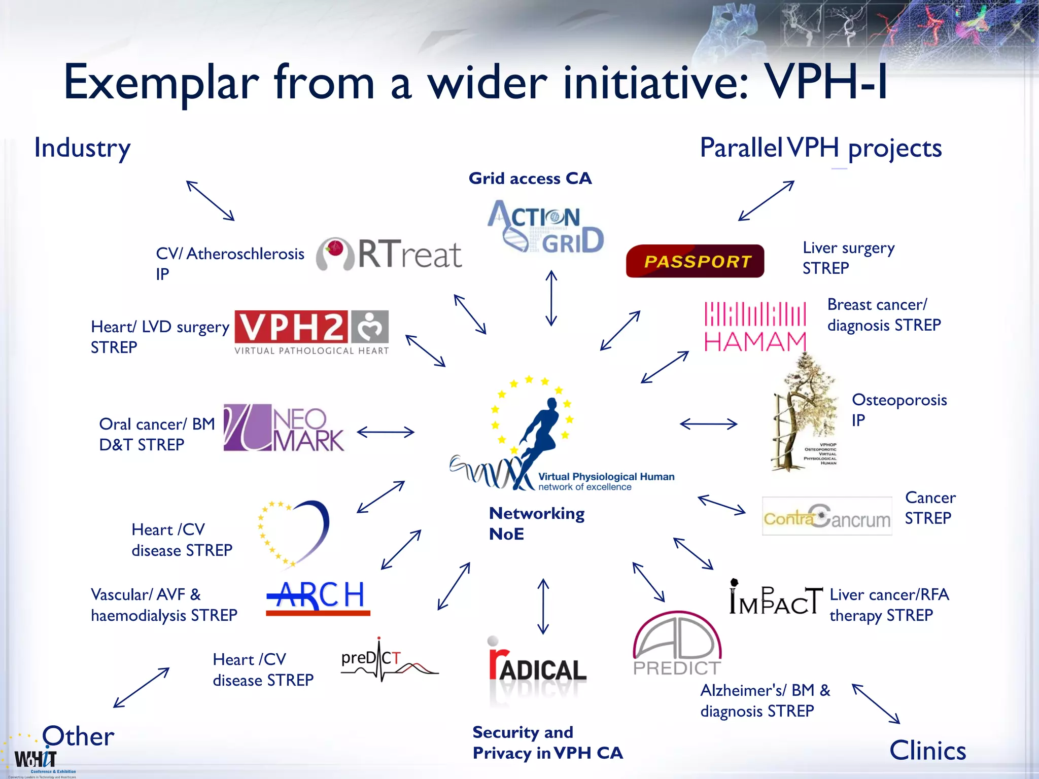
![Virtual Physiological Human (VPH)
or the Digital Me
A European Network of Excellence operated by 12 core EU institutions
“help support and progress
13 Core Partners
European research in
4 UK (UCL, UOXF, UNOTT, USFD)
biomedical modeling and 3 France (CNRS, INRIA, ERCIM)
simulation of the human 2 Spain (UPF, IMIM)
body.This will improve our Two important modeling issues 1 Germany1(EMBL [EBI]) Sweden (KI)
ability to predict, 1 Belgium (ULB)
diagnose and treat 1 New Zealand (UOA)
disease, and have a Model parameter personalization
dramatic impact on the
future of healthcare, the Populational inference of variability
pharmaceutical and
medical device Associate / General Members
industries.” 19 Candidate General Members
3 Candidate Associate Members
(organisations)
5 Candidate Associate Members (industry)
9 Associate Projects
www.vph-noe.eu … and growing
3](https://image.slidesharecdn.com/ps22-2-frangips22000-100323042045-phpapp01/75/ICT-for-a-global-infrastructure-for-health-research-VPH-Models-images-and-personalization-3-2048.jpg)
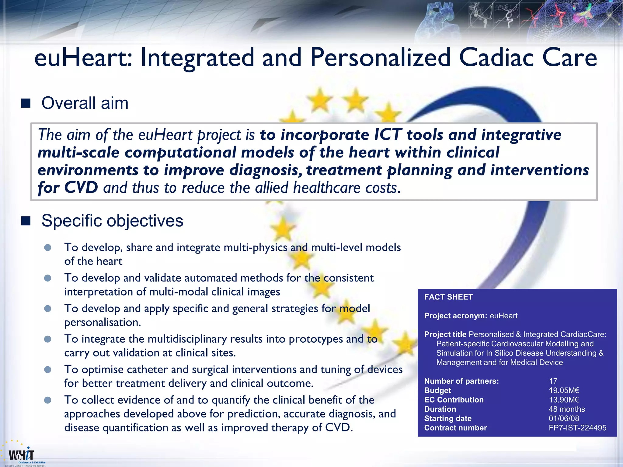
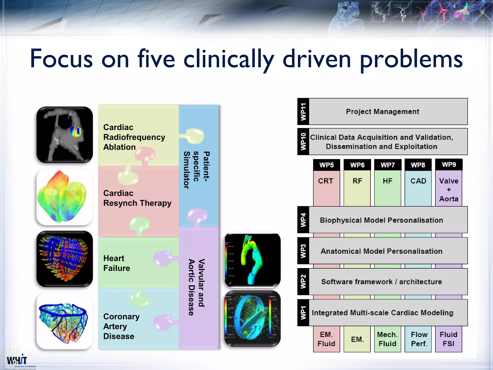

The document discusses the Virtual Physiological Human (VPH) project, aimed at improving diagnosis and treatment of cardiovascular diseases through a network of European institutions. It highlights the integration of ICT tools and multi-scale computational models within clinical environments as key objectives to optimize healthcare delivery. The project aims to utilize advanced biomedical imaging and personalized models for better patient outcomes in various cardiac conditions.


