This study investigated whether mild hypothermia (33°C core temperature) provides protection against neuronal apoptosis induced by anesthesia in neonatal mice. The researchers found that hypothermia to 33°C reduced anesthesia-induced neuroapoptosis in the cortex by approximately 30% compared to normothermia (37°C), and deeper hypothermia to 30°C provided further reduction in the cortex but not in the caudate-putamen region of the brain. However, the protection from hypothermia was only modest, and a safer intervention to reduce neuroapoptosis is still needed.
1. Hypothermia Provides Modest Protection against Anesthesia Induced Neuronal Apoptosis in Neonatal Mice
Leanne Cornel, Hikmatullah Arif, David Jardine
Department of Anesthesiology and Pain Medicine, Seattle Children’s Hospital, University of Washington, Seattle, WA
Background
Exposure to anesthetics increases apoptosis during rapid
brain growth in neonatal animals. Epidemiologic
investigations of human infants exposed to anesthesia
suggest that exposure may be associated with subtle
developmental deficits.
A recent report indicated that maintaining brain
temperature at 31.9°C may provide complete protection
against anesthesia induced neuroapoptosis in mouse
pups (1). Because deep hypothermia is difficult to safely
maintain in human patients, we elected to investigate
whether cooling to 33°C could provide equivalent
protection against neuroapoptosis.
IACUC approval was obtained. Mouse pups (P6) were
exposed to either 0.75% isoflurane in room air or room air
(no isoflurane) for 240 minutes in a temperature
controlled chamber. Rectal temperature was continuously
monitored and the chamber was adjusted to achieve the
target rectal temperature (30°C, 33°C, or 37°C). The mice
were sacrificed by intraperitoneal pentobarbital injection
and underwent trans-cardiac perfusion with PBS followed
by 4% paraformaldehyde for 7 minutes each. After 48
hours of fixation in 4% paraformaldehyde, the right
hemisphere of the brain was sectioned sagittally (50 µm
slices). Every 5th slice was collected for histology.
Staining was performed with an antibody to cleaved
caspase-3 (Asp175). Secondary staining was
accomplished with a fluorescent goat anti-rabbit IgG.
Stereological counting procedures (optical fractionator)
were used to count apoptotic cells in the cortex and
caudate-putamen regions. One way ANOVA analysis was
performed with the SPSS software.
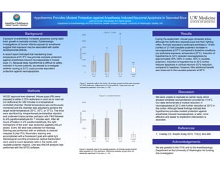
During the experiment, mouse pups remained active;
although the isoflurane exposed pups lost their righting
reflex. Animals exposed to isoflurane exhibited a 10 fold
(cortex) or 21 fold (caudate-putamen) increase in
neuroapoptosis at 37°C compared to baseline conditions
(no isoflurane exposure, temperature 37°C). Induction of
hypothermia to 33°C reduced neuroapoptosis by
approximately 30% (29% in cortex, 32% in caudate-
putamen). Induction of hypothermia to 30°C further
reduced apoptosis in cortical neurons (57% reduction
compared to baseline); however, little additional benefit
was observed in the caudate-putamen at 30°C.
We are grateful to the ITHS and to the Anesthesiology
Department at the University of Washington for funding
this investigation.
Methods
Results
Discussion
Acknowledgements
We were unable to replicate an earlier study which
showed complete neuroprotection provided by 31.9°C.
Our data demonstrate a modest reduction in
neuroapoptosis at 33°C with further reduction at 30°C in
the cortex. Although these findings indicate that
hypothermia provides modest protection against
anesthesia induced neuroapoptosis, a safer, more
effective and easier to implement intervention is
desirable.
UW Medicine
SCHOOL OF MEDICINE
Figure 1: Apoptotic cells in the cortex. All animals except controls were exposed
to 0.75% isoflurane. Differences of P<0.05 (ANOVA, Tukey post hoc) are
indicated by asterisks. Error bars: ± 1 SD.
1. Creeley CE. Anesth Analg 2010; 110(2): 442-448.
References
Figure 2: Apoptotic cells in the caudate-putamen. All animals except controls
were exposed to 0.75% isoflurane. Differences between groups were not
statistically significant. Error bars: ± 1 SD.
0
1000
2000
3000
4000
5000
6000
30° C
n=4
33° C
n=5
37° C
n=4
Control
n=4
ApoptoticCells
Figure 1: Cortex
*
*
**
**
***
***
0
1000
2000
3000
4000
30° C
n=4
33° C
n=5
37° C
n=4
Control
n=4
ApoptoticCells
Figure 2: Caudate-Putamen