1. Hypothermia Reduces Brain Injury in Rodent Newborn Hypoxic-Ischemic Injury:
Potential for Combinatorial Application of Human Neural Stem Cells
1
Department of Biological Science, California State
University, Fullerton, Fullerton, CA
2
Department of Pediatrics, Loma Linda University,
Loma Linda, CA
3
Stem Cell Center, University of California Riverside,
Riverside, CA
4
Claremont McKenna College, Claremont, CA
5
Program in Stem Cell & Regenerative Biology,
Sanford-Burnham Medical Research Institute, La
Jolla CA.
1,2,3
Brian McFadden
2
Beatriz Tone
2
Xiangpeng Yuan
2,4
Arielle Dennis
2
Nirmalya Ghosh
2,3
André Obenaus
5
Evan Snyder
2
Stephen Ashwal
I. INTRODUCTION
II. METHODS
III. RESULTS
Ÿ
Ÿ
Ÿ
Ÿ
Ÿ
HT treatment animals displayed less overall weight loss (Figure 4) when
compared to NT treated rat pups at 24hr after HT treatment; consistent with
improved health (greater body weight retention) than those treated with NT.
HT treated animal performed better on neurological assessments, such as
righting reflex, when compared to NTanimals (Figure 5).
Rat Pup Severity Score (RPSS), a measure of HII lesion severity, was
decreased 55.3% for animals treated with HT while NT animals treatment
showed a 35.3% increase (Figure 6).
HT treated animals have smaller lesions on neuroimaging than NT animals
(Figure 7).
HT treated animals have an overall reduction in lesion volume designated
as core compared to NT rat pups (Figure 8). This suggests which HT therapy
improves amount of unsalvagable tissues following neonatal HII.
KEYRESULTS
IV. CONCLUSIONS
Previous studies have independently shown
the beneficial effects of either hypothermia or
stem cells; but what is lacking are studies
evaluating the combination of these two
therapies. Thus, our findings of a
neuroprotective effect by HT following
neonatal HII provides the basis for combining
HT with hNSCs to recover viable tissues after
ischemic injury.
ACKNOWLEDGEMENTS
We thank Kamalakar Ambadipudi for
assistance with the MRI imaging and Ms.
Alena Yusof for data processing
Funding for these studies was provided by
NIH NINDS (1RO1NS059770-01A2) to Dr.
Stephen Ashwal. Mr. Brian McFadden was a
recipient of a California Institute of
Regenerative Medicine- Bridges to Stem
Cell Research (BSCR) funded scholarship.
Neonatal hypoxic ischemic injury (HII) affects 2-4/1000 live births. Cerebral
ischemic injuries, such as HII, occur as a result of the brain not receiving adequate
blood supply to function. Cell death then begins to occur leading to the
development of core (non-salvageable) and penumbral (salvageable) tissues.
Clinically, the sequelae of HII lead to devastating neurological outcomes. At
present time, hypothermia (HT) therapy has shown promise in minimizing injury in
children with HII. Indeed, HT in rat models of HII have shown to have a profound
neuroprotective effects. HT rescues vulnerable tissues after HII and reduces the
overall extent of tissue damage. But many of the molecular mechanisms involved
in the neuroprotective capabilities of HTare not fully examined.
Though HT is the clinical standard for short-term treatment of HII, there are no
known long term reparative treatments for HII. Implanting neuronal stem cells
(NSCs) is recently emerging as one such promising long-term therapeutic option
for neuroprotection after HII. Motivation of this work is that initial HT at the acute
HII phase has neuroprotective potential to create better viable tissue ambiance
for long-term NSC reparative activities. Hence, the purpose of this study is to
assess the therapeutic effectiveness of HT using diffusion weighted imaging
(DWI) to non-invasively monitor the progression and evolution of HII by lesion
volume combined with delineation of its core/penumbra tissues. Future studies
will use brain tissues to investigate levels of cytokine molecules, including
CXCL12/SDF-1á. These results will allow us to determine the interplay between
cell-cell signaling at the injury site and implanted stem cells.
In our present study we evaluated the effectiveness of HT treatment in
comparison to normothermia (NT) for HII in the neonatal brains. For this we have
used temporal trends of the following outcome measures: (1) weight-loss of the
pups, (2) righting reflex (a neurological score), (3) rat pup severity score (RPSS)
determined visually from magnetic resonance imaging (MRI), and (4) lesion
volume, including its core/penumbra proportions, automatically estimated by a
novel computational method, hierarchical region splitting (HRS) from MRI. These
data provide important background for future research investigating the long-term
reparative effects of HTin neonatal HII.
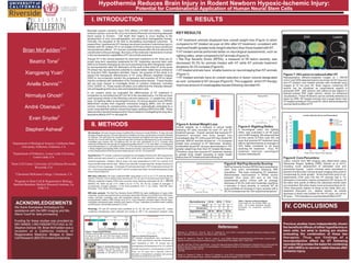
Figure 7: HII Lesion is reduced after HT
Representative diffusion-weighted images (b = 485.69
2
s/mm ) and HRS detected lesions are shown for NT (top row)
and HT (bottom row) treated animals with moderate injury
imaged at 72 hrs post HII. Brain regions containing HII
lesions can be visualized as hyperintense regions in
grayscale DWI (left column) and yellow-to-red regions in
pseudo-colored DWI (right column; shown for enhanced
visualization). These HII lesions can be correctly detected as
lesion by HRS. NT treated animals had a larger lesion than
HT treated animals at 72hrs post HII, which demonstrates the
neuroprotective effect of HT.
Figure 8: Core Penumbra:
Lesion volume from MR imaging was determined using
Hierarchal Region Splitting (HRS) (Ghosh et al 2011),
including novel assessment of estimated core and
penumbral tissues. Lesion volumes were normalized to
percent of entire brain volume at each imaging time-point to
compensate for brain growth. At the final time point of our
experiments (72hr post HII) the HT animals had a 1%
decrease in core lesion volume compared to NT rat pups.At
the other time points (0-48hrs) the (absolute) trends were
not consistent. But when lesion (core and penumbra) at 24-
72hrs time-points relative to those at the initial (0hr) pre-
treatment time-point (i.e., relative trends), an overall
decrease in lesion was evident for the HT pups, but not for
NTpups. This indicates an overt beneficial effect of HT.
Lesion Volume (Core and Penumbra)
Time Point: Hours Post Hypoxia
0 24 48 72
LesionVol.(%ofBrainVol.)
0
2
4
6
8
10
12
14
16
18
20
HT Core
HT Penumbra
NT Core
NT Penumbra
Rat Pup Severity Score
Time Post HII (hrs)
0 20 40 60 80
SeverityScore
0.0
0.5
1.0
1.5
2.0
2.5
3.0
3.5
HT Mild
HT Moderate
NT Mild
NT Moderate
Figure 6: Rat Pup Severity Scoring
Rat Pup Severity Scores (RPSS) were assessed at
each time point immediately following MRI
acquisition. Rat pups undergoing HT treatment
demonstrated improvement in RPSS scores
between 0hrs (post HII) and at the final
experimental period (72hrs) with an average
decrease of 55.3% (mild and moderate) signifying
a reduction in injury severity. In contrast, NT rat
pups exhibited an increase in injury severity with a
35.3% increase over the 72hr experimental period.
Figure 5: Righting Reflex
A neurological index, the righting
reflex, was evaluated in all HII pups
prior to MR imaging. We observed that
all time points (0-72hrs post HII) rat
pups that received HT treatment were
able to right themselves an average of
33% faster compared to rat pups
receiving NT treatment. Thus, HT
treatment improves neurological
function following HII.
Righting Reflex
Time Post HII (hrs)
0 20 40 60 80
Time(sec)
0.0
0.5
1.0
1.5
2.0
2.5
3.0
HT
NT
Figure 4:Animal Weight Loss
Animal weights, as a measure of animal health
following HII were recorded for both HT and NT
treatment groups. Overall, animals that received HT
treatment revealed less body weight loss
immediately after HT (24hrs post HII) than similar NT
animals. Mild NT animals showed an ~1% increased
weight loss compared to HT littermates. Similarly,
moderately injured NT rat pups demonstrated a ~5%
greater weight loss than their HT treated littermates.
Therefore, pups that received HT treatment retained
a greater body weight (equating to better health
status) than NTtreated animals following HII.
Weight Loss
WeightLoss(%ofpre-HIIweight)
-14
-12
-10
-8
-6
-4
-2
0
Mild Moderate
HT HTNT NT
0h 24h 48h 72h
MRI MRI MRI MRI
Histology
12h
Behavior
HII HT
Figure 1: Experimental Design
Animals received MRI scans immediately following HII
as well as at 24, 48, and 72hr time points. Animals
were also sacrificed and tissue was collected at the
same time points.
Figure 2: Animal Recovery Chamber
Animals were placed inside recovery
chamber where temperature was
controlled to 35°C(NT) or 30°C (HT)
Animal Temperature
Time Post HII (hrs)
0 5 10 15 20 25
0
TemperatureC
30
32
34
36
HT
NT
Figure 3:Animal Temperature
NT chamber temperatures were maintained at
35°C, while hypothermia chamber temperatures
were maintained at 30°C. NT animals had an
average basal core temperature of 34.7°C, while HT
animals experienced a core temperature of 32.8°C.
Thus, our HT pups had a requisite 2.0±0.1°C
decrease in temperature consistent with previous
hypothermia studies (Lee et al. 2010).
HII induction: HII was induced using a modified Rice-Vannucci model (RVM) in 10-day-old male
Sprague-Dawley rat pups. HII was induced by unilateral common carotid artery occlusion followed
by hypoxia. The right common carotid artery was exposed and ligated and pups were allowed to
recover for 2 hours with the dam. Hypoxia was then induced by placing pups in a jar containing a
humidified gas mixture (8% O2-balance N2) for 2hr and maintained at 37°C. Animals were
randomly divided into two groups for applying hypothermia (HT; n=12; see Table 1) or keeping at
normal temperature i.e. normothermia (NT; n=17) for the rest of experiment summarized in Figure
1. Body-weights and righting reflex (when placed on its back, how quick a pup returns to its usual
on-the-chest position) of the pups were measured at 0, 24, 48, 72 hrs post HII.
Hypothermia: HT animals underwent hypothermia immediately following 0hr MRI assessment.
Briefly animals were placed in a cooled (30°C) small animal hypothermic chamber (Figure 2)
(Harvard Apparatus, Holliston, MA) to lower core body temperature to 33°C for a period of 24
hours and then kept with the dam. While in the chamber, rat pups were fed Similac formula (0.4
ml) every 4 hours via gavage. Conversely, NT animals were kept in 35°C temperature during 0-24
hrs. Core body temperatures for all HT and NT animals were taken via rectal temperature probe
every 4 hours (Figure 3).
MRI data collection: HII pups underwent MRI using either a 4.7T or an 11.7T scanner (Bruker
Avance, Fremont, CA) to assess injury severity at 0, 24, 48, and 72 hrs post injury (see Figure 1).
Diffusion weighted imaging (DWI) were acquired using following parameters for
TR: 3000 ms (for 4.7T; 1096.5 ms for 11.7T); TE: 50 ms; number of averages: 2
(occasionally changed between 1-4 for time-constraint); FoV: 2 cm; matrix: 128x128; slice-
thickness: 1 mm; slices: 20 (contiguous).
MRI data analysis: The Rat Pup Severity Score (RPSS) for rapid stratification of injury (mild,
moderate, severe) was performed as previously described (Recker et al 2008) from DWI scans.
Lesion volumes, including core and penumbra tissues, were determined using our novel
computational method, HRS (Ghosh et al 2011). Injury detection included region of both hyper-
intensities (decreased water mobility) and regions of hypo- intensities (increased water mobility)
as DWI lesion contrast flips between 0-72 hrs.
Histology: HT and NT animals were sacrificed at 12, 24, 48, and 72 hrs post HII. Lesion
containing hemispheres were collected and stored at -80° C for subsequent analysis using
(b = 485.69
2
s/mm )
References
Obenaus, A. , Dilmac, N. , Tone, B. , Tian, H. , Hartman, R. , et al. (2011). Long-term magnetic resonance imaging of stem
cells in neonatal ischemic injury. Ann Neurol, 69(2), 282-291.
Recker, R. , Adami, A. , Tone, B. , Tian, H. , Lalas, S. , et al. (2009). Rodent neonatal bilateral carotid artery occlusion with
hypoxia mimics human hypoxic-ischemic injury. J Cereb Blood Flow Metab, 29(7), 1305-1316
Lee, BS, CW Woo, ST Kim, and KS Kim. “Long-Term Neuroprotective Effect of Postischemic Hypothermia in a Neonatal Rat
Model of Severe Hypoxic Ischemic Encephalopathy: A Comparative Study on the Duration and Depth of Hypothermia.”
Pediatric Research, 68.4 (2010):
303-308
Ghosh, N. , Recker, R., Shah, A. , Bhanu, B. , Ashwal, S. , et al. (2011). Automated ischemic lesion detection in a neonatal
model of hypoxic ischemic injury. Journal of Magnetic Resonance Imaging. 33 (4), 772-781
Normothermia 24 hr 48 hr 72 hr
Mild 3 3 2
Moderate 1 1 2
Hypothermia 24 hr 48 hr 72 hr
Mild 4 2 4
Moderate 2 3 2
Table 1: Number of Study Subjects
Count totals for all animals within our
study. All N totals represent animals
harvested for molecular cytokine
analysis.
DWI Lesion
NT
HT