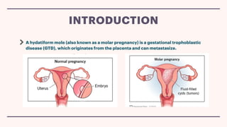
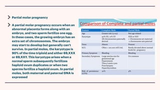
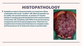
OBSTETRICS 403
Hina nazar
HYDATIDIFORM MOLE
Molar pregnancy
A hydatiform mole (also known as a molar pregnancy) is a gestational trophoblastic
disease (GTD), which originates from the placenta and can metastasize.
INTRODUCTION
What is gestational trophoblastic disease
It is a disease characterized by an abnormal
placental development that results in the
production of
f
luid-
f
illed grapelike clusters
(instead of normal placental tissue) and a
vast proliferation of trophoblastic tissue. It
is associated with loss of the pregnancy and
rarely, the development of cancer.
Complete molar pregnancy
In complete molar pregnancies, no embryo forms. It happens when a sperm fertilizes an
empty egg. Because the egg is empty, the embryo can’t grow. The placental tissue grows
but is abnormal and contains
f
luid-
f
illed cysts (or tumors). This tissue produces the
pregnancy hormone HCG (human chorionic gonadotropin), which is made by a
healthy placenta during pregnancy. This can make you feel like you’re pregnant and give
you a positive pregnancy test.In complete moles, the karyotype is 46,XX 90% of the time
and 46,XY 10% of the time. It arises when an enucleated egg is fertilized either by two
sperms or by a haploid sperm that then duplicates and therefore, only paternal DNA is
expressed
WHAT ARE THE TYPES OF MOLAR PREGNANCIES?
Partial molar pregnancy
A partial molar pregnancy occurs when an
abnormal placenta forms along with an
embryo, and two sperm fertilize one egg.
In these cases, the growing embryo has an
extra set of chromosomes. The embryo
may start to develop but generally can’t
survive. In partial moles, the karyotype is
90% of the time triploid and either 69,XXX
or 69,XXY. This karyotype arises when a
normal sperm subsequently fertilizes
haploid ovum duplicates or when two
sperms fertilize a haploid ovum. In partial
moles, both maternal and paternal DNA is
expressed
Hydatiform mole is characterized by an overgrown villous
trophoblast with cystic "swollen" villi. Macroscopically can
be visible, the second trimester, as clusters of vesicles
(similar to small grapes) developed from the transformation
of chorionic villi. Complete mole differ from partial mole by
cytogenetic and micrscopical appearance. Important is a
complete lack of embryonic/fetal tissue in complete moles
and the presence of-of embryonic tissue in partial moles
HISTOPATHOLOGY
Abnormal growth of the uterus, either bigger or smaller than usual.
Severe nausea and vomiting
Vaginal bleeding during the
f
irst 3 months of pregnancy
Symptoms of hyperthyroidism, including heat intolerance, loose stools, rapid heart
rate, restlessness or nervousness, warm and moist skin, trembling hands, or unexplained weight loss
Symptoms similar to preeclampsia that occur in the
f
irst trimester or early second trimester,
including high blood pressure and swelling in the feet, ankles, and legs (this is almost always a sign of
a hydatidiform mole, because preeclampsia is extremely rare this early in a normal pregnancy).
SYMPTOMS
An ultrasound of your uterus often shows several
f
luid-
f
illed sacs instead of a placenta.
There’s usually no embryo or fetus in your uterus, but sometimes pregnancy tissue
remains.
Ultrasound of complete mole shows :
no embryo or foetus, No amniotic
f
luid,A thick cystic placenta nearly
f
illing the
uterus,Ovarian cysts.
Ultrasound of partial molar pregnancy shows :
A foetus that’s smaller than expected,Low amniotic
f
luid,Placenta that appears unusual.
DIAGNOSIS
Signs of anaemia – including fatigue, breathlessness, dizziness and a fast heartbeat
HCG is produced at abnormally high levels. A blood test can detect these high levels
and suggest a molar pregnancy has occurred.
DIAGNOSIS
Dilation and curettage (D&C)
This procedure removes the molar tissue from the uterus. You lie on a table on your
back with your legs in stirrups. You receive medicine to numb you or put you to sleep.
After opening the cervix, the provider removes uterine tissue with a suction device.
A D&C for a molar pregnancy usually is done in a hospital or surgery center.
TREATMENT
Removal of the uterus : This occurs rarely if there's increased risk of gestational trophoblastic
neoplasia (GTN) and there's no desire for future pregnancies
HCG monitoring : After the molar tissue is removed, a provider keeps measuring the HCG level
until it goes down. A continuing high level of HCG in the blood might require more treatment.
TREATMENT
Occasionally, parts of the molar pregnancy remain in your uterus after surgical treatment or
miscarriage.
If this happens, abnormal cells may grow into the muscle layer around your uterus (also
called an invasive mole). This is rare and happens in less than 15% of people who’ve had a
molar pregnancy removed.
In very rare cases, molar pregnancy causes a type of cancer called choriocarcinoma.
Choriocarcinoma forms within your uterus and may spread to other parts of your body and
requires cancer treatments such as chemotherapy or radiation.
COMPLICATIONS
Other potential complications of a molar pregnancy include:
Infection of the blood (sepsis).
Uterine infection.
Preeclampsia (very high blood pressure).
Shock (very low blood pressure)
COMPLICATIONS
https://my.clevelandclinic.org/health/diseases/17889-molar-pregnancy
https://www.ncbi.nlm.nih.gov/books/NBK459155/#:~:text=Hydatiform mole (also
known as molar pregnancy) is a subcategory,rather than from maternal tissue
https://www.nhs.uk/conditions/molar-pregnancy/#:~:text=There are 2 types
of,cannot fully develop or survive
REFERENCES
THANK YOU

Hydatidiform mole gestational disease.pdf

  • 1.
  • 2.
    A hydatiform mole(also known as a molar pregnancy) is a gestational trophoblastic disease (GTD), which originates from the placenta and can metastasize. INTRODUCTION
  • 3.
    What is gestationaltrophoblastic disease It is a disease characterized by an abnormal placental development that results in the production of f luid- f illed grapelike clusters (instead of normal placental tissue) and a vast proliferation of trophoblastic tissue. It is associated with loss of the pregnancy and rarely, the development of cancer.
  • 4.
    Complete molar pregnancy Incomplete molar pregnancies, no embryo forms. It happens when a sperm fertilizes an empty egg. Because the egg is empty, the embryo can’t grow. The placental tissue grows but is abnormal and contains f luid- f illed cysts (or tumors). This tissue produces the pregnancy hormone HCG (human chorionic gonadotropin), which is made by a healthy placenta during pregnancy. This can make you feel like you’re pregnant and give you a positive pregnancy test.In complete moles, the karyotype is 46,XX 90% of the time and 46,XY 10% of the time. It arises when an enucleated egg is fertilized either by two sperms or by a haploid sperm that then duplicates and therefore, only paternal DNA is expressed WHAT ARE THE TYPES OF MOLAR PREGNANCIES?
  • 5.
    Partial molar pregnancy Apartial molar pregnancy occurs when an abnormal placenta forms along with an embryo, and two sperm fertilize one egg. In these cases, the growing embryo has an extra set of chromosomes. The embryo may start to develop but generally can’t survive. In partial moles, the karyotype is 90% of the time triploid and either 69,XXX or 69,XXY. This karyotype arises when a normal sperm subsequently fertilizes haploid ovum duplicates or when two sperms fertilize a haploid ovum. In partial moles, both maternal and paternal DNA is expressed
  • 6.
    Hydatiform mole ischaracterized by an overgrown villous trophoblast with cystic "swollen" villi. Macroscopically can be visible, the second trimester, as clusters of vesicles (similar to small grapes) developed from the transformation of chorionic villi. Complete mole differ from partial mole by cytogenetic and micrscopical appearance. Important is a complete lack of embryonic/fetal tissue in complete moles and the presence of-of embryonic tissue in partial moles HISTOPATHOLOGY
  • 7.
    Abnormal growth ofthe uterus, either bigger or smaller than usual. Severe nausea and vomiting Vaginal bleeding during the f irst 3 months of pregnancy Symptoms of hyperthyroidism, including heat intolerance, loose stools, rapid heart rate, restlessness or nervousness, warm and moist skin, trembling hands, or unexplained weight loss Symptoms similar to preeclampsia that occur in the f irst trimester or early second trimester, including high blood pressure and swelling in the feet, ankles, and legs (this is almost always a sign of a hydatidiform mole, because preeclampsia is extremely rare this early in a normal pregnancy). SYMPTOMS
  • 8.
    An ultrasound ofyour uterus often shows several f luid- f illed sacs instead of a placenta. There’s usually no embryo or fetus in your uterus, but sometimes pregnancy tissue remains. Ultrasound of complete mole shows : no embryo or foetus, No amniotic f luid,A thick cystic placenta nearly f illing the uterus,Ovarian cysts. Ultrasound of partial molar pregnancy shows : A foetus that’s smaller than expected,Low amniotic f luid,Placenta that appears unusual. DIAGNOSIS
  • 10.
    Signs of anaemia– including fatigue, breathlessness, dizziness and a fast heartbeat HCG is produced at abnormally high levels. A blood test can detect these high levels and suggest a molar pregnancy has occurred. DIAGNOSIS
  • 11.
    Dilation and curettage(D&C) This procedure removes the molar tissue from the uterus. You lie on a table on your back with your legs in stirrups. You receive medicine to numb you or put you to sleep. After opening the cervix, the provider removes uterine tissue with a suction device. A D&C for a molar pregnancy usually is done in a hospital or surgery center. TREATMENT
  • 12.
    Removal of theuterus : This occurs rarely if there's increased risk of gestational trophoblastic neoplasia (GTN) and there's no desire for future pregnancies HCG monitoring : After the molar tissue is removed, a provider keeps measuring the HCG level until it goes down. A continuing high level of HCG in the blood might require more treatment. TREATMENT
  • 13.
    Occasionally, parts ofthe molar pregnancy remain in your uterus after surgical treatment or miscarriage. If this happens, abnormal cells may grow into the muscle layer around your uterus (also called an invasive mole). This is rare and happens in less than 15% of people who’ve had a molar pregnancy removed. In very rare cases, molar pregnancy causes a type of cancer called choriocarcinoma. Choriocarcinoma forms within your uterus and may spread to other parts of your body and requires cancer treatments such as chemotherapy or radiation. COMPLICATIONS
  • 14.
    Other potential complicationsof a molar pregnancy include: Infection of the blood (sepsis). Uterine infection. Preeclampsia (very high blood pressure). Shock (very low blood pressure) COMPLICATIONS
  • 15.
    https://my.clevelandclinic.org/health/diseases/17889-molar-pregnancy https://www.ncbi.nlm.nih.gov/books/NBK459155/#:~:text=Hydatiform mole (also knownas molar pregnancy) is a subcategory,rather than from maternal tissue https://www.nhs.uk/conditions/molar-pregnancy/#:~:text=There are 2 types of,cannot fully develop or survive REFERENCES
  • 16.